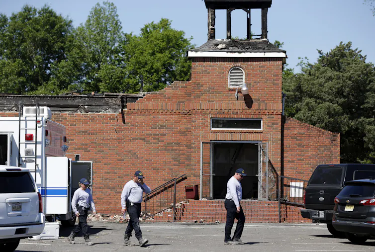
(RNS) — On Aug. 14, a man barged into New York City’s Central Synagogue during Shabbat services, striking a congregant, damaging religious property and headbutting a guard — leaving many Jewish Americans feeling vulnerable once again. The next day, an Islamic center under construction on Long Island was defaced with graffiti. “F— Islam” was written over a banner, which previously read, “NEW MOSQUE COMING SOON! Where Faith and Community Come Together!”
We — a rabbi and a Muslim educator — learned of the synagogue attack while attending a five-day interfaith retreat. We had spent days praying, studying our sacred texts and speaking honestly about grief, fear, hope and responsibility — not as representatives of competing communities but as faith leaders committed to the dignity of every human being. When we heard about the anti-Muslim vandalism after the retreat, it underscored the urgency of those conversations. The hate from these incidents was a brutal reminder of how much work we need to do.
Only months earlier, on May 18, worshippers at the Islamic Center of San Diego and its day school were targeted in a shooting that left three dead and shook Muslim communities across the country. A few weeks before that, on March 12, a vehicle-ramming and shooting attack occurred at Temple Israel, a Reform synagogue in West Bloomfield Township, Michigan.
Different houses of worship, different victims, the same message of hate and intimidation.
For too many American Jews and Muslims today, ordinary acts of faith have become calculations of risk. Is it safe for our children to visibly identify with their people? Should I wear my kippah walking to shul? Should I wear my hijab on the subway? Is it safe to send my child to Hebrew school or my daughter to Friday prayers? Will my synagogue need armed security? Will our mosque receive another bomb threat? These aren’t hypothetical questions — they’re daily realities.
America has seen one of the sharpest increases in both antisemitism and Islamophobia in modern history. Jewish institutions have faced violence, harassment, vandalism and threats. Muslim communities have endured deadly attacks, mosque vandalism and unprecedented levels of discrimination. National organizations that track hate have documented record or near-record numbers of incidents targeting both. Fear has become a common language for both.

Law enforcement escort families with children away from the Temple Israel synagogue Thursday, March 12, 2026, in West Bloomfield Township, Mich. (AP Photo/Paul Sancya)
The trauma following Hamas’ horrific Oct. 7 attack, Israel’s military campaign in Gaza, political polarization and increasingly inflammatory public rhetoric have fed a climate where people confuse individuals with governments, faith with geopolitics, neighbors with enemies. That confusion is dangerous.
American Jews are not responsible for every action of the Israeli government. American Muslims are not responsible for Hamas or any other armed group.
Yet both communities are increasingly judged through those lenses, and both are asking the same questions: Is it safe to gather for worship? Is it safe for our children to visibly identify with their people and practice their faith? Is it safe simply to be who we are?
In both communities, the fear is real, as is the pain. Perhaps the greatest tragedy is that this fear is so often carried alone, when it should be confronted together. As faith leaders, we know our communities don’t agree on every political issue. We have different histories, different narratives and often different views on the Middle East. Yet we also know this: No disagreement justifies hatred, no political conflict justifies the intimidation of American Jews or American Muslims and no child should grow up believing that their synagogue or mosque needs armed protection simply because of who they are.
There’s another truth our country too often overlooks. The ideology that threatens Jewish communities and the one that threatens Muslim communities are same. For generations, white supremacist movements have targeted Jews with conspiracy theories about power and control while portraying Muslims as dangerous outsiders with a hidden agenda — people who don’t belong in America.
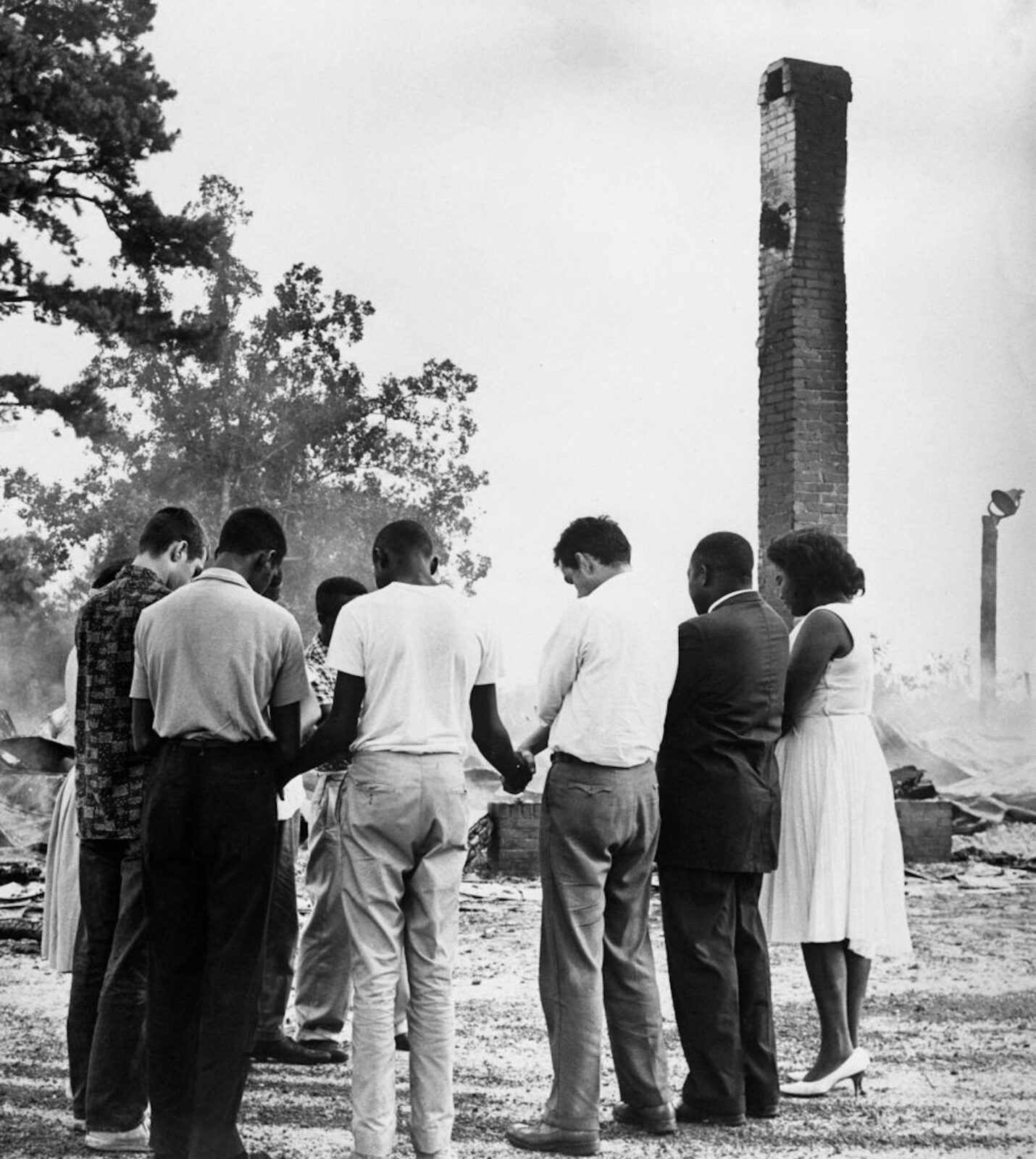
That same ideology has fueled attacks on Black churches, Sikh gurdwaras, immigrant communities and many others. It rejects the pluralistic democracy that both of our faiths depend on. White supremacy does not distinguish between the communities it seeks to intimidate. Our resistance shouldn’t distinguish between them, either.
Jewish and Muslim organizations have stood together after attacks on houses of worship. Rabbis have visited mosques after acts of anti-Muslim hatred; imams have stood beside Jewish congregations after antisemitic violence. Together, our communities have partnered on feeding the hungry, welcoming refugees, advocating for civil rights and strengthening our democracy.
Too much of our public conversation today rewards outrage over relationships. Social media amplifies the loudest voices. Political rhetoric often deepens suspicion instead of understanding. Every act of hate is met with more anger, feeding a cycle in which fear becomes normalized. It doesn’t have to be this way.
This moment calls for people of faith to have moral courage. Jewish leaders should continue speaking out unequivocally against Islamophobia — not because it’s politically convenient but because justice demands consistency. Muslim leaders should continue speaking out against antisemitism with equal clarity — not because every political disagreement has been resolved but because hatred toward Jews has no place in any movement for justice. And members of both communities must resist those who profit from keeping us divided, including news networks and nefarious interest groups.
We can mourn Palestinian lives and Israeli lives. We can condemn all forms of political violence — including violence committed by states, armed groups and extremists — and oppose the suffering of civilians. We can advocate for peace while rejecting hatred against our neighbors. Most importantly, we can refuse to let our grief become someone else’s weapon. Our interfaith retreat reminded us of our shared humanity. Because our faiths teach that every human being is created with inherent dignity.
Our call today is simple: Let us lower the temperature. Let us reject the politics of fear. Let us refuse the false choice that one community’s safety must come at another’s expense. And let us unite — Jews, Muslims, Christians and all people of conscience — to confront white supremacy and every ideology that seeks to divide Americans by race, religion or ethnicity.
The future of our communities won’t be secured by louder accusations. It will be secured by deeper relationships. The antidote to hate has never been silence — it has always been solidarity. If we defend one another with the same urgency we defend ourselves, we will not only make our synagogues and mosques safer. We will strengthen the promise of America itself.
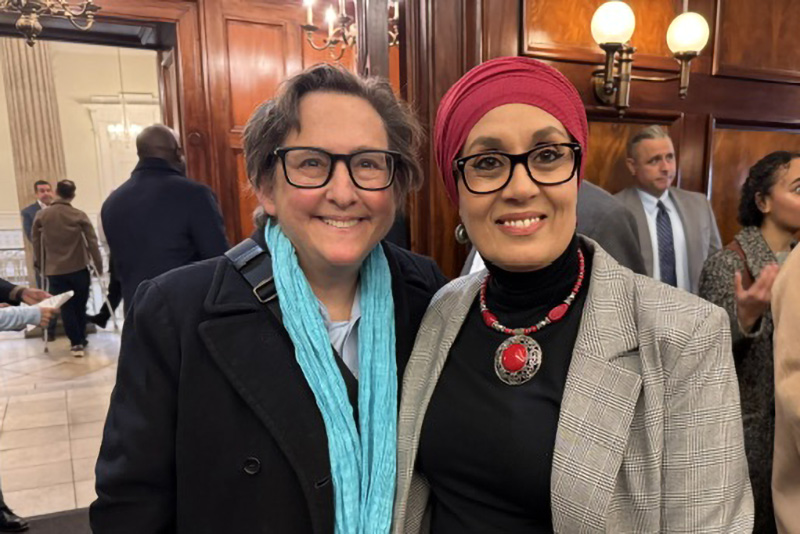
Rabbi Sharon Kleinbaum, left, and Debbie Almontaser. (Courtesy photo)
Debbie Almontaser is the founder and chief executive officer of Bridging Cultures Group and was the founding principal of Khalil Gibran International Academy in Brooklyn. Sharon Kleinbaum is senior rabbi emerita of Congregation Beit Simchat Torah in New York and co-founder and board member of New York Jewish Agenda.
Christian nationalists’ professed support for religious pluralism is often drowned out by perceptions that Christians are under threat
(The Conversation) — Christian nationalism, a worldview that sees the United States as a Christian nation and believes that Christianity should have a privileged role in American civic life, has been shown to be related to a host of social attitudes.
Research shows that people who hold Christian nationalistic beliefs tend to, on average, also show antipathy toward religious minorities and racial and ethnic minorities, as well as have sexist and anti-democratic attitudes.
Our research found that many Christian nationalists endorse religious pluralism, meaning that they value religious diversity – in principle. But we also found that this support for religious pluralism is often drowned out by perceptions of threats to Christians.
How we did the research
For our new research, published in the academic journal Politics & Religion in March 2026, we surveyed 1,500 American Christians in January 2024.
In healthy democracies, most citizens respect the civil liberties of others, including members of social groups they dislike. We asked our respondents which social group they liked the least, such as Christian fundamentalists, environmental activists, President Donald Trump supporters or atheists. We then asked them if they thought members of that group should be able to make public speeches, teach in public schools or run for political office, among others. The responses, taken together, showed respondents’ political tolerance for people who they dislike.
We also asked respondents how much they agreed with a series of statements commonly used to measure support for Christian nationalism, such as whether the federal government should declare the United States a Christian nation. On a scale of 0 to 1, the average Christian nationalism score was 0.64, indicating that American Christians tend to support the idea of a Christian America.
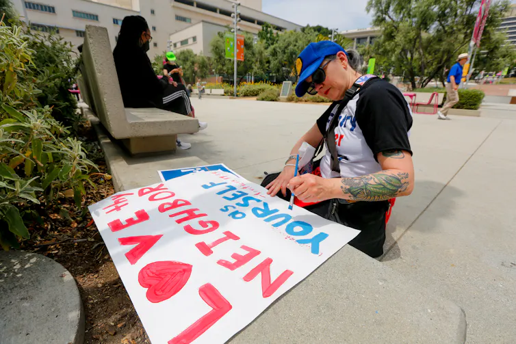
To capture ideas that run directly counter to Christian nationalism, we then asked about their support for religious pluralism. Respondents agreed or disagreed with statements such as “nations should not favor one religion over another,” and “we need to advocate the Christian values of loving your neighbor as yourself.” Together, these made up our anti-Christian nationalism measure.
On average, participants rated their agreement with these statements as 0.74, higher than the agreement with Christian nationalist ideas – suggesting that anti-Christian nationalist views were more widely supported by American Christians.
What the research found
Some of the survey questions measuring Christian nationalism and anti-Christian nationalism captured opposite ideas.
Without assigning respondents to any categories, we asked them if they agreed with statements like “The federal government should declare the United States a Christian nation” or “Nations should not favor one religion over another.” We found that people who agreed with one were likely to agree with the other. In other words, respondents who advocated for a Christian nation also affirmed religious freedom for all.
While this might seem surprising, social scientists have long known that people hold lots of conflicting arguments in their heads. American Christians have likely heard both Christian nationalist and anti-Christian nationalist messages through their congregations, media and social networks.
Our statistical models show that religious pluralism is related to tolerance differently depending on views on Christian nationalism. We isolated strong supporters and strong opponents of Christian nationalism. Predictably, among strong opponents of Christian nationalism, anti-Christian nationalism views were associated with increased tolerance. But, surprisingly, strong supporters of Christian nationalism maintained the same level of tolerance.
According to our data, people who agree with anti-Christian nationalist ideas, yet also embrace Christian nationalist ones, tend to have a fear of persecution. We asked respondents if their least-liked social group posed a threat to the American way of life and people’s freedom. Both groups had similar perceptions that the “American way of life” and everyday freedom were threatened.
This perception of threat was lower only among respondents who both strongly rejected Christian nationalism and embraced anti-Christian nationalism. In other words, support for religious pluralism, on its own, isn’t sufficient to reduce the sense of threat among those who hold Christian nationalist views.
Why it matters
Christian nationalism has emerged as a significant challenge to religious pluralism. Efforts to counter it can often be more credible if they came from other Christians. But for those efforts to work, they need to tackle Christian nationalism at its root.
Our findings indicate that the root of the problem is not a lack of support for religious pluralism or insufficient willingness to love thy neighbor. It is perceptions of threats posed by disliked groups.
We have observed that the Christian media ecosystem is saturated with messages that American Christians are excluded from public life – ostracized from social institutions like higher education, discriminated against in everyday life and only one election away from losing their Bibles or being jailed for their faith. This sense of threat can trigger authoritarian tendencies, including support for taking away civil liberties like the ability to give public speeches or run for political office from disliked others.
What still isn’t known
Political scientists need to know more about the contours of threat: By whom and what do some Christians feel threatened? Social scientists also do not fully understand how that sense of threat can be dialed down, not just in a lab experiment but also in Christians’ experiences.
Improving our understanding of threat could guide the work of faith leaders striving for a more tolerant, pro-democratic role of faith in the public sphere.
This research was funded by a Jack Shand Research Grant from the Society for the Scientific Study of Religion.
(Brooklyn Walker, Assistant Professor of Political Science, University of Tennessee. Paul Djupe, Director of the Data for Political Research Program, Denison University. The views expressed in this commentary do not necessarily reflect those of Religion News Service.)