Showing posts sorted by relevance for query CRISPR. Sort by date Show all posts

Tuesday, August 10, 2021

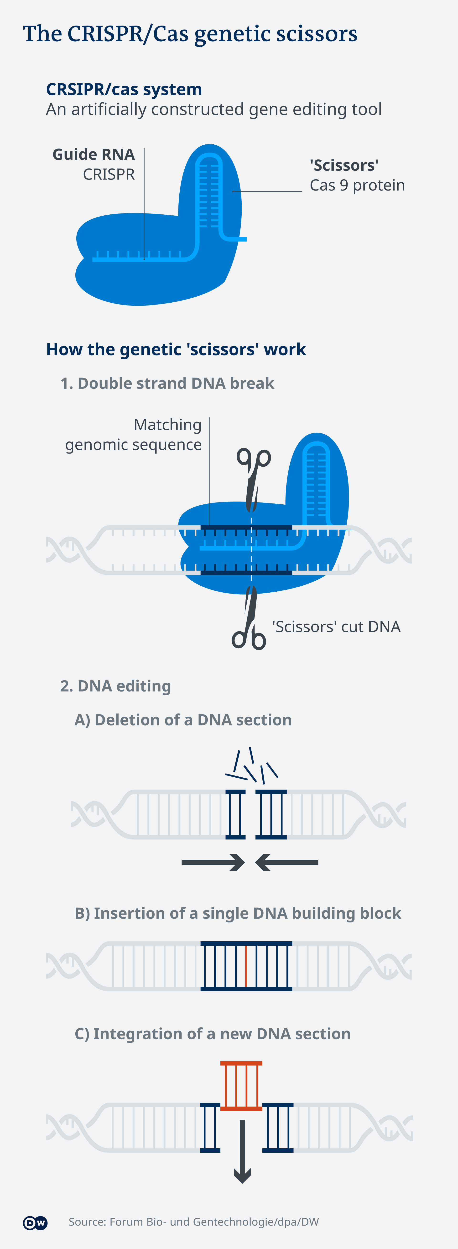
PETITE MIRICALE CRISPR CRITTER 

New CRISPR/Cas9 technique corrects cystic fibrosis in cultured human stem cells


Peer-Reviewed Publication

HUBRECHT INSTITUTE

Organoid swelling 

IMAGE: SWELLING RESPONSE OF PATIENT DERIVED MINI-GUTS. COLLAPSED ORGANOIDS (LEFT) SHOW ACTIVE SWELLING RESPONSE THAT IS MEDIATED BY THE CFTR ION CHANNEL AFTER ONE HOUR INCUBATION WITH FORSKOLIN (RIGHT). GREEN STAINING SHOWS COMPLETE CELLS (CALCEIN GREEN) AND DNA IS SHOWN IN BLUE. view more 

CREDIT: EYLEEN DE POEL, (C) UMC UTRECHT

Researchers from the group of Hans Clevers (Hubrecht Institute) corrected mutations that cause cystic fibrosis in cultured human stem cells. In collaboration with the UMC Utrecht and Oncode Institute, they used a technique called prime editing to replace the ‘faulty’ piece of DNA with a healthy piece. The study, published in Life Science Alliance on August 9th, shows that prime editing is safer than the conventional CRISPR/Cas9 technique. “We have for the first time demonstrated that this technique really works and can be safely applied in human stem cells to correct cystic fibrosis.”


Cystic fibrosis (CF) is one of the most prevalent genetic diseases worldwide and has grave consequences for the patient. The mucus in the lungs, throat and intestines is sticky and thick, which causes blockages in organs. Although treatments are available to dilute the mucus and prevent inflammations, CF is not yet curable. However, a new study from the group of Hans Clevers (Hubrecht Institute) in collaboration with the UMC Utrecht and Oncode Institute offers new hope.


Correcting CF mutations

The researchers succeeded in correcting the mutations that cause CF in human intestinal organoids. These organoids, also called mini-organs, are tiny 3D structures that mimic the intestinal function of patients with CF. They were previously developed by the same research group from stem cells of patients with CF and stored in a biobank in Utrecht. For the study, published in Life Science Alliance, a technique named prime editing was used to replace the piece of mutated DNA that causes CF with a healthy piece of DNA in these organoids.


Safer than CRISPR/Cas9

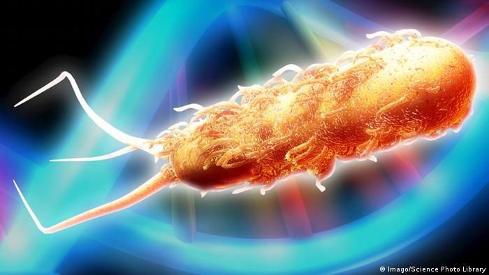
Prime editing is a newer version of the better-known gene editing technique CRISPR/Cas9. CRISPR/Cas9 cuts the DNA before correcting it. Although this corrects the mutated piece of DNA, it also causes damage in other regions in the genome. “In our study, prime editing proves to be a safer technique than the conventional CRISPR/Cas9. It can build in a new piece of DNA without causing damage elsewhere in the DNA. That makes the technique promising for application in patients,” says Maarten Geurts, first author on the publication.


Swelling

The mutations that cause CF are localized in the CFTR channel, which is present in the cells of various organs including the lungs. Due to the mutations, the channel does not function properly, leaving the layer of mucus that covers the cells with too little water: the mucus becomes sticky. The addition of a substance called forskolin causes healthy organoids to swell, but this does not happen in organoids with mutations in the CFTR channel. “We applied prime editing to the mutations, after which the treated organoids demonstrated the same response as the healthy organoids: they became swollen. That provided us with proof that our technique worked and replaced the mutated DNA,” Geurts explains.


Curing genetic diseases

Now that the researchers showed that the mutations that cause CF can be safely corrected, applications in the clinic come one step closer. “New variants of CRISPR/Cas9, such as prime editing, can safely correct mutations without causing damage in other regions of the DNA. This will hopefully enable us to cure or even prevent genetic diseases in the future.” But before that, some challenges still lie ahead for the researchers. The technique for example still needs to be adapted for safe use in humans. “But this is a great step towards successfully applying prime editing in the clinic,” Geurts concludes.

CAPTION

Cystic Fibrosis patient derived organoids do not show a swelling response. The swelling response is regained after prime-editing mediated repair of the CFTR channel.

CREDIT

Eyleen de Poel and Maarten Geurts, (c) UMC Utrecht and Hubrecht Institute

Publication

"Evaluating CRISPR-based Prime Editing for cancer modeling and CFTR repair in organoids". Maarten Geurts, Eyleen de Poel, Cayetano Pleguezuelos-Manzano, Rurika Oka, Léo Carrillo, Amanda Andersson-Rolf, Matteo Boretto, Jesse Brunsveld, Ruben van Boxtel, Jeffrey Beekman, and Hans Clevers, Life Science Alliance (2021).


Hans Clevers is group leader at the Hubrecht Institute for Developmental Biology and Stem Cell Research and at the Princess Máxima Center for Pediatric Oncology. He is also University Professor at the Utrecht University and Oncode Investigator.


About the Hubrecht Institute

The Hubrecht Institute is a research institute focused on developmental and stem cell biology. It encompasses 21 research groups that perform fundamental and multidisciplinary research, both in healthy systems and disease models. The Hubrecht Institute is a research institute of the Royal Netherlands Academy of Arts and Sciences (KNAW), situated on Utrecht Science Park. Since 2008, the institute is affiliated with the UMC Utrecht, advancing the translation of research to the clinic. The Hubrecht Institute has a partnership with the European Molecular Biology Laboratory (EMBL). For more information, visit http://www.hubrecht.eu.

Friday, November 12, 2021

CRISPR-CRITTERS
CRISPR-Cas9: A weapon against antibiotic-resistant superbugs?


Antibiotic-resistant bacteria are on the rise, causing hundreds of thousands of deaths each year. A technology to edit genes, called CRIPSR-Cas9, could help us eliminate these superbugs, a new study has found.


Genetically modified bacteria have shown antibacterial properties in a mouse model


Before the discovery of penicillin in 1928, even common infections such as strep throat could be a terminal diagnosis. Antibiotics gave us a great advantage in the battle against harmful bacteria. Since then, antibiotic medicine has improved a lot. But so have bacteria.

The fast rise in antibiotic resistance is one of the world's most concerning health issues. Antibiotic-resistant infections kill about 700,000 people each year, according to the Worl;d Health Organization (WHO). A 2018 study by the European Centre for Disease Control and Prevention found that these so-called superbugs were responsible for 33,000 deaths every year in the EU alone.

Scientists are struggling to keep up with resistant bacteria in developing new antibiotics.
Good bacteria against evil bacteria

In a recent study, researchers managed to kill resistant bacteria in mice guts. This method, which is still being researched, uses the famous Nobel Prize winning gene-editing technology, CRISPR-Cas9.

Scientists from the University of Sherbrooke in Canada used the technology, which works like "genetic scissors," to delve inside resistant bacteria and cut vital "genetic wires," which disabled the bacteria, killing them from within.

This result is not only promising because the bacteria were successfully killed, but also because CRISPR-Cas9 specifically targeted harmful bacteria, as opposed to killing off a whole bunch of good bacteria alongside the harmful bacteria — which is what antibiotics do.

What exactly is CRISPR-Cas9?

Imagine you have a book that you want a specific sentence taken out of. You have a little device that can search through the book for the exact sentence and take it out without damaging the book. On the computer, it would be like using the search function (control+F) to identify and delete the specified text. That's pretty much what CRISPR-Cas9 does, just on a much, much smaller scale — simply replace the book with a DNA or RNA sequence.


It's like a search-and-cut molecular machine. You give it a target DNA sequence, and it will cut precisely and only there. In this case, a DNA sequence belonging to an antibiotic resistant gene.

In theory, it seems simple. But getting this molecular machine inside the resistant bacteria is not easy. The Canada-based researchers managed to do so by exploiting a curious thing that bacteria can do. They can transfer genetic material between each other when they touch. The process is called conjugation. It's kind of like being able to send files from one smartphone to another when touching them.

These scientists used CRISPR-Cas9 to target an antibiotic-resistant gene, and modified it to be much more transferable between bacteria. Then, they put it inside harmless bacteria and fed them to the mice. Surprisingly, it eliminated more than 99.9% of the targeted antibiotic resistant bacteria after only four days.

Though CRISPR-Cas9 is a powerful and precise tool with the potential to be very helpful in eliminating antibiotic resistance, researchers don't yet know if bacteria are also capable of developing resistance to this technology, too.

But it's just one example of many studies being carried out in this area. Some research groups have used bacteria-attacking viruses, called bacteriophages, as vehicles, and others have targeted the bacterial RNA instead.


7 OF THE DEADLIEST SUPERBUGS
Klebsiella pneumoniae
Approximately 3-5% of the population carry Klebsiella pneumoniae. But most people can carry it without becoming sick. It's different for those with a weakened immune system or acute infections. They could suffer severe gastrointestinal infections, pneumonia, blood poisoning — it depends on where the bacteria settles. Klebsiella pneumoniae is a critical-priority drug-resistant bug, says the WHO.
1234567


Tuesday, May 07, 2024


Breakthrough paves the way for next generation of vision implants


CHALMERS UNIVERSITY OF TECHNOLOGY
Professor Maria Asplund 

IMAGE: 

MARIA ASPLUND, WHO LED THE TECHNOLOGY DEVELOPMENT PART OF THE PROJECT AND IS PROFESSOR OF BIOELECTRONICS AT CHALMERS UNIVERSITY OF TECHNOLOGY IN SWEDEN.

view more 

CREDIT: CHALMERS UNIVERSITY OF TECHNOLOGY | GABOR RICHTER


A group of researchers from Chalmers University of Technology in Sweden, University of Freiburg and the Netherlands Institute for Neuroscience have created an exceptionally small implant, with electrodes the size of a single neuron that can also remain intact in the body over time – a unique combination that holds promise for future vision implants for the blind.


Often when a person is blind, some or part of the eye is damaged, but the visual cortex in the brain is still functioning and waiting for input. When considering brain stimulation for sight restoration, there needs to be thousands of electrodes going into an implant to build up enough information for an image. By sending electrical impulses via an implant to the visual cortex of the brain, an image can be created, and each electrode would represent one pixel.


“This image would not be the world as someone with full vision would be able to see it. The image created by electrical impulses would be like the matrix board on a highway, a dark space and some spots that would light up depending on the information you are given. The more electrodes that ‘feed’ into it, the better the image would be,” says Maria Asplund, who led the technology development part of the project and is Professor of Bioelectronics at Chalmers University of Technology in Sweden.


The vision implant created in this study can be described as a ‘thread’ with many electrodes placed in a row, one after the other. In the long term you would need several threads with thousands of electrodes connected to each one, and the results of this study are a key step towards such an implant.


The future of vision implants

An electrical implant to improve vision in people with blindness is not a new concept. However, the implant technology currently being explored in human patients is from the 1990s and there are several factors that need to be improved, for example the bulky size, scarring in the brain due to their large size, materials corroding over time and materials being too rigid.


By creating a really small electrode the size of a single neuron, researchers have the potential to fit lots of electrodes onto a single implant and build up a more detailed image for the user. The unique mix of flexible, non-corrosive materials make this a long-term solution for vision implants.


“Miniaturisation of vision implant components is essential. Especially the electrodes, as they need to be small enough to be able to resolve stimulation to large numbers of spots in the ‘brain visual areas’. The main research question for the team was, ‘can we fit that many electrodes on an implant with the materials we have and make it small enough and also effective?’ and the answer from this study was – yes,” says Professor Asplund.


The smaller the size, the worse the corrosion

To create an electrical implant on such a small scale comes with its challenges, especially in a tough environment, such as the human body. The major obstacle is not to make the electrodes small, but to make such small electrodes last a long time in a moist, humid environment.  Corrosion of metals in surgical implants is a huge problem, and because the metal is the functional part, as well as the corroding part, the amount of metal is key. The electrical implant that Asplund and her team have created measures in at a miniscule 40 micrometers wide and 10 micrometers thick, like a split hair, with the metal parts being only a few hundred nanometers in thickness And since there is so little metal in the super tiny vision electrode, it cannot ‘afford’ to corrode at all, otherwise it would stop working.

In the past, this problem has not been possible to solve. But now, the research team have created a unique mix of materials layered up together that do not corrode. This includes a conducting polymer to transduce the electrical stimulation required for the implant to work, to electrical responses in the neurons. The polymer forms a protective layer on the metal and makes the electrode much more resilient to corrosion, essentially a protective layer of plastic covering the metal.


“The conducting polymer metal combination we have implemented is revolutionary for vision implants as it would mean they hopefully could remain functional for the entire implant life-time. We now know it is possible to make electrodes as small as a neuron (nerve cell) and keep this electrode effectively working in the brain over very long timespans, which is promising since this has been missing until now. The next step will be to create an implant that can have connections for 1000s of electrodes,” says Asplund, something that is currently explored within a larger team in the ongoing EU project Neuraviper.


More about: the study method

The method was implemented by the research collaborators at the Netherlands Institute for Neuroscience, where mice were trained to respond to an electrical impulse to the visual cortex of the brain. The study showed that not only could the mice learn to react to the stimulation applied via the electrodes in just a few sessions, but the minimal current threshold for which mice reported a perception was lower than standard metal-based implants. The research team further reported that the functionality of the implant stayed stable over time, for one mouse even until the end of its natural lifespan.


More about the research:

The research has been published in the article: "Flexible Polymer Electrodes for Stable Prosthetic Visual Perception in Mice" published in Advanced Healthcare Materials. It is written by Corinne Orlemann, Christian Boehler, Roxana N. Kooijmans, Bingshuo Li, Maria Asplund and Pieter R. Roelfsema. The authors are active at the Netherlands Institute for Neuroscience, University of Freiburg and Chalmers University of Technology.


For more information, please contact: 

Maria Asplund, Professor, Electronics Material and Systems, Microtechnology and Nanoscience, Chalmers University of Technology, Sweden maria.asplund@chalmers.se  +46 31 772 41 14


Pieter R. Roelfsema, Department of Vision and Cognition, Netherlands Institute for Neuroscience, Royal Netherlands Academy of Arts and Sciences, Amsterdam, The Netherlands p.roelfsema@nin.knaw.nl


The Chalmers contact person speaks English and Swedish, and is available for live and pre-recorded interviews. At Chalmers, we have podcast studios and broadcast filming equipment on site and would be able to assist a request for a television, radio or podcast interview.


Collage CAPTION: The exceptionally small vision implant created in this study uses electrical impulses to stimulate the visual cortex of the brain, where information is converted into visual impressions, like pixels on a highway matrix board.  Lower left corner: figure from the study - electrodes the size of a neuron are placed on implant “threads” as small as half a strand of hair. The implant is made of a non-corrosive material paving the way for a permanent – and more efficient - solution for the blind. 


Collage CREDIT: top left eye: Unsplash, lower left: Chalmers University of Technology\ Maria Asplund, right illustration: iStock


NOTE TO THE EDITOR - IMAGES:

The Chalmers Press portal can be accessed here and you can search for any images or videos. 

We kindly request credit to be given in the following format where possible:

Image/Graphic/Illustration: Chalmers University of Technology | Name Surname

Images provided in Chalmers University of Technology press releases are, unless specified otherwise, free for download and publication as long as credit is given to the University and the individual creator. Cropping and rescaling of the images is permitted when required for adaptation to the publication’s format, but modifications that would influence the message and content of the original are not. The material is primarily intended for journalistic and informative use, to assist in communication and coverage of Chalmers’ research and education. Commercial usage, for example the marketing of goods and services, is not permitted.

Participants of pioneering CRISPR gene editing trial see vision improve


OHSU scientist: Results show CRISPR can treat inherited retinal disease, support further research


OREGON HEALTH & SCIENCE UNIVERSITY


About 79% of clinical trial participants experienced measurable improvement after receiving experimental, CRISPR-based gene editing that is designed to fix a rare form of blindness, according to a paper published today in the New England Journal of Medicine.


Mark Pennesi, M.D., Ph.D. (OHSU)

“This trial shows CRISPR gene editing has exciting potential to treat inherited retinal degeneration,” said Mark Pennesi, M.D., Ph.D., a corresponding author on the paper, an ophthalmologist and Oregon Health & Science University’s lead scientist for the Phase 1/2 BRILLIANCE trial. “There is nothing more rewarding to a physician than hearing a patient describe how their vision has improved after a treatment. One of our trial participants has shared several examples, including being able to find their phone after misplacing it and knowing that their coffee machine is working by seeing its small lights.

“While these types of tasks might seem trivial to those who are normally sighted, such improvements can have a huge impact on quality of life for those with low vision.”

The BRILLIANCE trial evaluated the safety and effectiveness of EDIT-101, an experimental gene editing treatment developed by Editas Medicine that uses CRISPR technology. The experimental treatment was designed to edit a mutation in the CEP290 gene, which provides instructions to create a protein that is critical for sight.

People with this gene mutation have a rare condition that is commonly called Leber Congenital Amaurosis, or LCA, Type 10, for which there is currently no Food and Drug Administration-approved treatment. LCA’s various types occur in about 2 or 3 out of 100,000 newborns.

Learn more about how OHSU is a leader in eye health care and research.

The OHSU Casey Eye Institute treated the trial’s first participant in early 2020. That procedure also marked the first time that CRISPR had been used to edit genes within the human body, called in vivo gene editing.

The new paper describes the study’s findings through February 2023 and details how the trial’s 14 participants — 12 adults and two children — responded to receiving EDIT-101 in one eye. Key results include:

  • 11 participants, about 79%, showed improvement in at least one of four measured outcomes.
  • 6 participants, about 43%, showed improvement in two or more outcomes.
  • 6 participants, about 43%, reported improved vision-related quality of life.
  • 4 participants, about 29%, had clinically meaningful improvement in visual acuity, or how well they could identify objects or letters on a chart.
  • There were no serious adverse events related to the treatment.
  • Most adverse events were mild or moderate, and all have since been resolved.

Four specific outcomes were used to evaluate the experimental treatment’s effectiveness:

  • Visual acuity
  • How well participants saw colored points of light while looking into a specialized device, which scientists call a full-field test
  • How well participants navigated a research maze with physical objects and varying amounts of light
  • How much participants reported experiencing improved quality of life

Further research for a future treatment

In November 2022, trial sponsor Editas Medicine announced that it was pausing the trial’s enrollment and would seek another partner to continue the experimental therapy’s development. Pennesi and colleagues are exploring working with other commercial partners to conduct additional trials, in collaboration with Editas. The researchers hope future studies can examine ideal dosing, whether a treatment effect is more pronounced in certain age groups such as younger patients, and include refined endpoints to measure impacts on activities of daily living.

“This research demonstrates that CRISPR gene therapy for inherited vision loss is worth continued pursuit in research and clinical trials,” said Mass Eye & Ear ophthalmologist Eric Pierce, M.D., Ph.D., who is also a corresponding author. “While more research is needed to determine who may benefit most, we consider the early results promising. To hear from several participants how thrilled they were that they could finally see the food on their plates — that is a big deal. These were individuals who could not read any lines on an eye chart and who had no treatment options, which is the unfortunate reality for most people with inherited retinal disorders.”

“Our patients are the first congenitally blind children to be treated with gene editing, which significantly improved their daytime vision,” said the paper’s third corresponding author, Tomas S. Aleman, M.D., a pediatric ophthalmologist at the Children’s Hospital of Philadelphia and the University of Pennsylvania’s Scheie Eye Institute. “Our hope is that the study will pave the road for treatments of younger children with similar conditions and further improvements in vision. This trial represents a landmark in the treatment of genetic disease, in specific genetic blindness, by offering important alternative treatment when traditional forms of therapy, such as gene augmentation, are not an option.”

“The results from the BRILLIANCE trial provide proof of concept and important learnings for the development of new and innovative medicines for inherited retinal diseases. We’ve demonstrated that we can safely deliver a CRISPR-based gene editing therapeutic to the retina and have clinically meaningful outcomes,” said Editas Medicine Chief Medical Officer Baisong Mei, M.D., Ph.D.

The OHSU Casey Eye Institute is one of five clinical sites that recruited participants for the trial. The other sites are: Bascom Palmer Eye Institute in Miami, Florida; Mass Eye and Ear in Boston, Massachusetts; Scheie Eye Institute at the University of Pennsylvania and Children’s Hospital of Philadelphia; and Kellogg Eye Center in Ann Arbor, Michigan.

More information is also available from Mass Eye and Ear and Penn Medicine.

REFERENCE: Eric A. Pierce, Tomas S. Aleman, Kanishka T. Jayasundera, Bright S. Ashimatey, Keunpyo Kim, Alia Rashid, Michael Jaskolka, Rene L. Myers, Bryon L. Lam, Steven T. Bailey, Jason I. Commander, Andreas K. Lauer, Albert M. Maguire, Mark E. Pennesi, Gene-editing for CEP290-associated Retinal Degeneration, New England Journal of Medicine, May 6, 2023, DOI: 10.1056/NEJMoa2309915, https://www.nejm.org/doi/full/10.1056/NEJMoa2309915.

This research was supported by Editas Medicine, the National Institutes of Health’s National Eye Institute (grants P30 EY014104 and P30 EY010572), Malcolm M. Marquis MD Endowed Fund for Innovation, Research to Prevent Blindness (unrestricted grants to OHSU Casey Eye Institute and University of Pennsylvania’s Scheie Eye Institute), the Irene Heinz Given and John LaPorte Given Endowment, and Hope for Vision.

This content is solely the responsibility of the authors and does not necessarily represent the official views of the National Institutes of Health.

In the interest of ensuring the integrity of our research and as part of our commitment to public transparency, OHSU actively regulates, tracks and manages relationships that our researchers may hold with entities outside of OHSU. In regards to this research, Dr. Mark Pennesi has received payments for consulting from Editas Medicine. Review details of OHSU's conflict of interest program to find out more about how we manage these business relationships.