Necrophagy by insects in Oculudentavis and other lizard body fossils preserved in Cretaceous amber
- Mónica M. Solórzano‑Kraemer,
- Enrique Peñalver,
- Mélanie C. M. Herbert,
- Xavier Delclòs,
- Brian V. Brown,
- Nyi Nyi Aung &
- Adolf M. Peretti
Scientific Reports volume 13, Article number: 2907 (2023) Cite this article
When a vertebrate carcass begins its decay in terrestrial environments, a succession of different necrophagous arthropod species, mainly insects, are attracted. Trophic aspects of the Mesozoic environments are of great comparative interest, to understand similarities and differences with extant counterparts. Here, we comprehensively study several exceptional Cretaceous amber pieces, in order to determine the early necrophagy by insects (flies in our case) on lizard specimens, ca. 99 Ma old. To obtain well-supported palaeoecological data from our amber assemblages, special attention has been paid in the analysis of the taphonomy, succession (stratigraphy), and content of the different amber layers, originally resin flows. In this respect, we revisited the concept of syninclusion, establishing two categories to make the palaeoecological inferences more accurate: eusyninclusions and parasyninclusions. We observe that resin acted as a “necrophagous trap”. The lack of dipteran larvae and the presence of phorid flies indicates decay was in an early stage when the process was recorded. Similar patterns to those in our Cretaceous cases have been observed in Miocene ambers and actualistic experiments using sticky traps, which also act as “necrophagous traps”; for example, we observed that flies were indicative of the early necrophagous stage, but also ants. In contrast, the absence of ants in our Late Cretaceous cases confirms the rareness of ants during the Cretaceous and suggests that early ants lacked this trophic strategy, possibly related to their sociability and recruitment foraging strategies, which developed later in the dimensions we know them today. This situation potentially made necrophagy by insects less efficient in the Mesozoic. details
Figure 1
From: Necrophagy by insects in Oculudentavis and other lizard body fossils preserved in Cretaceous amber

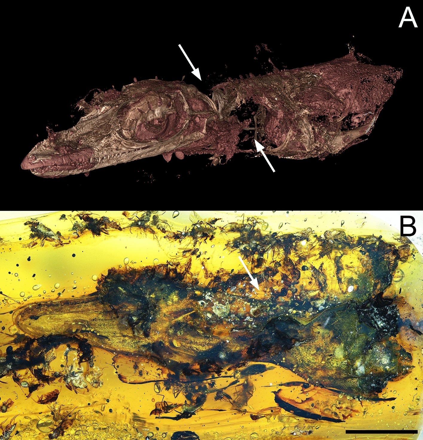
Piece GRS-Ref-28627 with Oculudentavis naga Bolet et al.39. (A) Virtual representation of Oculudentavis in frontolateral view (arrows show the place where the soft tissues were already partially consumed). (B) Photograph of Oculudentavis in ventrolateral view (arrow shows the place where most of the flies were trapped into the resin), as observed in side A. Scale bar 1 mm. Arnau Bolet provided high resolution photo for A.
No comments:
Post a Comment