Oh, yes, there is (slightly NSFW) video footage.
JENNIFER OUELLETTE - 12/26/2019
Video courtesy of Improbable Research
There's rarely time to write about every cool science-y story that comes our way. So this year, we're once again running a special Twelve Days of Christmas series of posts, highlighting one science story that fell through the cracks each day, from December 25 through January 5. Today: celebrating the 20-year anniversary of the most viewed article (and accompanying video) in the history of the British Medical Journal.
Christmas just wouldn't be the same for lovers of science without the annual Christmas issue of the British Medical Journal (BMJ). The tradition began in 1982, originally as a one-off attempt to bring a bit of levity to the journal for the holidays. While the papers selected for inclusion evinced a quirky sense of humor, they were also peer-reviewed and scientifically rigorous.
Some of the more notable offerings over the last 37 years included the side effects of sword-swallowing; a thermal imaging study on reindeer offering a possible explanation for why Rudolph's nose was so red; and an analysis of the superior antioxidant properties of martinis that are shaken, not stirred. (Conclusion: "007's profound state of health may be due, at least in part, to compliant bartenders.")
But by far the most widely read Christmas issue paper was a 1999 study that produced the very first MRI images of a human couple having sex. In so doing, the researchers busted a couple of long-standing myths about the anatomical peculiarities of the male and female sexual organs during sex. Naturally, the study was a shoo-in for the 2000 Ig Nobel Prize for Medicine.
Leonardo da Vinci famously studied cadavers to learn about human anatomy. He even drew an anatomical image of a man and woman in flagrante delicto, noting "I expose to men the origin of their first, and perhaps second, reason for existing." However, "The Copulation" (circa 1493) was based not on his own studies, but on ancient Greek and Arabic texts, with, shall we say, some highly dubious assumptions. Most notably, Leonardo depicted a ramrod straight penis within the vagina.
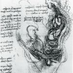
The Copulation as imagined
by Leonardo da Vinci.
The Royal Collection via MBJ
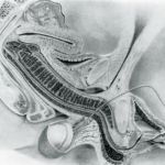
Midsagittal image of the anatomy of
sexual intercourse envisaged by
RL Dickinson and drawn by
RS Kendall
W.W. Schultz et al./BMJ (1999)
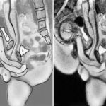
Left: Midsagittal image of the
anatomy of sexual intercourse
(labelled). Right: Midsagittal
image of the anatomy of sexual
intercourse.
W.W. Schultz et al./BMJ (1999)
The modern era of the science of sex arguably began in the 1930s with gynecologist Robert Dickinson, who sought to dispel the absurd notion of "coital interlocking," whereby the penis penetrates the cervix like a key fitting into a lock. Dickinson's experiments involved inserting a penis-sized glass tube into the vaginas of female subjects aroused via clitoral stimulation, as well as making plaster casts of women's vulvas and vaginas. (His work influenced Alfred Kinsey, whose own research produced the famous Kinsey scale to describe sexual orientation.)
Masters and Johnson revolutionized the field in the 1960s with their own provocative series of experiments on sexual response. Volunteers would have sex while hooked up to instruments in the lab. Masters and Johnson famously participated personally in their experiments, becoming lovers—a development that inspired the 2013 Showtime series Masters of Sex. They identified four stages of the Human Sexual Response Cycle: excitement, plateau, orgasmic, and resolution. Most relevant for the current discussion: they used an artificial penis for some experiments, noting among their findings that the volume of a woman's uterus increased by as much as 50-100 percent during orgasm. However, they acknowledged this might not be accurate due to the artificial nature of their experiments.
The 1999 study was the brainchild of Dutch physiologist Pek van Andel, who co-invented the artificial cornea and hence had a solid academic reputation that enabled him to persuade a hospital in Groningen, the Netherlands, to let him use an MRI machine after hours. But his original intention was to produce a piece of "body art." He'd been inspired by the image of an MRI cross-section of a singer's mouth and throat as she sang and thought it might be possible to use an MRI to take images of human coitus.
His co-authors included gynecologist William Schultz, radiologist Eduard Mooyaart, and anthropologist Ida Sabelis. Sabelis was an actual participant in the first experiment, conducted with her then-boyfriend, identified only as "Jupp." (Others would follow in her pioneering footsteps, including popular science author Mary Roach, who memorably persuaded her husband to participate in an ultrasound imaging sex study for her 2008 book, Bonk.)
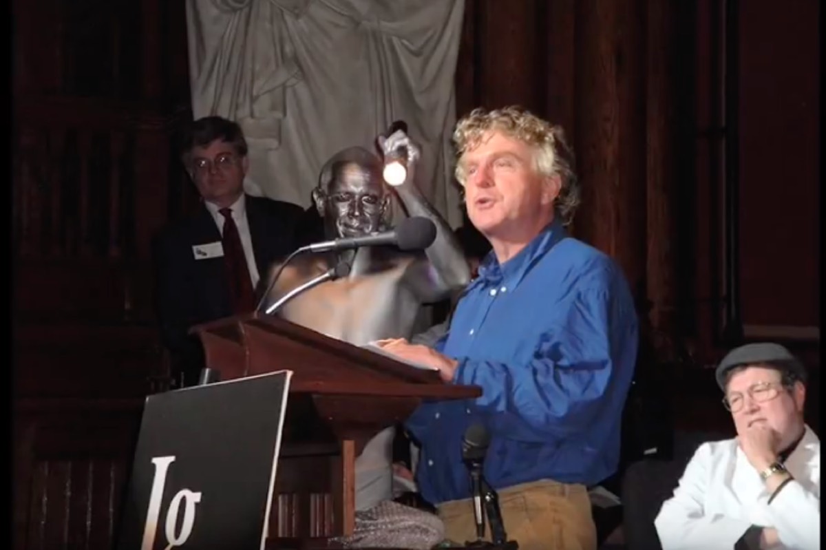
Enlarge / Co-author Pek van Andel accepting an Ig Nobel prize for his 1999 MRI study.
YouTube/Improbable Research
The first experiment, with Sabelis and Jupp, took place in 1992. While the latter had some concerns about whether he'd be able to adequately perform while being packed into a noisy metal tube with his partner, he managed to rise to the occasion. The experiment lasted 45 minutes, and the incredible detail of the resulting images rendered Sabelis momentarily speechless. Van Andel was delighted to note that the penis took on a boomerang shape during sex—disproving what Leonardo had depicted in his sketch centuries before. The images also disproved Masters and Johnson's finding that female sexual arousal increases the size of the uterus.
Convinced the images were scientifically relevant, van Andel submitted their findings to Nature. The journal rejected the paper outright. Controversy erupted when the Dutch tabloids got wind of the experiment, and the hospital balked at letting him use the MRI for additional experiments, although van Andel persuaded them to let him continue in the end. Between then and 1999, eight couples and three single women participated in 13 experiments. (Sabelis recently boasted to Vice that she and Jupp were the only couple who'd managed the feat without the aid of Viagra.)
The paper finally found a publisher in the British Medical Journal, whose editors thought the study made a fine addition to their annual Christmas issue. (In the final paper, the authors thanked “those hospital officials on duty who had the intellectual courage to allow us to continue this search despite obtrusive and sniffing press hounds.") Former BMJ editor Tony Delamothe recalled the thought process behind the decision:
Twenty years on, any paperwork relating to the decision to publish is gone, and the memories of editorial staff are hazy (one remembered discussion about how thin the participants must have been to manage intercourse in a 50cm diameter tube). It was possibly the close fit with the da Vinci drawing that swung the decision. Nobody thought the study was particularly useful clinically or scientifically, but it contained “a striking image using a new technology, and everyone agreed that readers might be interested to see it.” Sandy Goldbeck-Wood, the journal’s paper editor at the time, said: “I remember our conversation about it as serious (if wry), rather than ribald. I think the Christmas issue was the only place it would properly have fitted.”
Delamothe puzzles over the enduring appeal of the paper, and he seems to conclude that it's mostly due to the public's prurient interest at "seeing coitus on screen (for free)." But there is now tons of free pornography all over the Internet, yet these images continue to attract interest, and even admiration. Van Andel created a timeless piece of "body art" after all. The sexual act is, as Leonardo observed, "the origin of [our] first, and perhaps second, reason for existing"—and hence a thing of beauty.
DOI: British Medical Journal, 2019. https://doi.org/10.1136/bmj.l6654 (About DOIs).
DOI: British Medical Journal, 1999. 10.1136/bmj.319.7225.1596 (About DOIs).
Listing image by W.W. Schultz et al./BMJ (1999)
No comments:
Post a Comment