Thursday, November 11, 2021

'Cannibal' sun eruption gives departing astronauts their best aurora views yet


By Tereza Pultarova 

Astronauts get treated to mesmerizing polar lights fairly regularly, so this one must have been quite something.
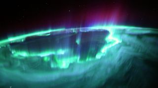
Astronauts at the International Space Station enjoyed the most stunning display of aurora borealis thanks to a cannibal coronal mass ejection.o (Image credit: ESA/Thomas Pesquet)

Astronauts bidding farewell to the International Space Station enjoyed the most spectacular aurora display of their entire mission over the weekend after a massive blast of material from the sun reached our planet.

The sun has been acting out lately, waking up to its new period of activity after years of quietness. This variation is part of the sun's regular 11-year cycle, the little understood ebb and flow of sunspots and solar flares that is next expected to peak in 2025.

"We were treated to the strongest auroras of the entire mission, over North America and Canada," European Space Agency's astronaut Thomas Pesquet tweeted with a mesmerizing photo of greenish glow. "Amazing spikes higher than our orbit. Star-struck, and we flew right above the centre of the ring, rapid waves and pulses all over

The spectacle must have been quite something since astronauts do get treated to aurora displays fairly regularly. Pesquet himself has shared many images of the magnificent polar lights on his Flickr account since his arrival at the orbital outpost with SpaceX Crew-2 in April.

The latest aurora display was triggered by a series of coronal mass ejections, bursts of magnetized plasma that the sun blasted out last week within a short period of time. The second outburst, travelling a bit faster than the first one, cannibalized its predecessor on the way, resulting in a much more powerful plasma cloud than originally expected.

Auroras occur in Earth's atmosphere when magnetized plasma particles from the sun hit Earth's magnetic field, creating a temporary magnetic havoc around the planet. In addition to providing the glowing spectacle the magnetic storms can damage satellites and knock out power grids. The worst geomagnetic storm in recorded history, the so-called Carrington Event of 1859, disabled telegraph networks all over Europe and North America.

For Pesquet and his Crew-2 companions, NASA astronaut Shane Kimbrough and Megan McArthur, and Japan's Akihiko Hoshide, the latest aurora provided a memorable conclusion to their six-month orbital adventure. Crew-2 is set to return to Earth today. You can watch their departure from the orbital outpost aboard SpaceX's Dragon capsule here.

A view of the Gulf of Morbihan, France
Thomas Pesquet: The best pictures from French astronaut's space missionUpdated: 09/11/2021By James Thomas
France's Thomas Pesquet and three other astronauts have landed back on Earth after a six-month mission aboard the International Space Station (ISS).
Pesquet, representing the European Space Agency, and his crewmates landed in the Gulf of Mexico off the coast of Florida on Monday
Pesquet was known for his active social media presence while in space, regularly posting pictures of his life aboard the ISS, breathtaking views of the Earth and stars and also the occasional meme
To mark his return, we've chosen some of his most popular and striking images from his time in orbit.
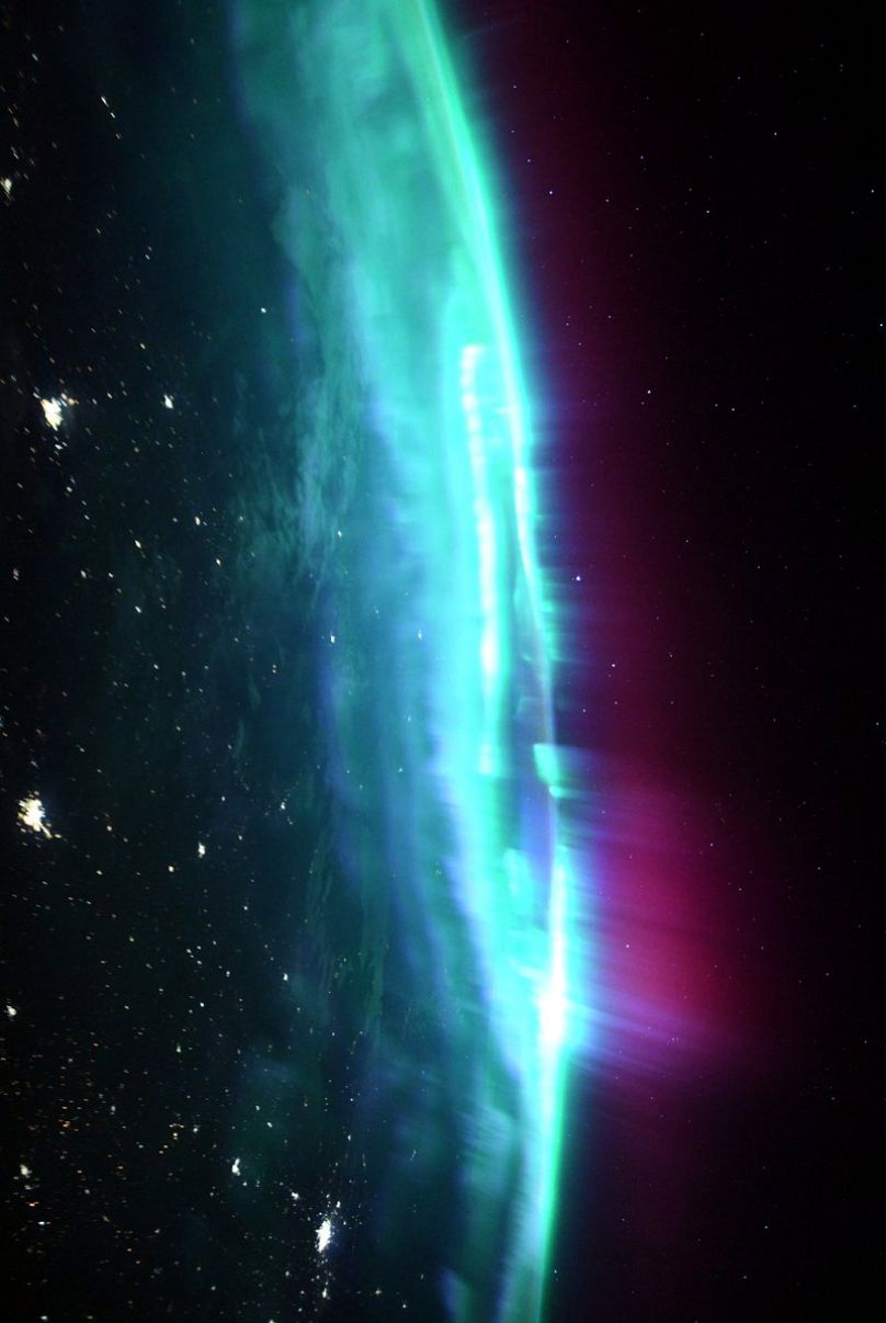
A view of the Northern Lights from the International Space Station. Pesquet described this particular sighting on 6 November as “the most intense” of the whole mission.Credit: European Space Agency
A view of the space station in orbit.Credit: European Space Agency
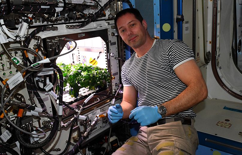
The crew grows chilli peppers for the first time in outer space.
Credit: European Space Agency
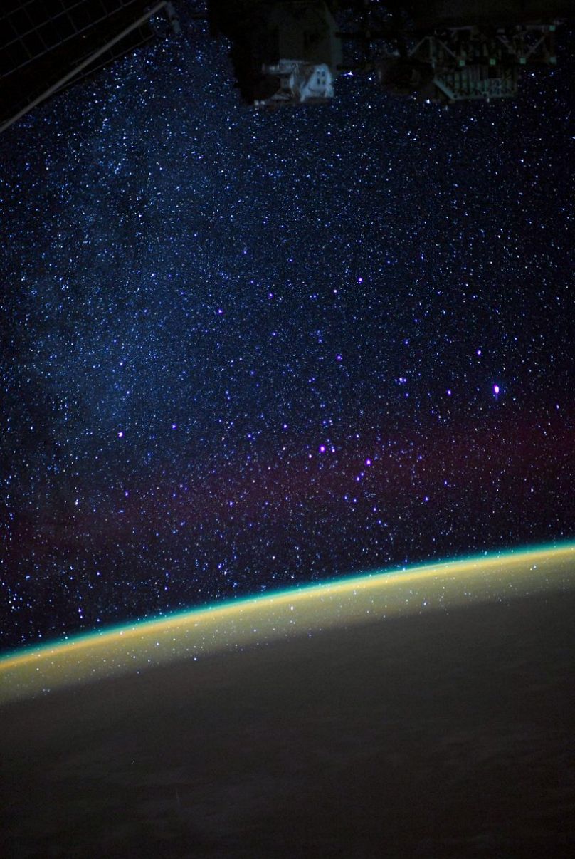
A view of the stars from the space station.Credit: European Space Agency
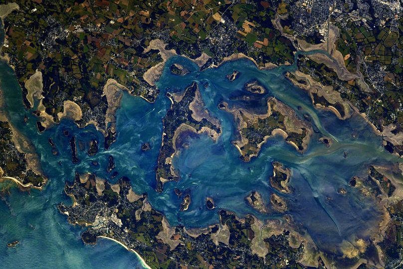
A view of the Gulf of Morbihan, FranceCredit: European Space Agency
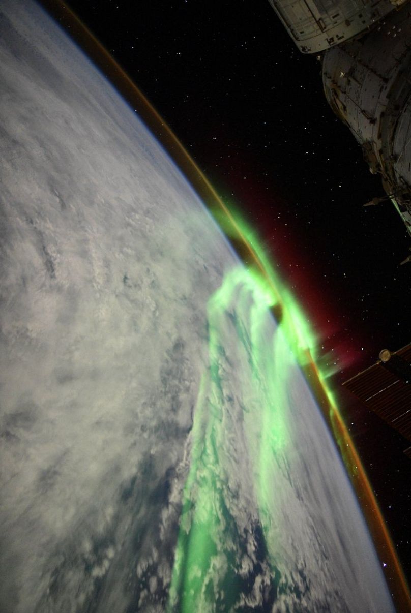
The Northern Lights.Credit: European Space Agency
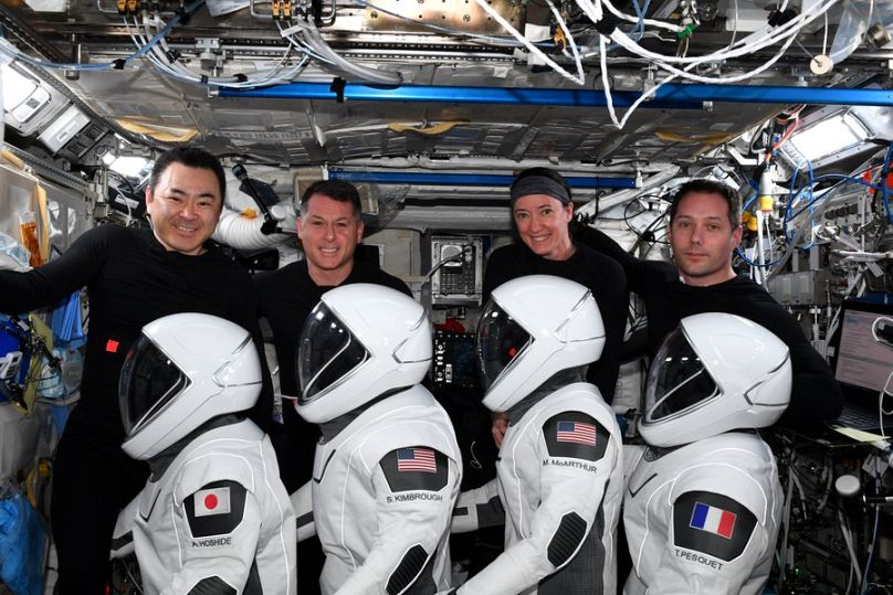
The four astronauts conduct their final checks before their return to Earth.Credit: European Space Agency
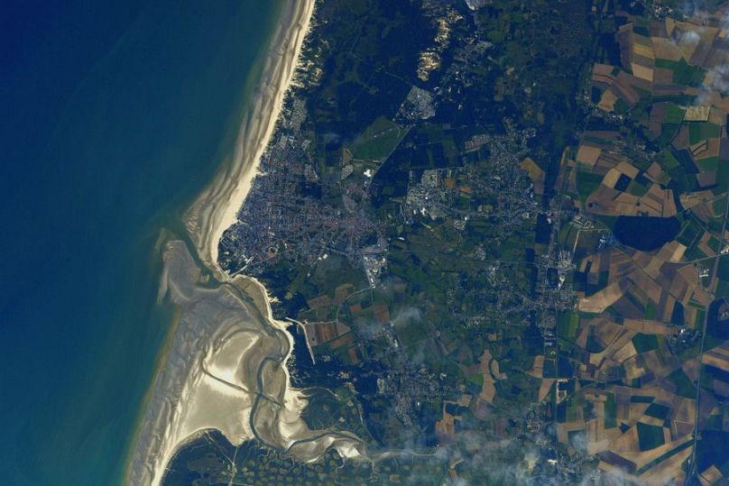
A view of Berck, France, near where Pesquet went to flight school.Credit: European Space Agency
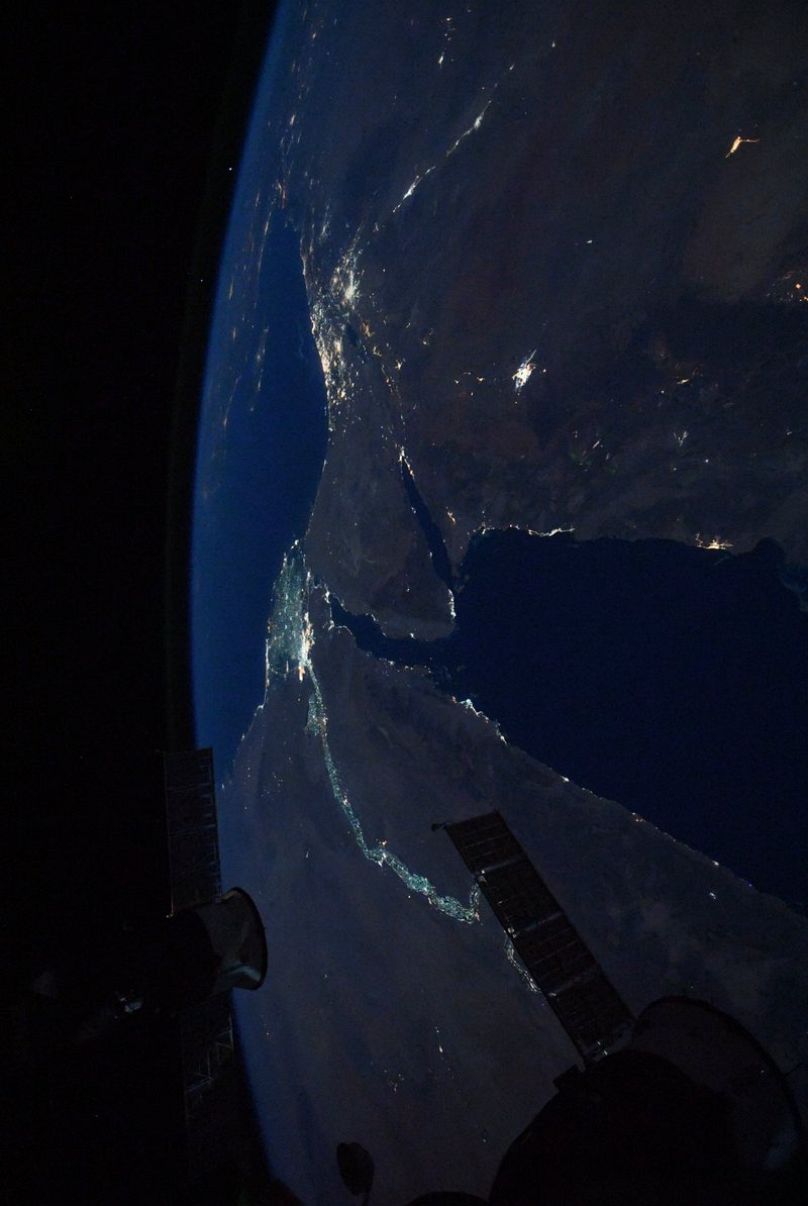
A view of Egypt by night.Credit: European Space Agency
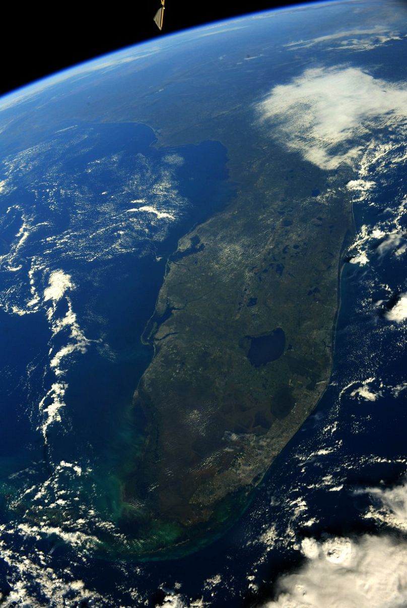
A view of Florida. Pesquet notes that unlike other forms of travel, space travel allows you to see your destination before you depart.Credit: European Space Agency

Modified silk cloth keeps skin cooler than cotton

Credit: DOI: 10.1038/s41565-021-00987-0

A team of researchers affiliated with a host of entities in China and one in the U.S. has developed a modified textile that can keep skin cooler than materials made of cotton. In their paper published in the journal Nature Nanotechnology, the group describes their approach to developing garments that are cooler when worn in outdoor conditions.

Humans have been wearing clothes for hundreds of thousands of years, and over that time, have been refining them to suit the needs of their environments, which are mostly cold environments. In this new effort, the researchers wondered if it might be possible to create a type of material that would be cooler to wear than other  and or bare  under direct sunlight. To find out, they started with silk fabric, a material that has been used for thousands of years because of its looks and comfort.

The researchers noted that silk does a good job of reflecting sunlight in the mid-infrared range, which suggests it could be suitable as a cooling garment material. But because it is made by spiders, it contains a protein component that tends to absorb , making the material and its wearer grow hotter under .

To make the silk material UV reflective, the researchers dipped a standard piece of silk fabric into a liquid solution containing highly refractive inorganic oxide nanoparticles. These adhered to the  fabric, allowing it to become evenly saturated throughout the material. They allowed the fabric to dry and then tested it to see if the addition of the nanoparticles made the material more UV reflective. They found that under peak sunlight conditions, the temperature under the material was approximately 3.5 degrees Celsius cooler than the ambient air temperature. Next, they placed the material on a patch of simulated skin and found the skin temperature was approximately 8 degrees Celsius cooler than the same type of simulated skin without the material covering. They also found that it kept the artificial skin approximately 12.5 degrees Celsius cooler than standard cotton material. Further testing showed that the material was able to reflect approximately 95% of , preventing it from passing through to the skin underneath.Microfiber-based metafabric provides daytime radiative cooling

More information: Bin Zhu et al, Subambient daytime radiative cooling textile based on nanoprocessed silk, Nature Nanotechnology (2021). DOI: 10.1038/s41565-021-00987-0

Journal information: Nature Nanotechnology 

© 2021 Science X Network

POSTMODERN ALCHEMY

Going for gold to reduce antibiotic resistance

Going for gold to reduce antibiotic resistance
The gold nanoclusters in their "molecular envelope." The ligands in blue are the zwitterionic ones while those in red are positively charged ones. They are bound to the Au25 cluster (in brown) via thiol molecules (yellow). Credit: University of Leeds

Tiny particles of gold could be the new weapon in the fight against bacterial antibiotic resistance, according to research just published.

Scientists have been investigating the use of gold nanoclusters—each made up of about 25 atoms of gold—to target and disrupt , making them more susceptible to standard antibiotic treatments.

A report from the World Health Organization last year said, "Antibiotic resistance is rising to dangerously high levels in all parts of the world," and called for greater investment in ways to tackle the problem.

For several years, researchers have recognized the antimicrobial properties of specially-adapted gold nanoparticles, but they have struggled to find a way of getting the nanoparticles to the site of a bacterial infection without harming healthy host mammalian .

Now a study by an international team of scientists from the University of Leeds, Southern University of Science and Technology in Shenzhen and Fudan University, Shanghai, both in China, has identified a way of packaging the gold nanoclusters in a molecular envelope that makes them less toxic to healthy tissue without affecting their antibacterial properties.

Laboratory studies have shown that the approach has had a "strong effect" in terms of killing a range of bacteria, some linked to hospital acquired infections and resistant to standard antibiotic treatments.

The findings, which are based on laboratory investigations and not patient trials, have been published in the journal Chemical Science.

Forces of nature

The scientists' solution exploits electrostatic forces in nature.

Bacterial cell walls are more strongly negatively charged than the cells of mammals. Using the idea that opposite charges attract, the gold nanoclusters are wrapped in a molecule called a ligand that is positively charged. Like a carrier pigeon, it finds and delivers the nanoclusters to the wall of bacteria cells, where they disrupt the bacterial cell membrane.

The disruption to the cell membrane increases the permeability of the bacterial cell to standard antibiotic treatments, giving a new lease of life to  that are either ineffective or have waning effectiveness against resistant bacteria.

The problem, though, is the positively charged molecule wrapped around each  is also toxic to healthy host mammalian cells.

Reducing toxicity

To protect host cells, the scientists have added a second ligand to the envelope around each nanocluster. These molecules have both positive and negative charges and are called zwitterionic groups, which are also found in the lipids of cell membranes in mammals. This makes the gold nanoclusters more compatible with host mammalian cells, and easier for the gold nanoclusters to pass through the kidney and be excreted from the body.

In , the scientists investigated whether the gold nanoclusters would be effective in reducing the defenses of the bacterial cells—and make them more susceptible to antibiotic treatment.

They used a bacterial strain called methicillin resistant Staphylococcus epidermidis (MRSE), which is responsible for some hospital-acquired infections.

They tested three antibiotics—each representing a class of antibiotics—against MRSE with and without the gold nanoclusters.

In those cases where the antibiotic was used in combination with the gold nanoclusters, there was an improved antimicrobial effect. With one class of antibiotics, there was a 128-fold decrease in the amount of antibiotic needed to inhibit growth of MRSE.

Dejian Zhou, Professor at Nanochemistry at the University of Leeds and one of the supervisors of the research, said, "Despite extensive research in antibacterial nanomaterials, most of the research has only focused on boosting antibacterial potency without considering their biocompatibility, stability and ability to be excreted from the body. These are essential requirements for clinical approval. As a result, many of the promising antibacterial nanomaterials will not progress to become therapeutic agents to be used in medicine.

"By systematically tuning the ratio of the two ligands, we have identified a way of using gold nanoclusters not only to act as effective antimicrobial agents, but as a mechanism to enhance the potency of antibiotics which have become ineffective because of bacterial drug resistance.

"The research has a significance on the way we should be thinking about responding to antimicrobial resistance."

Professor Zhou hopes that the research findings will be picked up by the pharmaceutical industry. He believes combing  nanoclusters with existing antibiotics will be a faster and cheaper alternative to developing a host of new antibiotics in response to .Atomically precise noble metal nanoclusters

More information: Zeyang Pang et al, Controlling the pyridinium–zwitterionic ligand ratio on atomically precise gold nanoclusters allowing for eradicating Gram-positive drug-resistant bacteria and retaining biocompatibility, Chemical Science (2021). DOI: 10.1039/D1SC03056F

Journal information: Chemical Science 

Provided by University of Leeds 

Sponge Cells Hint On Evolution Of Nervous System

November 10, 2021

Sponges are simple multicellular organisms, though they are skillful filter feeders, filtering tens of thousands of liters of water through their bodies each day to get food. Their ability for this complicated behavior is more exceptional as they do not have a brain or a single neuron.

According to a study published in Science, sponges use a complex cell communication system to regulate their feeding and potentially annihilate invading bacteria. This is exciting research that enables us to look at sponges in a whole new way. According to Casey Dunn, an evolutionary biologist at Yale University, Connecticut, the results could benefit us in understanding the evolution of animals’ nervous systems.
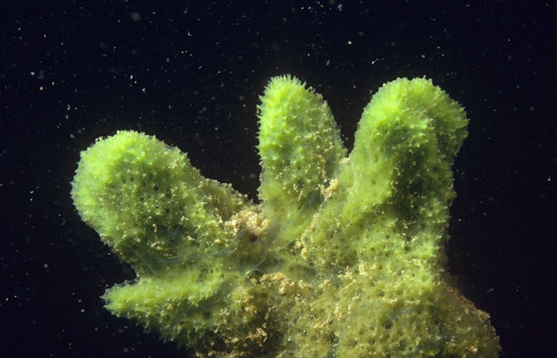
This freshwater sponge (Spongilla lacustris) may hold clues about the evolution of the nervous system. Image Credits: Willem Kolvoort/Nature Picture Library

Neurons interact with one another by transferring electrical or chemical signals via small and targeted connections termed synapses. Despite the animals’ lack of neurons, earlier research discovered that sponges have genes encoding proteins that support synapses function.

Detlev Arendt, an evolutionary biologist, EMBL, Germany, and team sequenced the RNA in several separate cells from a freshwater sponge (Spongilla lacustris) to
determine which cells expressed these genes.

They observed that the sponge has 18 distinguished cell types. Synaptic genes were active in some of them, which were grouped near the digestive cells of the sponges. This implies that some kind of cellular interaction may coordinate the animal’s filter-feeding behavior.

The team then studied one of these cell types, which they named secretory neuroid cells, using X-ray imaging and electron microscopy. The study uncovered that neuroids extend long arms to reach choanocytes, a type of cell with hair-like projections that power sponges’ water-flow systems and capture the majority of their food.


Nervous-system precursor

The team believes that these arms enable neuroids to interact with choanocytes, allowing them to halt the water-flow system and clean out any detritus or foreign microorganisms, based on the proximity of the two cell types and the expression of genes that may allow for chemical secretion. Though these neuroid cells are not nerves, and there is no evidence of the synapses that allow neurons to interact so fast.

Jacob Musser, an evolutionary biologist, EMBL, and co-author of the study, stated that this cell type could be an evolutionary precursor to an actual nervous system.

He further added that they are at an intermediary position, where they have gone from having all these independent pieces to bringing them together more extensively, yet all the interconnectivity required to form a fast synapse is not obtained.

Some experts believe that referring to these cells as nervous system precursors is a stretch. Linda Holland, an evolutionary developmental biologist, UC San Diego, stated that it’s intriguing but not conclusive. She commented that it would be challenging to determine whether the evolution of nervous systems happened from this cellular communication system or arose earlier or even several times, as suggested by some experts. According to Sally Leys, a marine biologist, the University of Alberta, Canada, several other organisms, like unicellular eukaryotes, have the same synaptic genes.

April Hill, a developmental geneticist, Bates College, Maine, believes that this research and its methods will serve as a “launchpad” for further studies into this ubiquitous sponge. She further stated that whether other sponges utilize a comparable cellular interaction system is an important unanswered mystery.

What sponges can tell us about the evolution of the brain

More than a gut reaction
Sponge neuroid cell (orange) extends arms that enwrap the feeding apparatus of a 
sponge digestive cell (green) to create a link for targeted communication. 
The image was taken using electron microscopy. Credit: Jacob Musser, Giulia Mizzon, 
Constantin Pape, Nicole Schieber / EMBL

Despite its central importance, the brain's origins have not yet been uncovered. The first animal brains appeared hundreds of millions of years ago. Today, only the most primitive animal species, such as aquatic sponges, lack brains. Paradoxically, these species may hold the key to unlock the mystery of how neurons and brains first evolved.

Individual neurons in a  communicate via synapses. These connections between  lie at the heart of brain function and are regulated by a number of different genes. Sponges do not have these synapses, but their genome still encodes many of the synaptic genes. EMBL scientists asked the question why this might be the case. Their latest findings are published today in the journal Science.

"We know that these synaptic genes are involved in neuronal function in higher . Finding them in primitive species like sponges begs the question: if these animals don't have brains, what is the role of these genes?" said Detlev Arendt, EMBL group leader and senior scientist at EMBL Heidelberg. "As simple as that sounds, answering this question was beyond our technological abilities so far."

To study the role of these synaptic genes in sponges, the Arendt lab established microfluidic and genomic technologies in the freshwater sponge Spongilla lacustris. Using these techniques, the scientists captured  from several  inside microfluidic droplets and then profiled each cell's genetic activity.

Neuroid cells (purple and red) send cellular arms to contact and communicate with specific digestive cells in the sponge digestive chamber (blue, green, yellow). Other digestive chamber cells are shown in grey. Credit: Jacob Musser, Giulia Mizzon, Constantin Pape, Nicole Schieber / EMBL

"We showed that certain cells in the sponge digestive chambers activate the synaptic genes. So even in a primitive animal lacking synapses, the synaptic genes are active in specific parts of its body," said Jacob Musser, research scientist in the Arendt group and lead author on the study.

Sponges use their digestive chambers to filter out food from the water and interact with environmental microbes. To understand what the cells expressing synaptic  do, the Arendt group joined forces with six EMBL teams as well as collaborators in Europe and worldwide. Working with EMBL's Electron Microscopy Core Facility, Yannick Schwab's team and Thomas Schneider's group operating synchrotron beamlines at EMBL Hamburg the researchers developed a new correlative imaging approach. "By combining  with X-ray imaging on a synchrotron beamline we were able to visualize the stunning behavior of these cells," Dr. Schwab explained.

The scientists captured three-dimensional snapshots of cells crawling throughout the digestive chamber to clear out bacterial invaders and sending out long arms that enwrap the feeding apparatus of specific digestive cells. This behavior creates an interface for targeted cell-cell communication, as it also happens across synapses between neuronal cells in our brains.

"Our results point to the cells regulating feeding and controlling the microbial environment as possible evolutionary precursors for the first animal brains," Dr. Musser said.Microglia pruning brain synapses captured on film for the first time

More information: Jacob Musser et al, Profiling cellular diversity in sponges informs animal cell type and nervous system evolution, Science (2021). DOI: 10.1126/science.abj2949. www.science.org/doi/10.1126/science.abj2949

Journal information: Science 

Provided by European Molecular Biology Laboratory 


Sponge Genes Hint at the Origins of Neurons and Other Cells

A new study of gene expression in sponges reveals the complex diversity of their cells as well as some possibly ancient connections between the nervous, immune and digestive systems.


A new atlas of gene expression in the sponge Spongilla has revealed surprising levels of cellular diversity in these primitive animals.
Allexxandar / Dreamstime.com


Viviane Callier

November 4, 2021

When the first sponge genomes were sequenced in the early 2000s, researchers were surprised to find that sponges not only have roughly as many genes as humans and other complex creatures but also have many of the same genes. Sponges are among the earliest branching lineages on the evolutionary tree of animal life; their simple bodies don’t even have a pattern of symmetry or a set number of parts. The presence of those genes implied that the genetic information for functions like muscle contraction and the differentiation of neurons was much more ancient than muscles or nervous systems themselves.

But what were those genes doing in an animal without neurons or muscles? Researchers could only make educated guesses and investigate expression patterns on a painstaking gene-by-gene basis.

Today, however, a new study taking advantage of rapid advances in genomic technologies has illuminated where about 26,000 genes are expressed in the freshwater sponge Spongilla. This atlas of gene expression reveals the genetic configuration of cell types throughout the sponge’s body, including some cell types never described before. It offers important hints about how cell types evolved in the first place, and it may help to settle a long, thorny debate about whether neurons evolved just once or many times. The study appears in the latest issue of Science.

This ambitious paper “leapfrogs” over previous work, according to Scott Nichols, who studies sponge evolution at the University of Denver. “What is extraordinary about it is that really fascinating hypotheses have emerged from this data set,” he said. “But I would emphasize strongly that they need to be experimentally tested.”

The most exciting hypothesis concerns cells inside the sponge’s digestive chambers. The chambers are lined with distinctive cells called choanocytes, which have a collar of fingerlike protrusions (microvilli) and a flagellum. The choanocytes beat their flagella to regulate the flow of water through the digestive chamber, all the while feeding on small particles and debris the water carries. The digestive chambers also contain mobile “neuroid” cells that were described years ago, although their identity and function were mysterious.

Using high-throughput single-cell RNA sequencing technology, Detlev Arendt’s team at the European Molecular Biology Laboratory in Heidelberg discovered that choanocytes express genes that in neurons produce the postsynaptic “scaffolding” involved in receiving and responding to neurotransmitters. They also discovered that the mobile neuroid cells express a suite of genes that are typically active in the presynaptic bulb of a neuron. This led the researchers to hypothesize that the neuroid cells might be talking to the choanocytes, and that the neuroid cells’ job might be to patrol the microbial environment in the digestive chamber and regulate the choanocytes’ feeding behaviors accordingly.


Sponges have digestive chambers lined with cells called choanocytes. Waving their flagella to propel water through the chambers, the choanocytes digest small particles in the flow.

Caterina Longo, Bari University; source: doi.org/10.1371/journal.pone.0042392.g005

When Jacob Musser, the postdoctoral fellow in Arendt’s lab who led the project, stained the sponge to look at where exactly the pre- and postsynaptic genes were being expressed, he saw that the neuroid cells expressing presynaptic genes were indeed near the choanocytes expressing postsynaptic genes. In fact, the neuroid cells reached out pseudopod arms that seemed to touch the choanocytes.

“This was obviously really tantalizing,” Musser said. “But you can’t really tell what is going on.”

To get a more detailed picture of what the cells were doing, Musser and the team used focused ion beam electron microscopy at the X-ray synchrotron facility in Hamburg to get very high-resolution 3D images of the cells, which could distinguish cellular features as small as 15 nanometers, roughly the size of many folded proteins. They saw that projections from the neuroid cells enveloped the choanocytes’ microvilli collar and flagellum, and that the neuroid cells held vesicles like those in the presynaptic bulb of a neuron. They suspect the vesicles are probably releasing glutamate, a neurotransmitter.

But tempting as it is to imagine these sponges as having primitive synapses, the researchers never observed direct, stable contacts between the neuroid cells and choanocytes. The connections between the cells instead seem to be transient. Furthermore, the DNA of sponges lacks genes for some of the key ion channels needed to create an action potential — the sharp electrical signal that stimulates the release of neurotransmitters in neurons.

Nevertheless, because sponges have always been thought to lack anything even resembling a nervous system, the suggestion that they have cellular mechanisms with a deep evolutionary relationship to neurons “is an exciting path forward to connect sponge biology to neural cell biology, to understand where neuronal signaling came from at all in animals,” Nichols said.


A colorized micrograph of the cells in a sponge digestive chamber (left) reveals the interaction of a neuroid cell (magenta) with a choanocyte (green). In a magnified detail (right), the transient contact between the two cells could be suggestive of the synaptic contact between neurons.
Quanta Magazine; source: Jacob Musser, Giulia Mizzon, Constantin Pape, Nicole Schieber / EMBL

The origin of neurons and nervous systems — and in particular, the question of whether neurons arose once or multiple times — is one of the most contentious topics in the field of evolutionary developmental biology, according to Maria Antonietta Tosches, who studies the evolution of cell types in vertebrates at Columbia University and previously trained in Arendt’s lab. The findings from this new study seem to bear on that mystery because the researchers found presynaptic gene sets expressed in neuroid cells and postsynaptic genes expressed in choanocytes. (Both sets of genes were active in other cell types as well.) That fact suggests that the genetic modules responsible for both the sending and receiving ends of cell-cell communication systems were deployed in various types of ancestral animal cells. Neurons could therefore have evolved repeatedly and independently through different applications of these gene modules, Tosches said.

In fact, many multifunctional cells in sponges express modules of genes usually associated with specialized cells in more complex animals like vertebrates. For example, sponge neuroid cells not only express some of the presynaptic machinery of neurons, but also express immune genes. (It’s possible that if neuroid cells monitor the microbial content of the digestive chambers for sponges, these immune genes assist in that role.) Sponges also have cells called pinacocytes that contract in unison like muscle cells to squeeze the animal and expunge waste or unwanted debris; pinacocytes have some sensory machinery that responds to nitric oxide, a vasodilator.

“Nitric oxide is what relaxes our smooth muscle in our blood vessels, so when our blood vessels expand, that’s nitric oxide causing that relaxation,” Musser said. “And we’ve actually shown through experiments in the paper that nitric oxide is also regulating contractions in this sponge.” Like glutamate, nitric oxide might have been part of an early signaling mechanism to coordinate primitive behaviors in the sponge, he suggests.


EVOLUTION
Scientists Debate the Origin of Cell Types in the First Animals

JULY 17, 2019


“Our data are very consistent with this notion that a large number of important functional pieces of machinery existed early in animal evolution,” Musser said. “And a lot of early animal evolution was about starting to subdivide this out to different cells. But likely these very first cell types were very multifunctional, and they had to do multiple things.” The earliest animal cells, like their close relatives the protozoans, probably had to be cellular Swiss Army knives. As multicellular animals evolved, their cells may have taken on different roles, a division of labor that may have led to more specialized cell types. But different lineages of animals may have divvied things up differently and to different degrees.

If the mixing and matching of genetic modules was a crucial theme of early animal evolution, then comparing the arrangement and expression of those modules in different species could tell us about their history — and about possible limitations on how haphazardly they can be shuffled. One researcher looking for those answers is Arnau Sebé-Pedrós, who studies cell type evolution at the Center for Genomic Regulation in Barcelona and who published the first atlases of cell types in sponges, placozoans and comb jellies in 2018.

Sebé-Pedrós thinks that the spatial configuration of the genes along the chromosomes could be revelatory because genes located together can share regulatory machinery. “I’m absolutely shocked by the degree of conservation of the gene orders in animal genomes,” he said. He suspects that the need to co-regulate sets of functionally related genes keeps them in the same chromosomal neighborhood.

Scientists are still in the early days of learning how cell types evolve and relate to one another. But as important as it is to clarify the muddy origins of animal evolution, sponge cell atlases are also making a major contribution by revealing the possibilities in animal cell biology. “It is not just important for us to understand the very origin of animals,” Sebé-Pedrós said, “but also to understand things that may be radically different from anything else that we know about other animals.”
A big discovery of a tiny critter


Water bear fossil found in 16-million-year-old amber is the third tardigrade species ever discovered

Amber specimen including tardigrades and other invertebrates.


Image by Phil Barden/NJIT

BY Juan Siliezar Harvard Staff Writer

DATE November 5, 2021

Good luck finding an animal tougher than a tardigrade.

These tiny creatures are famous for their ability to survive in the most extreme conditions, including boiling water, freezing water, and even the vacuum of space. Called water bears or moss piglets because of their appearance under a microscope, tardigrades are the smallest-known animals with legs. They have a pudgy body — no larger than a pencil point — their eight legs have several pointed claws at the end, and they have a spear-like sucker that extends from their mouth.

Tardigrades are found on all the continents (basically wherever there is water) and have survived on Earth for more than 500 million years. Despite such a long evolutionary history and global presence, the fossil record on tardigrades is thin, with only two clear examples identified as separate species ever found. But thanks to a 16-million-year-old piece of amber discovered in the Dominican Republic, scientists can now add a third — a discovery immortalized in word and song.


Magnified example of a tardigrade, with its pudgy body that is no larger than a pencil point.

Credit: Phil Barden/NJIT


The researchers from Harvard and the New Jersey Institute of Technology who made the discovery describe their finding in a new paper in the Proceedings of the Royal Society B. The ultra-rare fossil can help shed new light on these ancient animals and provide new evolutionary insight into how the 1,300 tardigrade species that exist today evolved.

The specimen is the first water bear fossil ever recovered from the Cenozoic era, which started 66 million years ago and encompasses the planet’s current geological era. The researchers believe this is the best-imaged tardigrade fossil to date. In fact, they were able to describe microscopic details on parts of the mouth and needle-like claws that are about 20 to 30 times finer than a human hair. They were also able to get an unprecedented look at the internal anatomy of its foregut, which played the key role in identifying the fossil as a new genus and species.

“We could see it had this unique foregut organization that warranted for us to formalize a new genus within this extant group of tardigrade superfamilies,” said Marc A. Mapalo, a Ph.D. candidate who does his work in the Department of Organismic and Evolutionary Biology, Harvard’s Graduate School of Arts and Sciences, and is lead author of the study. “We saw characters that are not observed in extant species, but are observed in the fossils. This helps us understand what changes in the body occurred across millions of years.”

The researchers were able to examine these distinct anatomical features with a confocal laser microscope, a piece of equipment that uses a laser instead of visible light to peer into the specimen and produce a high-quality image. Usually, it is used to see biological and molecular processes such as cell division 

.
Reconstruction of tardigrades from the Miocene of Dominican Republic. Artwork by Holly Sullivan

“Using this high-powered technique that is usually employed for studying cell biology, it was possible to obtain extremely detailed anatomical information,” said Javier Ortega-Hernández, an assistant professor in OEB and curator of invertebrate paleontology in the Museum of Comparative Zoology. “We saw the whole animal in way better detail than previously possible using conventional light microscopy.”

The researchers called the new species Paradoryphoribius chronocaribbeus. The name uses the Greek word for time, “chrono,” and refers to the Caribbean region where it was found, “caribbeus.” The new species is a relative of the modern living family of tardigrades known as Isohypsibioidea.

The other two fully described unequivocal tardigrade fossils are Milnesium swolenskyi and Beorn leggi, both known from Cretaceous-age amber in North America. This makes the new Dominican species the first water bear fossil to be found outside that region.

Co-author Phillip Barden from the New Jersey Institute of Technology introduced the fossil to Ortega-Hernández and Mapalo in 2019 after visiting the Museum of Comparative Zoology as a guest speaker. Mapalo, who was new to the lab, is a specialist in tardigrades and took the lead in analyzing the fossil using confocal microscopes in the Harvard Center for Biological Imaging.

Mapalo and Ortega-Hernández hope their success with the confocal microscope will inspire other researchers to use it to examine their own amber samples. The pair will also continue to employ the technology to study other tardigrades fossils trapped in amber.

Meanwhile, Mapalo has written a song (as one does) to celebrate number three.


How studying fossilized parasites can contribute to knowledge of infectious diseases

Expert comment: How studying fossilized parasites can contribute to knowledge of infectious diseases
Examples of parasite–host interactions preserved on marine animal host skeletons. 
Credit: University of Missouri

Over the last decade, John Huntley, a paleontologist and an associate professor of geological sciences at the University of Missouri, has studied the history of parasite-host interactions. These interactions can occur either outside a host's body, such as a tick, or inside a host's body, such as a flatworm.

Recently, Huntley and his colleagues developed the first known database of parasite-host interactions among animals living in the ocean, including the fossilized ancestors of today's crabs, shrimp and oysters. Their results were recently published in the journal Philosophical Transactions of the Royal Society B.

Huntley explains how studying fossilized parasites can contribute to our knowledge of infectious diseases.

How can studying fossil parasites contribute to our knowledge of infectious diseases?

We're learning that paleontological research is more than a purely academic undertaking. Paleontologists have the privilege of studying ancient life and the environments in which these animals lived. By better understanding how parasite-host interactions have occurred in the past, we now have the primary evidence for how life has responded to a variety of calamities over hundreds of millions of years.

This insight can help us better understand the evolution of biodiversity over time, and give us greater context to modern problems such as the COVID-19 pandemic and climate change.

What insights have you developed from your recent research on how parasite-host relationships have changed over time?

The first known occurrence of parasitism among animals occurred about 520 million years ago. Since that time, the occurrence of parasitism and the percentage of individuals affected by parasites has dramatically increased. In particular, the last 65 million years have seen an intensification in parasitism. This is consistent with my earlier studies of predator-prey interactions.

In general, we've found the world has become a more  for animals in the oceans over the last half a billion years. There are a variety of competing, but not mutually exclusive, ideas for why this has occurred, and arguments generally center around food chain processes, and changing nutrient and habitat availability.

What can these parasite-host relationships tell us about biodiversity and the health of ecosystems throughout the history of life on Earth?

We've found a strong correlation between parasitism and biodiversity. At the broadest scale, parasites are more common when there are more species. This makes sense because more species can mean more chances for developing these interactions. We also compared parasitism to the rate at which species originate and go extinct and found negative relationships, which suggests parasite-host interactions flourish when higher and more stable levels of diversity are present.

Therefore, we've seen evidence that parasites can positively stabilize coastal ecosystems that provide food and other services to millions of people in today's world. Even though parasites harm the individual hosts that they infest, evidence shows they make the overall ecosystem more stable because of their actions.Birds' eye size offers clues to coevolutionary arms race between brood parasites, hosts

More information: Kenneth De Baets et al, Phanerozoic parasitism and marine metazoan diversity: dilution versus amplification, Philosophical Transactions of the Royal Society B: Biological Sciences (2021). DOI: 10.1098/rstb.2020.0366

Journal information: Philosophical Transactions of the Royal Society B 

Provided by University of Missouri 

Scientists Say The Deepest Earthquake Ever Detected Should’ve Been ‘Impossible’ Which Can Only Mean One Thing

I

STOCKPHOTO


  • Scientists are baffled by an earthquake that set a record for the deepest seismic disturbance ever recorded
  • Experts say the earthquake—which was detected off of Japan—was theoretically impossible based on widely-accepted research

It’s been almost two years since most of us got a crash course in what it’s like to be in a movie that kicks off with the bulk of the world opting to ignore murmurings concerning a mysterious airborne disease responsible for sparking a health crisis in the Chinese city that was placed on lockdown in the hopes of stopping the spread (a strategy that—as we know all too well by now—didn’t exactly pan out as hoped).

Based on what we learned from that situation, I feel like it no longer hurts to err on the side of caution in the hopes we aren’t doomed to repeat a similarly catastrophic scenario—which is why I feel like we might want to pay attention to a new report from Live Science concerning an earthquake that occurred off the coast of Japan in 2015.

In June of that year, researchers who were monitoring a 7.9-magnitude seismic event that hit the Bonin Islands detected a minor aftershock that originated 467 miles below the surface of the planet—a depth that set a new record for the deepest earthquake ever observed. While it may not seem like an incredibly newsworthy development, it came as a bit of a surprise to experts who say the disturbance was theoretically impossible based on everything that was previously known about the conditions required for such a disturbance to occur

I’d recommend checking out the aforementioned article if you’re looking for an in-depth explanation of their reasoning, which centers around the long-held belief that the nature of rocks located in Earth’s lower mantle (where the earthquake originated) makes them impervious to the effects of water that triggers the quakes that occur closer to the surface by leaking into more porous objects and weakening their structure.

As things currently stand, there are a couple of operating theories. One is that researchers incorrectly estimated the depth of the boundary between the upper and lower mantle, while the other rests upon the assumption that certain minerals found at the depth were exposed to unexpected conditions that made them susceptible to cracking.

Of course, there’s also the possibility that the initial earthquake roused one of the Great Old Ones from their eternal slumber and it’s only a matter of time until a vast, indescribable horror beyond the comprehension of all but those unfortunate enough to witness its unbridled wrath and might rises from the depths of the Pacific Ocean, but I guess we’ll just have to wait and see.


The Deepest Earthquake Ever Recorded Happened 467 Miles Underground, Surprising Scientists

Because of intense heat and pressure, quakes are rare beyond 186 miles deep beneath Earth’s crust


Rasha Aridi

SMITHSONIAN
Daily Correspondent
November 8, 2021
In 2015, a 7.9 magnitude earthquake struck beneath Japan's Bonin Islands.
 Lee Render via Flickr

Between 1976 and 2020, nearly 57,000 earthquakes rattled our planet. The bulk of them were shallow, and only a mere four percent occurred beyond 186 miles deep, which was thought to be the maximum depth for what scientists call "deep earthquakes," reports Maya Wei-Haas for National Geographic.

Now, a team of researchers has zeroed in on what could be the deepest earthquake ever detected, shaking up scientists' understanding of them. In 2015, a 7.9 magnitude earthquake struck beneath Japan's Bonin Islands. One of the aftershocks occurred deeper than the original earthquake itself, at 467 miles. It's so deep that it nears the layer of Earth known as the lower mantle, reports Andrei Ionescu for Earth.com.

"This is by far the best evidence for an earthquake in the lower mantle," Douglas Wiens, a seismologist at Washington University in St. Louis who was not involved in the study, tells National Geographic.

The study, published in the journal Geophysical Research Letters, used measurements collected by the High Sensitivity Seismograph Network, a string of stations across Japan that record seismic data. They were able to trace the origin of the seismic waves produced by the 7.9 magnitude earthquake and its aftershocks, according to a press release.

But what puzzled this team is that the shock erupted in the lower mantle, closer to Earth's core. There, temperatures can exceed 6,000 degrees Fahrenheit and the pressure is 1.3 million times the atmospheric pressure.

Deep earthquakes occur at subduction zones, where two tectonic plates collide and one is forced below the other, sending shockwaves through the Earth, National Geographic reports. But in such intense elements, rock tends to bend instead of break, begging the question: How did this earthquake even happen?

The researchers introduced a few possibilities. First, the molecular structure of minerals becomes unstable as pressure increases further into the mantle. That deformation could leave weak spots in the rock, causing earthquakes. Another theory is that the larger earthquake caused a torn slab of the seafloor to shift, and even a miniscule shift is enough to cause an earthquake, reports National Geographic.

This discovery throws a wrench in what geologists thought they knew about earthquakes in the lower mantle. They were surprised that one could occur so deep in the Earth, raising questions about the mechanisms at play beneath our feet.

Deepest earthquake ever detected should have been impossible


By Stephanie Pappas 

The quake occurred in the lower mantle, well deeper than previous quakes.

The Bonin Islands are part of a geologic arc called Izu-Bonin-Mariana Arc. The arc sits above the subduction zone, where the Pacific plate is slowly diving beneath the Philippine Sea Plate. (Image credit: pianoman555 via Getty Images)


Scientists have detected the deepest earthquake ever, a staggering 467 miles (751 kilometers) below the Earth's surface.

That depth puts the quake in the lower mantle, where seismologists expected earthquakes to be impossible. That's because under extreme pressures, rocks are more likely to bend and deform than they are to break with a sudden release of energy. But minerals don't always behave precisely as expected, said Pamela Burnley, a professor of geomaterials at the University of Nevada, Las Vegas, who was not involved in the research. Even at pressures where they should transform into different, less quake-prone states, they may linger in old configurations.

"Just because they ought to change doesn't mean they will," Burnley told Live Science. What the earthquake may reveal, then, is that the boundaries within Earth are fuzzier than they're often given credit for.

Crossing the boundary

The quake, first reported in June in the journal Geophysical Research Letters, was a minor aftershock to a 7.9-magnitude quake that shook the Bonin Islands off mainland Japan in 2015. Researchers led by University of Arizona seismologist Eric Kiser detected the quake using Japan's Hi-net array of seismic stations. The array is the most powerful system for detecting earthquakes in current use, said John Vidale, a seismologist at the University of Southern California who was not involved in the study. The quake was small and couldn't be felt at the surface, so sensitive instruments were needed to find it.

The depth of the earthquake still needs to be confirmed by other researchers, Vidale told Live Science, but the finding looks reliable. "They did a good job, so I tend to think it's probably right," Vidale said.

The deepest earthquake ever, which occurred off Japan in 2015, reached into Earth's lower mantle.


This makes the quake something of a head-scratcher. The vast majority of earthquakes are shallow, originating within the Earth's crust and upper mantle within the first 62 miles (100 km) under the surface. In the crust, which extends down only about 12 miles (20 km) on average, the rocks are cold and brittle. When these rocks undergo stress, Burnley said, they can only bend a little before breaking, releasing energy like a coiled spring. Deeper in the crust and lower mantle, the rocks are hotter and under higher pressures, which makes them less prone to break. But at this depth, earthquakes can happen when high pressures push on fluid-filled pores in the rocks, forcing the fluids out. Under these conditions, rocks are also prone to brittle breakage, Burnley said.

These kinds of dynamics can explain quakes as far down as 249 miles (400 km), which is still in the upper mantle. But even before the 2015 Bonin aftershock, quakes have been observed in the lower mantle, down to about 420 miles (670 km). Those quakes have long been mysterious, Burnley said. The pores in the rocks that hold water have been squeezed shut, so fluids are no longer a trigger.

"At that depth, we think all of the water should be driven off, and we're definitely far, far away from where we would see classic brittle behavior," she said. "This has always been a dilemma."

Changing minerals


The problem with earthquakes deeper than around 249 miles has to do with the ways the minerals behave under pressure. Much of the planet's mantle is made up of a mineral called olivine, which is shiny and green. Around 249 miles down, the pressures caused olivine's atoms to rearrange into a different structure, a blue-ish mineral called wadsleyite. Another 62 miles (100 km) deeper, wadsleyite rearranges again into ringwoodite. Finally, around 423 miles (680 km) deep into the mantle, ringwoodite breaks down into two minerals, bridgmanite and periclase. Geoscientists can't probe that far into the Earth directly, of course, but they can use lab equipment to recreate extreme pressures and create these changes at the surface. And because seismic waves move differently through different mineral phases, geophysicists can see signs of these changes by looking at vibrations caused by large earthquakes.

That last transition marks the end of the upper mantle and the beginning of the lower mantle. What's important about these mineral phases is not their names, but that each behaves differently. It's similar to graphite and diamonds, said Burnley. Both are made of carbon, but in different arrangements. Graphite is the form that's stable at Earth's surface, while diamonds are the form that's stable deep in the mantle. And both behave very differently: Graphite is soft, gray and slippery, while diamonds are extremely hard and clear. As olivine transforms into its higher-pressure phrases, it becomes more likely to bend and less likely to break in a way that triggers earthquakes.

Geologists were puzzled by earthquakes in the upper mantle until the 1980s, and still don't all agree on why they occur there. Burnley and her doctoral advisor, mineralogist Harry Green, were the ones to come up with a potential explanation. In experiments in the 1980s, the pair found that olivine mineral phases were not so neat and clean. In some conditions, for example, olivine can skip the wadsleyite phase and head straight to ringwoodite. And right at the transition from olivine to ringwoodite, under enough pressure, the mineral could actually break instead of bending.

"If there was no transformation happening in my sample, it wouldn't break," Burnley said. "But the minute I had transformation happening and I was squishing it at the same time, it would break."

Burnley and Green reported their finding in 1989 in the journal Nature, suggesting that this pressure in the transition zone could explain earthquakes below 249 miles.

Going deeper


The new Bonin earthquake is deeper than this transition zone, however. At 467 miles down, it originated in a spot that should be squarely in the lower mantle.

One possibility is that the boundary between the upper and lower mantle is just not exactly where seismologists expect it to be in the Bonin region, said Heidi Houston, a geophysicist at the University of Southern California who was not involved in the work. The area off the Bonin island is a subduction zone where a slab of oceanic crust is diving beneath a slab of continental crust. This sort of thing tends to have a warping effect.

"It's a complicated place, we don't know exactly where this boundary between the upper and lower mantle is," Houston told Live Science.

The paper's authors argue that the subducting slab of crust may have essentially settled onto the lower mantle firmly enough to put the rocks there under a tremendous amount of stress, generating enough heat and pressure to cause a very unusual break. Burnley, however, suspects the most likely explanation has to do with minerals behaving badly — or at least oddly. The continental crust that plunges toward the center of the Earth is much cooler than the surrounding materials, she said, and that means that the minerals in the area might not be warm enough to complete the phase changes they are supposed to at a given pressure.

Again, diamonds and graphite are a good example, Burnley said. Diamonds aren't stable at Earth's surface, meaning they wouldn’t form spontaneously, but they don't degrade into graphite when you stick them into engagement rings. That's because there's a certain amount of energy the carbon atoms need to rearrange, and at Earth's surface temperatures, that energy isn't available. (Unless someone zaps the diamond with an X-ray laser.)

Something similar may happen at depth with olivine, Burnley said. The mineral might be under enough pressure to transform into a non-brittle phase, but if it's too cold — say, because of a giant slab of chilly continental crust all around it — it might stay olivine. This could explain why an earthquake could originate in the lower crust: It's just not as hot down there as scientists expect it to be.
Advertisement

"My general thinking is that if the material is cold enough to build up enough stress to release it suddenly in an earthquake, it's also cold enough for the olivine to have been stuck in its olivine structure," Burnley said.

Whatever the cause of the quake, it's not likely to be repeated often, Houston said. Only about half of subduction zones around the world even experience deep earthquakes, and the kind of large quake that preceded this ultra-deep one only occurs every two to five years, on average.

"This is a pretty darn rare occurrence," she said.


Originally published on Live Science.

Stephanie Pappas Live Science Contributor

Stephanie Pappas is a contributing writer for Live Science covering topics from geoscience to archaeology to the human brain and behavior. A freelancer based in Denver, Colorado, she also regularly contributes to Scientific American and The Monitor, the monthly magazine of the American Psychological Association. Stephanie received a bachelor's degree in psychology from the University of South Carolina and a graduate certificate in science communication from the University of California, Santa Cruz.