Monday, January 20, 2020

First Living Robots Created by Assembling Living Cells From Frogs Into Entirely New Life-Forms
Xenobots
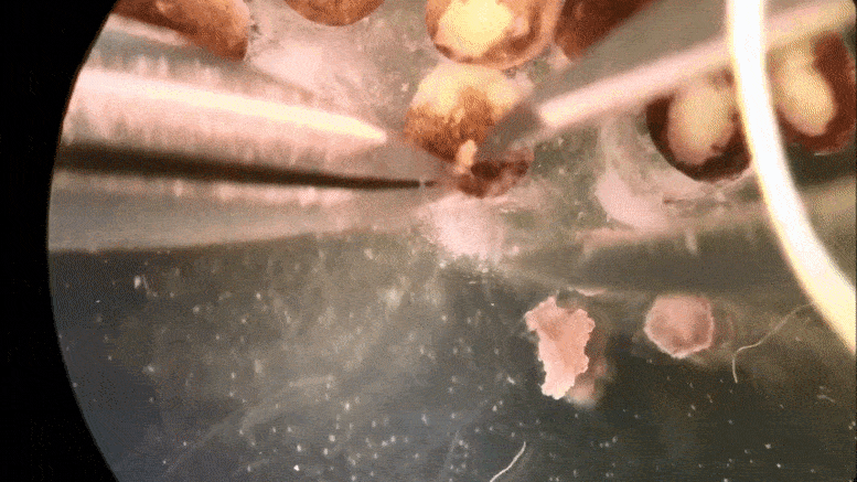
Cells being manipulated and assembled. Credit: Douglas Blackiston, Tufts University

Tiny ‘xenobots’ assembled from cells promise advances from drug delivery to toxic waste clean-up.

A book is made of wood. But it is not a tree. The dead cells have been repurposed to serve another need.
Now a team of scientists has repurposed living cells—scraped from frog embryos—and assembled them into entirely new life-forms. These millimeter-wide “xenobots” can move toward a target, perhaps pick up a payload (like a medicine that needs to be carried to a specific place inside a patient)—and heal themselves after being cut.
“These are novel living machines,” says Joshua Bongard, a computer scientist and robotics expert at the University of Vermont who co-led the new research. “They’re neither a traditional robot nor a known species of animal. It’s a new class of artifact: a living, programmable organism.”
The new creatures were designed on a supercomputer at UVM—and then assembled and tested by biologists at Tufts University. “We can imagine many useful applications of these living robots that other machines can’t do,” says co-leader Michael Levin who directs the Center for Regenerative and Developmental Biology at Tufts, “like searching out nasty compounds or radioactive contamination, gathering microplastic in the oceans, traveling in arteries to scrape out plaque.”
The results of the new research were published on January 13, 2020, in the Proceedings of the National Academy of Sciences.
A team of scientists at the University of Vermont and Tufts University designed living robots on a UVM supercomputer. Then, at Tufts, they re-purposed living frog cells — and assembled them into entirely new life-forms. These tiny ‘xenobots’ can move on their own, circle a target and heal themselves after being cut. These novel living machines are neither a traditional robot nor a known species of animal. They’re a new class of artifact: a living, programmable organism. They could, one day, be used for tasks as varied as searching out radioactive contamination, gathering microplastic in the oceans, or traveling in human arteries to scrape out plaque.

Bespoke Living Systems

People have been manipulating organisms for human benefit since at least the dawn of agriculture, genetic editing is becoming widespread, and a few artificial organisms have been manually assembled in the past few years—copying the body forms of known animals.
But this research, for the first time ever, “designs completely biological machines from the ground up,” the team writes in their new study.
With months of processing time on the Deep Green supercomputer cluster at UVM’s Vermont Advanced Computing Core, the team—including lead author and doctoral student Sam Kriegman—used an evolutionary algorithm to create thousands of candidate designs for the new life-forms. Attempting to achieve a task assigned by the scientists—like locomotion in one direction—the computer would, over and over, reassemble a few hundred simulated cells into myriad forms and body shapes. As the programs ran—driven by basic rules about the biophysics of what single frog skin and cardiac cells can do—the more successful simulated organisms were kept and refined, while failed designs were tossed out. After a hundred independent runs of the algorithm, the most promising designs were selected for testing.
Then the team at Tufts, led by Levin and with key work by microsurgeon Douglas Blackiston—transferred the in silico designs into life. First they gathered stem cells, harvested from the embryos of African frogs, the species Xenopus laevis. (Hence the name “xenobots.”) These were separated into single cells and left to incubate. Then, using tiny forceps and an even tinier electrode, the cells were cut and joined under a microscope into a close approximation of the designs specified by the computer.
A time-lapse recording of cells being manipulated and assembled, using in silico designs to create in vivo living machines, called xenobots. These novel living robots were created by a team from Tufts University and the University of Vermont.
Assembled into body forms never seen in nature, the cells began to work together. The skin cells formed a more passive architecture, while the once-random contractions of heart muscle cells were put to work creating ordered forward motion as guided by the computer’s design, and aided by spontaneous self-organizing patterns—allowing the robots to move on their own.
These reconfigurable organisms were shown to be able move in a coherent fashion—and explore their watery environment for days or weeks, powered by embryonic energy stores. Turned over, however, they failed, like beetles flipped on their backs.
Later tests showed that groups of xenobots would move around in circles, pushing pellets into a central location—spontaneously and collectively. Others were built with a hole through the center to reduce drag. In simulated versions of these, the scientists were able to repurpose this hole as a pouch to successfully carry an object. “It’s a step toward using computer-designed organisms for intelligent drug delivery,” says Bongard, a professor in UVM’s Department of Computer Science and Complex Systems Center.

Living Technologies

Many technologies are made of steel, concrete or plastic. That can make them strong or flexible. But they also can create ecological and human health problems, like the growing scourge of plastic pollution in the oceans and the toxicity of many synthetic materials and electronics. “The downside of living tissue is that it’s weak and it degrades,” say Bongard. “That’s why we use steel. But organisms have 4.5 billion years of practice at regenerating themselves and going on for decades.” And when they stop working—death—they usually fall apart harmlessly. “These xenobots are fully biodegradable,” say Bongard, “when they’re done with their job after seven days, they’re just dead skin cells.”
Joshua Bongard, University of Vermont
Robotics expert Joshua Bongard, a computer scientist at the University of Vermont, co-led new research that led to the creation of a new class of artifact: a living, programmable organism a called xenobot. Credit: Joshua Brown, UVM
Your laptop is a powerful technology. But try cutting it in half. Doesn’t work so well. In the new experiments, the scientists cut the xenobots and watched what happened. “We sliced the robot almost in half and it stitches itself back up and keeps going,” says Bongard. “And this is something you can’t do with typical machines.”

Cracking the Code

Both Levin and Bongard say the potential of what they’ve been learning about how cells communicate and connect extends deep into both computational science and our understanding of life. “The big question in biology is to understand the algorithms that determine form and function,” says Levin. “The genome encodes proteins, but transformative applications await our discovery of how that hardware enables cells to cooperate toward making functional anatomies under very different conditions.”
To make an organism develop and function, there is a lot of information sharing and cooperation—organic computation—going on in and between cells all the time, not just within neurons. These emergent and geometric properties are shaped by bioelectric, biochemical, and biomechanical processes, “that run on DNA-specified hardware,” Levin says, “and these processes are reconfigurable, enabling novel living forms.”
The scientists see the work presented in their new PNAS study—”A scalable pipeline for designing reconfigurable organisms,”—as one step in applying insights about this bioelectric code to both biology and computer science. “What actually determines the anatomy towards which cells cooperate?” Levin asks. “You look at the cells we’ve been building our xenobots with, and, genomically, they’re frogs. It’s 100% frog DNA—but these are not frogs. Then you ask, well, what else are these cells capable of building?”
“As we’ve shown, these frog cells can be coaxed to make interesting living forms that are completely different from what their default anatomy would be,” says Levin. He and the other scientists in the UVM and Tufts team—with support from DARPA’s Lifelong Learning Machines program and the National Science Foundation—believe that building the xenobots is a small step toward cracking what he calls the “morphogenetic code,” providing a deeper view of the overall way organisms are organized—and how they compute and store information based on their histories and environment.

Future Shocks

Many people worry about the implications of rapid technological change and complex biological manipulations. “That fear is not unreasonable,” Levin says. “When we start to mess around with complex systems that we don’t understand, we’re going to get unintended consequences.” A lot of complex systems, like an ant colony, begin with a simple unit—an ant—from which it would be impossible to predict the shape of their colony or how they can build bridges over water with their interlinked bodies.
“If humanity is going to survive into the future, we need to better understand how complex properties, somehow, emerge from simple rules,” says Levin. Much of science is focused on “controlling the low-level rules. We also need to understand the high-level rules,” he says. “If you wanted an anthill with two chimneys instead of one, how do you modify the ants? We’d have no idea.”
“I think it’s an absolute necessity for society going forward to get a better handle on systems where the outcome is very complex,” Levin says. “A first step towards doing that is to explore: how do living systems decide what an overall behavior should be and how do we manipulate the pieces to get the behaviors we want?”
In other words, “this study is a direct contribution to getting a handle on what people are afraid of, which is unintended consequences,” Levin says—whether in the rapid arrival of self-driving cars, changing gene drives to wipe out whole lineages of viruses, or the many other complex and autonomous systems that will increasingly shape the human experience.
“There’s all of this innate creativity in life,” says UVM’s Josh Bongard. “We want to understand that more deeply—and how we can direct and push it toward new forms.”
Reference: “A scalable pipeline for designing reconfigurable organisms” by Sam Kriegman, Douglas Blackiston, Michael Levin and Josh Bongard, 13 January 2020, Proceedings of the National Academy of Sciences.
DOI: 10.1073/pnas.1910837117

CHINESE TAOIST ALCHEMY

Newly Sequenced Genome of ‘Sacred Lotus’ May Hold Anti-Aging Secrets

Scientists Sequence Genome of Sacred Lotus
Nelumbo nucifera from China, more commonly known as the ‘sacred lotus’. Credit: Jane Shen-Miller /UCLA
Scientists have sequenced the genome of the ‘sacred lotus,” a finding that could reveal many secrets about aging and genetic defects.
A team of 70 scientists from the U.S., China, Australia and Japan today reports having sequenced and annotated the genome of the “sacred lotus,” which is believed to have a powerful genetic system that repairs genetic defects, and may hold secrets about aging successfully. The scientists sequenced more than 86 percent of the nearly 27,000 genes of the plant, Nelumbo nucifera, which is revered in China and elsewhere as a symbol of spiritual purity and longevity.
“The lotus genome is an ancient one, and we now know its ABCs,” said Jane Shen-Miller, one of three corresponding authors of the research and a senior scientist with UCLA’s Center for the Study of Evolution and the Origin of Life. “Molecular biologists can now more easily study how its genes are turned on and off during times of stress and why this plant’s seeds can live for 1,300 years. This is a step toward learning what anti-aging secrets the sacred lotus plant may offer.”
Shen-Miller said the lotus’ genetic repair mechanisms could be very useful if they could be transferred to humans or to crops — such as rice, corn and wheat — whose seeds have life spans of only a few years. “If our genes could repair disease as well as the lotus’ genes, we would have healthier aging. We need to learn about its repair mechanisms, and about its biochemical, physiological and molecular properties, but the lotus genome is now open to everybody.”
In the early 1990s, Shen-Miller led a UCLA research team that recovered a viable lotus seed that was almost 1,300 years old from a lake bed in northeastern China. It was a remarkable discovery, given that many other plant seeds are known to remain viable for just 20 years or less.
In 1996, Shen-Miller led another visit to China. Working in Liaoning province, her team collected about 100 lotus seeds — most were approximately 450 to 500 years old — with help from local farmers. To the researchers’ surprise, more than 80 percent of the lotus seeds that were tested for viability germinated. That indicated that the plant must have a powerful genetic system capable of repairing germination defects arising from hundreds of years of aging, Shen-Miller said.
Understanding how the lotus repair mechanism works — and its possible implications for human health — is essentially a three-step process, said Crysten Blaby-Haas, a UCLA postdoctoral scholar in chemistry and biochemistry and co-author of the research. “Knowing the genome sequence was step one. Step two would be identifying which of these genes contributes to longevity and repairing genetic damage. Step three would be potential applications for human health, if we find and characterize those genes. The genome sequence will aid in future analysis.
“The next question is what are these genes doing, and the biggest question is how they contribute to the longevity of the lotus plant and its other interesting attributes,” Blaby-Haas said. “Before this, when scientists studied the lotus, it’s almost as if they were blind; now they can see. Once you know the repertoire of genes, you have a foundation to study their functions.”
The genome sequence reveals that, when compared with known gene sequences of dozens of other plants, the lotus bears the closest resemblance to the ancestor of all eudicots, a broad category of flowering plants that includes the apple, peanut, tomato, cotton, cactus and tobacco plants.
The lotus forms a separate branch of the eudicot family tree; it lacks a signature triplication of the genome seen in most other members of this family, said Ray Ming, professor of plant biology at the University of Illinois at Urbana-Champaign, who led the analysis with Shen-Miller and Shaohua Li, director of the Wuhan Botanical Garden at the Chinese Academy of Sciences.
Whole-genome duplications — the doubling or tripling of an organism’s entire genetic endowment — are important events in plant evolution, Ming said. Some of the duplicated genes retain their original structure and function, and others gradually adapt and take on new functions. If those changes are beneficial, the genes persist; if they’re harmful, they disappear from the genome.
Many agricultural crops, including watermelon, sugar cane and wheat, benefit from genome duplications, said Robert VanBuren, a graduate student in Ming’s laboratory and a co-author of the study.
The genome of most other eudicots triplicated 100 million years ago, but the researchers found that the lotus experienced a separate, whole-genome duplication about 65 million years ago.
Shen-Miller said experts in aging and stress will be eager to study the lotus genes because of the plant’s extraordinary longevity. “The lotus can age for 1,000 years, and even survives freezing weather,” she said. “Its genetic makeup can combat stress. Most crops don’t have a very long shelf life. But starches and proteins in lotus seeds remain palatable and actively promote seed germination, even after centuries of aging.”
The lotus’ unusual genetics give it some unique survival skills. Its leaves repel grime and water, its flowers generate heat to attract pollinators and the coating of lotus fruit is covered with antibiotics and wax that ensure the viability of the seed it contains.
Blaby-Haas studied lotus gene families potentially involved in how plants metabolize metals. One family, in particular, caught her attention. “We found that the lotus has 16 of these genes, while most plants have only one or two,” Blaby-Haas said. “Either this is an extremely important protein in the lotus, which is why it needs so many copies, or the duplication allows a novel function to arise; we don’t know which is correct.”
These genes may be related to the unique environment of the lotus, which grows with its roots submerged in water, she said. (Lotus was a land plant that adapted to the water.)
The sacred lotus is known from the geologic record as early as 135 million years ago, when dinosaurs roamed the Earth, Shen-Miller said. It has been grown for at least 4,000 years in China, where every part of the plant has long been used in food and medicine.
Co-author Sabeeha Merchant is a UCLA professor of biochemistry whose laboratory studies the biology of metals like iron, copper and zinc. Other co-authors include J. William Schopf, director of UCLA’s Center for the Study of Evolution and the Origin of Life and a professor in the department of Earth and space sciences, who studied the geology of the lotus burial lake; and Steven Karpowicz, a former UCLA graduate student in Merchant’s laboratory who is currently at Eastern Oregon University.
Publication: Ray Ming, et al., “Genome of the long-living sacred lotus (Nelumbo nucifera Gaertn.),” Genome Biology 2013, 14:R41; doi:10.1186/gb-2013-14-5-r41
Image: Jane Shen-Miller /UCLA

Revealed: The Mysterious, Legendary Giant Squid’s Genome


Giant Squid Illustration
How did the monstrous giant squid – reaching school-bus size, with eyes as big as dinner plates and tentacles that can snatch prey 10 yards away – get so scarily big?
Today, important clues about the anatomy and evolution of the mysterious giant squid (Architeuthis dux) are revealed through publication of its full genome sequence by a University of Copenhagen-led team that includes scientist Caroline Albertin of the Marine Biological Laboratory (MBL), Woods Hole.
Giant squid are rarely sighted and have never been caught and kept alive, meaning their biology (even how they reproduce) is still largely a mystery. The genome sequence can provide important insight.
Giant Squid Captures Sailor
The giant squid has long been a subject of horror lore. In this original illustration from Jules Verne’s ‘20,000 Leagues Under the Sea,’ a giant squid grasps a helpless sailor. Credit: Alphonse de Neuville
“In terms of their genes, we found the giant squid look a lot like other animals. This means we can study these truly bizarre animals to learn more about ourselves,” says Albertin, who in 2015 led the team that sequenced the first genome of a cephalopod (the group that includes squid, octopus, cuttlefish, and nautilus).
Led by Rute da Fonseca at University of Copenhagen, the team discovered that the giant squid genome is big: with an estimated 2.7 billion DNA base pairs, it’s about 90 percent the size of the human genome.
Albertin analyzed several ancient, well-known gene families in the giant squid, drawing comparisons with the four other cephalopod species that have been sequenced and with the human genome.
She found that important developmental genes in almost all animals (Hox and Wnt) were present in single copies only in the giant squid genome. That means this gigantic, invertebrate creature – long a source of sea-monster lore – did NOT get so big through whole-genome duplication, a strategy that evolution took long ago to increase the size of vertebrates.
So, knowing how this squid species got so giant awaits further probing of its genome.
“A genome is a first step for answering a lot of questions about the biology of these very weird animals,” Albertin said, such as how they acquired the largest brain among the invertebrates, their sophisticated behaviors and agility, and their incredible skill at instantaneous camouflage.
“While cephalopods have many complex and elaborate features, they are thought to have evolved independently of the vertebrates. By comparing their genomes we can ask, ‘Are cephalopods and vertebrates built the same way or are they built differently?'” Albertin says.
Albertin also identified more than 100 genes in the protocadherin family – typically not found in abundance in invertebrates – in the giant squid genome.
“Protocadherins are thought to be important in wiring up a complicated brain correctly,” she says. “They were thought they were a vertebrate innovation, so we were really surprised when we found more than 100 of them in the octopus genome (in 2015). That seemed like a smoking gun to how you make a complicated brain. And we have found a similar expansion of protocadherins in the giant squid, as well.”
Lastly, she analyzed a gene family that (so far) is unique to cephalopods, called reflectins. “Reflectins encode a protein that is involved in making iridescence. Color is an important part of camouflage, so we are trying to understand what this gene family is doing and how it works,” Albertin says.
“Having this giant squid genome is an important node in helping us understand what makes a cephalopod a cephalopod. And it also can help us understand how new and novel genes arise in evolution and development.”
Reference: “A draft genome sequence of the elusive giant squid, Architeuthis dux” by Rute R da Fonseca, Alvarina Couto, Andre M Machado, Brona Brejova, Carolin B Albertin, Filipe Silva, Paul Gardner, Tobias Baril, Alex Hayward, Alexandre Campos, Ângela M Ribeiro, Inigo Barrio-Hernandez, Henk-Jan Hoving, Ricardo Tafur-Jimenez, Chong Chu, Barbara Frazão, Bent Petersen, Fernando Peñaloza, Francesco Musacchia, Graham C Alexander, Jr, Hugo Osório, Inger Winkelmann, Oleg Simakov, Simon Rasmussen, M Ziaur Rahman, Davide Pisani, Jakob Vinther, Erich Jarvis, Guojie Zhang, Jan M Strugnell, L Filipe C Castro, Olivier Fedrigo, Mateus Patricio, Qiye Li, Sara Rocha, Agostinho Antunes, Yufeng Wu, Bin Ma, Remo Sanges, Tomas Vinar, Blagoy Blagoev, Thomas Sicheritz-Ponten, Rasmus Nielsen and M Thomas P Gilbert, 16 January 2020, GigaScience.
DOI: 10.1093/gigascience/giz152

Study Reveals That Giant Squid Throughout the World Are Genetically Similar


Study Reveals Population Structure of the Giant Squid Architeuthis
Study reveals that giant squid such as this one are genetically similar throughout the world. David Paul/Museum Victoria
In a newly published study, researchers examine the mitochondrial genome diversity of 43 giant squid samples collected from across the range of the species, finding that there is only one global species of giant squid, Architeuthis.
The giant squid is one of the most enigmatic animals on the planet. It is extremely rarely seen, except as the remains of animals that have been washed ashore, and placed in the formalin or ethanol collections of museums. But now, researchers at the University of Copenhagen leading an international team, have discovered that no matter where in the world they are found, the fabled animals are so closely related at the genetic level that they represent a single, global population, and thus despite previous statements to the contrary, a single species worldwide. Thus the circle, that was first opened in 1857 by the famous Danish naturalist Japetus Steenstrup as he first described the animal, can be closed. It was Steenstrup that realized this beast was the same animal that in the past gave rise to centuries of sailors tails, and even in more recent became immortalized by writers such as Jules Verne and Herman Melville, by demonstrating that the monster was based in reality, and gave it the latin name Architeuthis dux.
It was less than 1 year ago, that the giant squid, Architeuthis dux, was first filmed alive in its natural element. Taken at a depth of 630m and after 100 missions and 400 hours of filming, the footage was captured by a small submarine lying off the Japanese island of Chichi Jima – near to the famous Iwo Jima that was the scene of some of the bloodiest fighting between Japan and the USA in the Second World War.
Now, PhD student Inger Winkelmann and her supervisor Professor Tom Gilbert, from the Basic Research Center in GeoGenetics at the Natural History Museum of Denmark, Copenhagen University, have managed to place new bricks into the puzzle of this giant 10 armed invertebrate, that is credibly believed to grow up to 13 meters long and way over 900 kg.
And the two scientists conclusions are: No matter what a sample looks like, its one species all over the deep oceans of the planet.
Sinking to the depths
PhD student Inger Winkelmann says about these findings, that are published in the esteemed British journal, the Proceedings of the Royal Society B:
– We have analysed DNA from the remains of 43 giant squid collected from all over the world. The results show, that the animal is genetically nearly identical all over the planet, and shows no evidence of living in geographically structured populations. We suggest that one possible explanation for this is that although evidence suggests the adults remain in relatively restricted geographic regions, the young that live on the ocean’s surfaces must drift in the currents globally. Once they reach a large enough size to survive the depths, we believe they dive to the nearest suitable deep waters, and there the cycle begins again. Nevertheless, we still lack a huge amount of knowledge about these creatures. How big a range to they really inhabit as adults? Have they in the past been threatened by things such as climate change, and the populations of their natural enemies, such as the planet’s largest toothed whale, the sperm whale that can grow up to 20 m in length and 50 tons? And at an even more basic level…how old do they even get and how quickly do they grow?
The kraken and the seamonk
These new results about the mysterious giant squid are released, fittingly enough, on the 200th anniversary of the Danish naturalist and polymath, Japetus Steenstrup (born in 1813).
At the age of 44, in 1857, it was Steenstrup who saw that many of the monsters of sea-legend were related to fragments that he had been sent of what appeared to be a giant squid, and in doing so described the species for the first time and removed any hope that sea monsters such as the Kraken and sea-monk really existed (although nevertheless, similar monsters still inspired beasts in literature and even films throughout the 20th century, including Tolkein’s Lord of the Rings in 1957).
Professor Tom Gilbert, who lead the team that undertook the research, says:
– It has been tremendous to apply the latest techniques in genetic and computational analyses, to follow up on Steenstrup’s scientific research 146 years after he started it. But its also been a fantastic experience to work with the giant squid as a species, because of its legendary status as a seamonster. But despite our findings, I have no doubt that these myths and legends will continue get today’s children to open their eyes up – so they will be just as big as the real giant squid is equipped with to navigate the depths.
The work was undertaken in collaboration with researchers around the world, including scientists in Australia, New Zealand, Japan, Spain, Portugal, USA and Ireland.
Publication: Inger Winkelmann, et al., “Mitochondrial genome diversity and population structure of the giant squid Architeuthis: genetics sheds new light on one of the most enigmatic marine species,” Proc. R. Soc. B 22 May 2013 vol. 280 no. 1759; doi: 10.1098/rspb.2013.0273
Image: David Paul/Museum Victoria


After 15 Years, Scientists Finally Solve Huygens Landing Spin Mystery

Airflow Across Huygens Replica
As part of the international Cassini mission, ESA’s Huygens probe made history on January 14, 2005, when it became the first probe to successfully land on another world in the outer Solar System. However, during its descent, the probe began spinning the ‘wrong’ way – and new results confirm why.
The image, produced in the wind tunnel at the PRISME Laboratory at the University of Orléans, France, shows how air flows across a 1:3 scale replica of Huygens – as visualized using white smoke. It was taken as part of subsonic testing performed from 2017 to 2019 to determine how ESA’s Huygens probe spun during its descent to Titan.
Huygens was released from Cassini spinning anti-clockwise but, approximately 10 minutes after entering Titan’s atmosphere, the probe’s spin unexpectedly reversed to become clockwise. It kept spinning this way for the rest of the descent; luckily, the magnitude of this reversed spin was similar to that expected by the researchers, meaning that the unexpected flip affected the timing of the planned observations, but did not dramatically affect their quality.
The recent tests now confirm the cause of this flip in spin direction. While the probe was equipped with vanes to regulate its spin, other appendages on the spacecraft produced a torque in the opposite direction; this was only exacerbated by the way in which these vanes redirected the gas flow around the body of the probe, so that an overall ‘negative’, or clockwise, spin effect was created. There are also indications that the booms of the Huygens Atmospheric Structure Instrument (HASI) might have not been fully or symmetrically deployed during descent; this effect is under further investigation.
Credit: CNRS/LPC2E/PRISME
Fifteen years ago, ESA’s Huygens probe made history when it descended to the surface of Saturn’s moon Titan and became the first probe to successfully land on another world in the outer Solar System. However, during its descent, the probe began spinning the wrong way – and recent tests now reveal why.
Launched in 1997, the NASA/ESA/ASI Cassini-Huygens mission remains iconic and has contributed an enormous amount to our understanding of Saturn and its moon Titan since its arrival at the ringed planet in late 2004.
The mission comprised an orbiter, Cassini, which went on to orbit Saturn for over 13 years after becoming the first spacecraft to do so, and a small atmospheric probe, ESA’s Huygens lander, which headed down to explore the physical properties and atmosphere of Titan on 14 January 2005.
Huygens’ risky descent lasted for 2 hours and 27 minutes, and the data the small probe gathered went on to facilitate a wealth of discoveries about this fascinating moon.
A rendering of Huygens descent and touchdown created using real data recorded by the probe’s instruments as it descended to the surface of Titan, Saturn’s largest moon, on January 14, 2005.
The animation takes into account Titan’s atmospheric conditions, including the Sun and wind direction, the behavior of the parachute (with some artistic interpretation only on the movement of the ropes after touchdown), and the dynamics of the landing itself. Even the stones immediately facing Huygens were rendered to match the photograph of the landing site returned from the probe, which is revealed at the end of the animation.
The lander returned the first in situ measurements of Titan’s atmosphere, determining its pressure, density and temperature from an altitude of 1400km down to the surface. The probe’s Doppler Wind Experiment (DWE) spotted strong east-west winds in the moon’s atmosphere, some of which rotated faster than the moon itself. It shed light on why Titan’s atmosphere contains methane, nitrogen, and tiny aerosols, and in what quantities, and detected signs of geological processes and features in the moon’s interior such as cryovolcanism and, potentially, a large subsurface ocean.
Predicted and Actual Spin Rates of Huygens
This graph shows the ‘spin profile’ of ESA’s Huygens probe as it descended to the surface of the Saturnian moon Titan on January 14, 2005: the dotted line shows the predicted profile, while the solid line shows the actual profile as tracked by the probe’s onboard engineering sensors. The horizontal axis indicates UTC time and the vertical axis the spin rate (in revolutions per minute).
As part of the international Cassini mission, Huygens made history when it became the first probe to successfully land on another world in the outer Solar System. However, during its descent, the probe began spinning the ‘wrong’ way – as this graph illustrates. Huygens was released from the Cassini orbiter spinning anti-clockwise at a rate of 7.5 rotations per minute. This direction of spin was intended to continue throughout the descent, with the spin rate controlled and influenced by the deployment of landing parachutes.
These results were obtained via subsonic wind tunnel testing at the PRISME Laboratory at the University of Orléans, France.
Credit: Reproduced from Lebreton et al. (2005)
By cutting through and exploring the thick haze that enshrouds the moon, the probe also helped scientists to visualize the surface of Titan, returning evidence of past watery activity, such as dried up riverbeds and drainage networks and long-empty lake basins, and observations of the vast dunes of sand and ice.
However, one thing remained a mystery: why Huygens spun in the ‘wrong’ direction during its descent. The probe was released from Cassini spinning anti-clockwise at a rate of 7.5 rotations per minute. Due to the design of the probe, its spin rate helped to keep Huygens stable firstly as it spent three weeks coasting down to Titan, and then as it eventually entered the moon’s atmosphere.
This movie was produced in the wind tunnel at the PRISME Laboratory at the University of Orléans, France, where subsonic testing was performed from 2017 to 2019 to determine how ESA’s Huygens probe spun during its descent to Titan.
Although Huygens initially behaved as anticipated, during descent the probe’s spin rate decreased far more rapidly than expected, before reversing after approximately 10 minutes to adopt a clockwise direction.
It kept spinning this way for the remaining 2 hours and 15 hours of descent; luckily, the magnitude of this reversed spin was similar to that expected by the researchers, meaning that the unexpected flip affected the timing of the planned observations, but did not dramatically affect their quality.
Huygens Probe in PRISME Laboratory Wind Tunnel
This image shows a replica of ESA’s Huygens probe, as used in recent wind tunnel testing to characterize how the spacecraft spun as it descended to the surface of Titan – an event that made history on 14 January 2005. This mock-up is at a scale of 1/3 – the real Huygens probe, and appendages, was 3 times bigger.
Various booms and appendages can be seen around the probe, including four flat Radar Altimeter antennas, two deployable booms for the Huygens Atmospheric Structure Instrument (HASI), different telemetry and sensor heads, pressure and temperature sensors, and the mechanism by which the heat shield and coverings would detach after atmospheric entry. This side of the spacecraft would be the side to touch down on the surface of Titan; the probe’s descent parachute is stored on the side not in view.
In this image, the mock-up is deployed in the test section of the wind tunnel at the PRISME Laboratory at the University of Orléans, France, where subsonic testing was performed from 2017 to 2019 to determine the spin profile of Huygens during its descent.
Credit: CNRS/LPC2E/PRISME; ESA
Previous studies have investigated this behavior (for example a study conducted by Vorticity in 2014–2015) and recent subsonic wind tunnel testing at the PRISME Laboratory at the University of Orléans, France, now confirms the main cause. The study was carried out from 2017 to 2019 under an ESA contract with LPC2E/CNRS-University of Orléans.
Huygens was equipped with 36 angled vanes that were used to control the spin of the descent module. However, two of the probe’s main appendages, the Separation Subsystem (SEPS) and the Radar Altimeter (RA) antennae, actually produced an unexpected torque opposite to that produced by the vanes. This effect was amplified as the vanes altered the gas flow around the descent module in a way that enhanced the amplitude of the ‘negative torque’ – the effect that made Huygens flip its direction of spin – until it exceeded the influence of the vanes.
The resolution of this engineering mystery will help inform the design of entry probes in the future, furthering our exploration of the Solar System.