Sunday, August 30, 2020

Extensive Search for COVID-19 Drugs Finds Promising Compounds Originally Developed for SARS
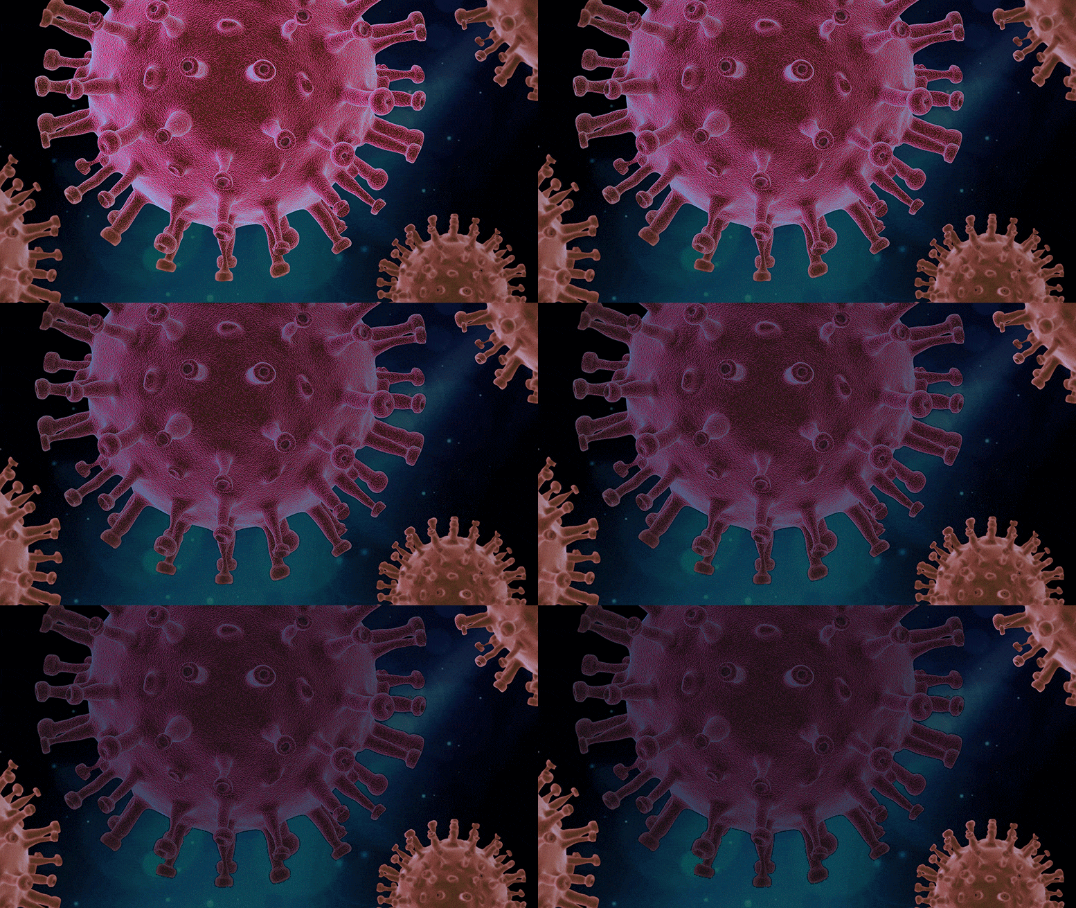
COVID-19 Achille's Heel
An extensive search and testing of current drugs and drug-like compounds has revealed compounds previously developed to fight SARS might also work against COVID-19.
Using the National Drug Discovery Centre, researchers from the Walter and Eliza Hall Institute identified drug-like compounds that could block a key coronavirus protein called PLpro. This protein, found in all coronaviruses, is essential for the virus to hijack and multiply within human cells, and disable their anti-viral defenses.
Initially developed as potential treatments for SARS, the compounds prevented the growth of the SARS-CoV-2 virus (which causes COVID-19) in the laboratory.
The discovery, published yesterday in The EMBO Journal, was led by Professor David Komander, Professor Marc Pellegrini, Professor Guillaume Lessene, and Dr. Theresa Klemm.

At a glance

  • Australian researchers have identified a molecular target for potential new COVID-19 treatments
  • A chemical compound, originally discovered to inhibit SARS, shows promise for halting the growth of the COVID-19 virus (SARS-CoV-2)
  • The discoveries were made by leveraging the capabilities of the National Drug Discovery Centre and ANSTO’s Australian Synchrotron, and may underpin the development of new drugs for COVID-19

Targeting a key viral protein

Coronaviruses, including the viruses that cause COVID-19 and SARS, all contain a protein called PLpro, which allows the virus to hijack human cells and disable their anti-viral defenses.
Professor Komander said PLpro belonged to a family of proteins called ‘deubiquitinases’, which his team had studied for the last 15 years in a range of diseases.
“When we looked at how SARS-CoV-2 functions, it became clear that the PLpro deubiquitinase was a key component of the virus — as it is in other coronaviruses, including the SARS-CoV-1 virus, which causes SARS,” he said.
“We quickly established the VirDUB program to investigate how PLpro functions and what it looks like. These are critical first steps towards discovering new drugs that could be potential therapies for COVID-19.”
Using ANSTO’s Australian Synchrotron, the VirDUB team rapidly ascertained how PLpro interacts with human proteins — homing in on a target that could be blocked by new drugs.

Discovering new medicines

The National Drug Discovery Centre was critical to rapidly search for drugs that could block PLpro.
“We scanned thousands of currently listed drugs, as well as thousands of drug-like compounds, to see if they were effective in blocking the SARS-CoV-2 PLpro,” Professor Komander said.
“While existing drugs were not effective in blocking PLpro, we discovered that compounds developed in the last decade against SARS, could prevent the growth of SARS-CoV-2 in pre-clinical testing in the laboratory.”
The next step is to turn these compounds into drugs that could be used to treat COVID-19, Professor Komander said.
“We now need to develop the compounds into medicines, and make sure they are safe for patients.
“Importantly, drugs that are able to inactivate PLpro may be useful not just for COVID-19 but may also work against other coronavirus diseases, as they emerge in the future.”
###
Reference: “Mechanism and inhibition of the papain‐like protease, PLpro, of SARS‐CoV‐2” by Theresa Klemm, Gregor Ebert, Dale J Calleja, Cody C Allison, Lachlan W Richardson, Jonathan P Bernardini, Bernadine GC Lu, Nathan W Kuchel, Christoph Grohmann, Yuri Shibata, Zhong Yan Gan, James P Cooney, Marcel Doerflinger, Amanda E Au, Timothy R Blackmore, Gerbrand J van der Heden van Noort, Paul P Geurink, Huib Ovaa, Janet Newman, Alan Riboldi‐Tunnicliffe, Peter E Czabotar, Jeffrey P Mitchell, Rebecca Feltham, Bernhard C Lechtenberg, Kym N Lowes, Grant Dewson, Marc Pellegrini, Guillaume Lessene and David Komander, 26 August 2020, The EMBO Journal.
DOI: 10.15252/embj.2020106275
The publication in The EMBO Journal research was a multidisciplinary collaboration of research teams at the Walter and Eliza Hall Institute of Medical Research, the National Drug Discovery Centre, ANSTO’s Australian Synchrotron, the Commonwealth Scientific and Industrial Research Organisation (CSIRO), the Oncode Institute and Department of Cell and Chemical Biology (Leiden University, The Netherlands).
The research was funded by Hengyi Pacific Pty Ltd, the Australian National Health and Medical Research Council and Medical Research Future Fund, the Nederlandse Organisatie voor Wetenschappelijk Onderzoek (NWO), and the Victorian Government.

New Internet Speed World Record: 178 Terabits a Second

Fast Internet Concept
The world’s fastest data transmission rate has been achieved by a team of University College London engineers who reached an internet speed a fifth faster than the previous record.
Working with two companies, Xtera and KDDI Research, the research team led by Dr. Lidia Galdino (UCL Electronic & Electrical Engineering), achieved a data transmission rate of 178 terabits a second (178,000,000 megabits a second) – a speed at which it would be possible to download the entire Netflix library in less than a second.
The record, which is double the capacity of any system currently deployed in the world, was achieved by transmitting data through a much wider range of colors of light, or wavelengths, than is typically used in optical fiber. (Current infrastructure uses a limited spectrum bandwidth of 4.5THz, with 9THz commercial bandwidth systems entering the market, whereas the researchers used a bandwidth of 16.8THz.)
To do this, researchers combined different amplifier technologies needed to boost the signal power over this wider bandwidth and maximized speed by developing new Geometric Shaping (GS) constellations (patterns of signal combinations that make best use of the phase, brightness and polarisation properties of the light), manipulating the properties of each individual wavelength. The achievement is described in a new paper in IEEE Photonics Technology Letters.
The benefit of the technique is that it can be deployed on already existing infrastructure cost-effectively, by upgrading the amplifiers that are located on optical fiber routes at 40-100km intervals. (Upgrading an amplifier would cost £16,000, while installing new optical fibers can, in urban areas, cost up to £450,000 a kilometer.)
The new record, demonstrated in a UCL lab, is a fifth faster than the previous world record held by a team in Japan.
Dr. Lidia Galdino
Dr. Lidia Galdino (UCL Electronic & Electrical Engineering). Credit: UCL
At this speed, it would take less than an hour to download the data that made up the world’s first image of a black hole (which, because of its size, had to be stored on half a ton of hard drives and transported by plane). The speed is close to the theoretical limit of data transmission set out by American mathematician Claude Shannon in 1949.
Lead author Dr. Galdino, a Lecturer at UCL and a Royal Academy of Engineering Research Fellow, said: “While current state-of-the-art cloud data-center interconnections are capable of transporting up to 35 terabits a second, we are working with new technologies that utilize more efficiently the existing infrastructure, making better use of optical fiber bandwidth and enabling a world record transmission rate of 178 terabits a second.”
Since the start of the COVID-19 crisis, demand for broadband communication services has soared, with some operators experiencing as much as a 60% increase in internet traffic compared to before the crisis. In this unprecedented situation, the resilience and capability of broadband networks has become even more critical.
Dr. Galdino added: “But, independent of the Covid-19 crisis, internet traffic has increased exponentially over the last 10 years and this whole growth in data demand is related to the cost per bit going down. The development of new technologies is crucial to maintaining this trend towards lower costs while meeting future data rate demands that will continue to increase, with as yet unthought-of applications that will transform people’s lives.”
This work is funded by the Royal Academy of Engineering, The Royal Society Research grant, and the EPSRC program grant TRANSNET (EP/R035342/1).

Evidence of Hibernation-Like State Discovered in Tusks of Strange 250-Million-Year-Old Antarctic Creature

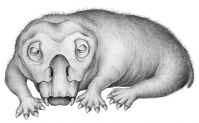
Torpor in Lystorsaurus
Life restoration of Lystrosaurus in a state of torpor. Credit: Crystal Shin
Researchers discover Fossil evidence of ‘hibernation-like’ state in tusks of 250-million-year-old Antarctic animal.
Among the many winter survival strategies in the animal world, hibernation is one of the most common. With limited food and energy sources during winters — especially in areas close to or within polar regions — many animals hibernate to survive the cold, dark winters. Though much is known behaviorally on animal hibernation, it is difficult to study in fossils.
According to new research, this type of adaptation has a long history. In a paper published on August 27, 2020, in the journal Communications Biology, scientists at Harvard University and the University of Washington report evidence of a hibernation-like state in an animal that lived in Antarctica during the Early Triassic, some 250 million years ago.
The creature, a member of the genus Lystrosaurus, was a distant relative of mammals. Lystrosaurus were common during the Permian and Triassic periods and are characterized by their turtle-like beaks and ever-growing tusks. During Lystrosaurus‘ time, Antarctica lay largely within the Antarctic Circle and experienced extended periods without sunlight each winter.
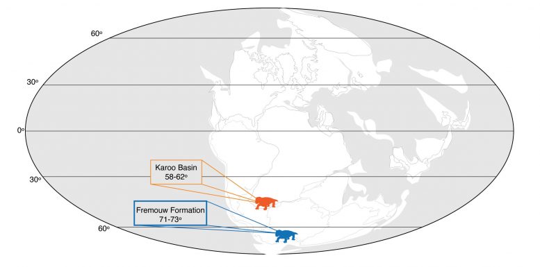
Pangea Map Early Triassic
A map of Pangea during the Early Triassic, showing the locations of the Antarctic (blue) and South African (orange) Lystrosaurus populations compared in this study. Credit: Megan Whitney/Christian Sidor
“Animals that live at or near the poles have always had to cope with the more extreme environments present there,” said lead author Megan Whitney, a postdoctoral researcher at Harvard University in the Department of Organismic and Evolutionary Biology, who conducted this study as a UW doctoral student in biology. “These preliminary findings indicate that entering into a hibernation-like state is not a relatively new type of adaptation. It is an ancient one.”
The Lystrosaurus fossils are the oldest evidence of a hibernation-like state in a vertebrate animal and indicate that torpor — a general term for hibernation and similar states in which animals temporarily lower their metabolic rate to get through a tough season — arose in vertebrates even before mammals and dinosaurs evolved.
Lystrosaurus arose before Earth’s largest mass extinction at the end of the Permian Period — which wiped out 70% of vertebrate species on land — and somehow survived. It went on to live another 5 million years into the Triassic Period and spread across swathes of Earth’s then-single continent, Pangea, which included what is now Antarctica. “The fact that Lystrosaurus survived the end-Permian mass extinction and had such a wide range in the early Triassic has made them a very well-studied group of animals for understanding survival and adaptation,” said co-author Christian Sidor, a UW professor of biology and curator of vertebrate paleontology at the Burke Museum.
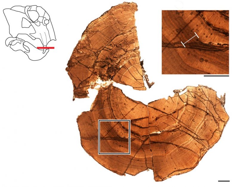
Antarctic Lystrosaurus Tusk
This thin-section of the fossilized tusk from an Antarctic Lystrosaurus shows layers of dentine deposited in rings of growth. The tusk grew inward, with the oldest layers at the edge and the youngest layers near the center, where the pulp cavity would have been. At the top right is a close-up view of the layers, with a white bar highlighting a zone indicative of a hibernation-like state. Scale bar is 1 millimeter. Credit: Megan Whitney/Christian Sidor
Today, paleontologists find Lystrosaurus fossils in India, China, Russia, parts of Africa and Antarctica. The creatures grew to be 6 to 8 feet long, had no teeth, but bore a pair of tusks in the upper jaw. The tusks made Whitney and Sidor’s study possible because, like elephants, Lystrosaurus tusks grew continuously throughout their lives. Taking cross-sections of the fossilized tusks revealed information about Lystrosaurus metabolism, growth and stress or strain. Whitney and Sidor compared cross-sections of tusks from six Antarctic Lystrosaurus to cross-sections of four Lystrosaurus from South Africa. During the Triassic, the collection sites in Antarctica were roughly 72 degrees south latitude — well within the Antarctic Circle. The collection sites in South Africa were more than 550 miles north, far outside the Antarctic Circle.
The tusks from the two regions showed similar growth patterns, with layers of dentine deposited in concentric circles like tree rings. The Antarctic fossils, however, held an additional feature that was rare or absent in tusks farther north: closely-spaced, thick rings, which likely indicate periods of less deposition due to prolonged stress, according to the researchers. “The closest analog we can find to the ‘stress marks’ that we observed in Antarctic Lystrosaurus tusks are stress marks in teeth associated with hibernation in certain modern animals,” said Whitney.
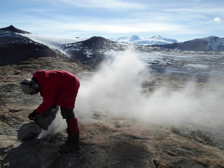
Paleontologist Christian Sidor
University of Washington paleontologist
Christian Sidor excavating fossils in Antarctica in 2017. Credit: Megan Whitney
The researchers cannot definitively conclude that Lystrosaurus underwent true hibernation. The stress could have been caused by another hibernation-like form of torpor, such as a more short-term reduction in metabolism. Lystrosaurus in Antarctica likely needed some form of hibernation-like adaptation to cope with life near the South Pole, said Whitney. Though Earth was much warmer during the Triassic than today — and parts of Antarctica may have been forested — plants and animals below the Antarctic Circle would still experience extreme annual variations in the amount of daylight, with the sun absent for long periods in winter.
Many other ancient vertebrates at high latitudes may also have used torpor, including hibernation, to cope with the strains of winter, Whitney said. But many famous extinct animals, including the dinosaurs that evolved and spread after Lystrosaurus died out, don’t have teeth that grow continuously.
Paleontologist Megan Whitney
Megan Whitney, then a University of
Washington doctoral student, excavating fossils in
Antarctica in 2017. Whitney is now a paleontologist at
Harvard University. Credit: Christian Sidor
“To see the specific signs of stress and strain brought on by hibernation, you need to look at something that can fossilize and was growing continuously during the animal’s life,” said Sidor. “Many animals don’t have that, but luckily Lystrosaurus did.” If analysis of additional Antarctic and South African Lystrosaurus fossils confirms this discovery, it may also settle another debate about these ancient, hearty animals. “Cold-blooded animals often shut down their metabolism entirely during a tough season, but many endothermic or ‘warm-blooded’ animals that hibernate frequently reactivate their metabolism during the hibernation period,” said Whitney. “What we observed in the Antarctic Lystrosaurus tusks fits a pattern of small metabolic ‘reactivation events’ during a period of stress, which is most similar to what we see in warm-blooded hibernators today.” If so, this distant cousin of mammals is a reminder that many features of life today may have been around for hundreds of millions of years before humans evolved to observe them.
Reference: “Evidence of torpor in the tusks of Lystrosaurus from the Early Triassic of Antarctica” by Megan R. Whitney and Christian A. Sidor, 27 August 2020, Communications Biology.
DOI: 10.1038/s42003-020-01207-6
The research was funded by the National Science Foundation. Grant numbers: PLR-1341304, DEB-1701383.

Scientists Use Fruit Peel to Turn Old Lithium-Ion Batteries Into New

NTU Scientists Fruit Peel Waste
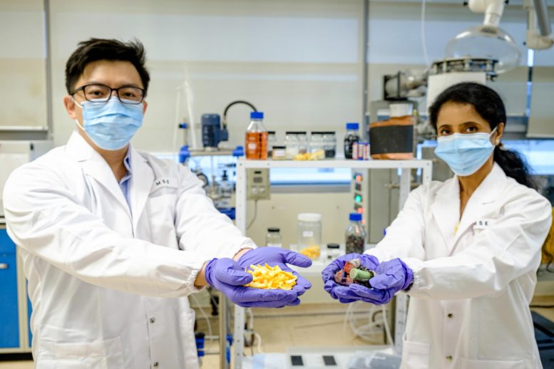
A team of scientists led by NTU has developed a novel method of using fruit peel waste to extract and reuse precious metals from spent lithium-ion batteries in order to create new batteries. L-R: Asst Prof Dalton Tay, Prof Madhavi Srinivasan. Credit: NTU Singapore
Scientists led by Nanyang Technological University, Singapore (NTU Singapore) have developed a novel method of using fruit peel waste to extract and reuse precious metals from spent lithium-ion batteries in order to create new batteries.
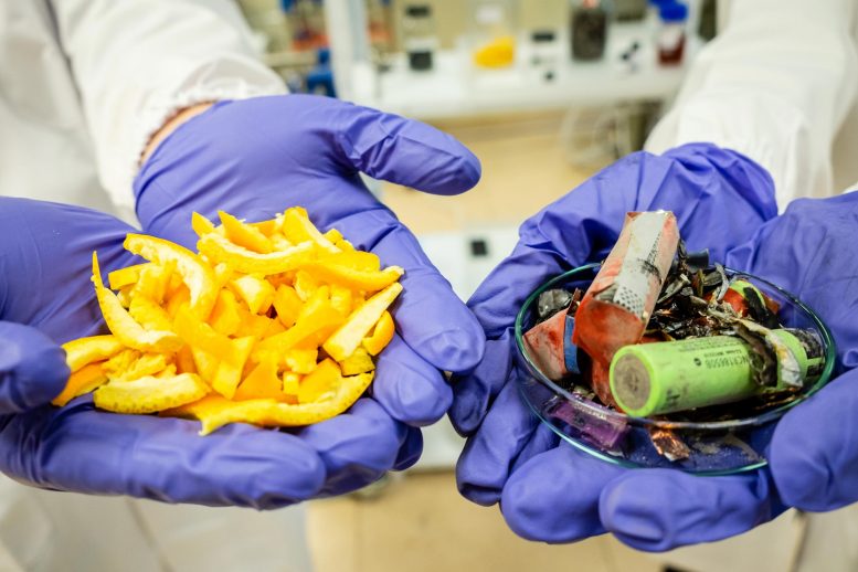
The team demonstrated their concept using orange peel, which recovered precious metals from battery waste efficiently. They then made functional batteries from these recovered metals, creating minimal waste in the process.
The scientists say that their waste-to-resource approach tackles both food waste and electronics waste,  supporting the development of a circular economy with zero waste, in which resources are kept in use for as long as possible. An estimated 1.3 billion tonnes of food waste and 50 million tonnes of e-waste are generated globally each year.
Spent batteries are conventionally treated with extreme heat (over 500°C) to smelt valuable metals, which emits hazardous toxic gases. Alternative approaches that use strong acid solutions or weaker acid solutions with hydrogen peroxide to extract the metals are being explored, but they still produce secondary pollutants that pose health and safety risks, or rely on hydrogen peroxide which is hazardous and unstable.
NTU Fruit Peel Waste
The team demonstrated their concept using orange peel, which recovered precious metals from battery waste efficiently. They then made functional batteries from these recovered metals, creating minimal non-toxic waste in the process. Credit: NTU Singapore
Professor Madhavi Srinivasan, co-director of the NTU Singapore-CEA Alliance for Research in Circular Economy (NTU SCARCE) lab, said: “Current industrial recycling processes of e-waste are energy-intensive and emit harmful pollutants and liquid waste, pointing to an urgent need for eco-friendly methods as the amount of e-waste grows. Our team has demonstrated that it is possible to do so with biodegradable substances.
“These findings build on our existing body of work at SCARCE under NTU’s Energy Research Institute (ERI@N). The SCARCE lab was set up to develop greener ways of recycling e-waste. It is also part of the NTU Smart Campus initiative, which aims to develop technologically advanced solutions for a sustainable future.”
Assistant Professor Dalton Tay of the NTU School of Materials Science and Engineering and School of Biological Sciences said: “In Singapore, a resource-scarce country, this process of urban mining to extract valuable metals from all kinds of discarded electronics becomes very important. With this method, we not only tackle the problem of resource depletion by keeping these precious metals in use as much as possible, but also the problem of e-waste and food waste accumulation – both a growing global crisis.”
The findings were published in the scientific journal Environmental Science & Technology in July.

A low-cost, sustainable approach

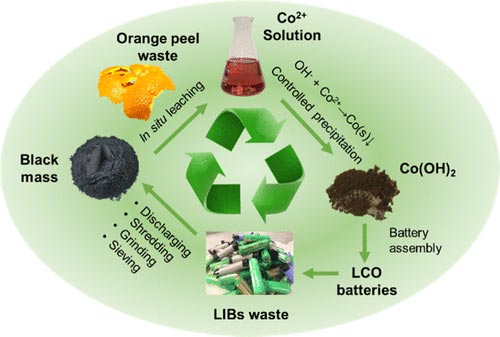
With industrial approaches to recycling battery waste generating harmful pollutants, hydrometallurgy – using water as a solvent for extraction – is increasingly being explored as a possible alternative. This process involves first shredding and crushing used batteries to form a crushed material called black mass. Researchers then extract valuable metals from black mass by dissolving it in a mix of strong acids or weak acids plus other chemicals like hydrogen peroxide under heat, before letting the metals precipitate.
While relatively more eco-friendly than conventional methods, the use of such strong chemicals on an industrial scale could generate a substantial amount of secondary pollutants, posing significant safety and health risks, said Asst Prof Tay.
Recycling Batteries
A waste-to-resource approach to recycling batteries. Credit: NTU Singapore
The NTU team found that the combination of orange peel that has been oven-dried and ground into powder, and citric acid, a weak organic acid found in citrus fruits, can achieve the same goal. 
In lab experiments, the team found that their approach successfully extracted around 90 percent of cobalt, lithium, nickel, and manganese from spent lithium-ion batteries – a comparable efficacy to the approach using hydrogen peroxide.
Asst Prof Tay explained: “The key lies in the cellulose found in orange peel, which is converted into sugars under heat during the extraction process. These sugars enhance the recovery of metals from battery waste. Naturally-occurring antioxidants found in orange peel, such as flavonoids and phenolic acids, could have contributed to this enhancement as well.”
Importantly, solid residues generated from this process were found to be non-toxic, suggesting that this method is environmentally sound, he added.
From the recovered materials, they then assembled new lithium-ion batteries, which showed a similar charge capacity to commercial ones. Further research is underway to optimize the charge-discharge cycling performance of these new batteries made from recovered materials.
This suggests that this new technology is “practically feasible for recycling spent lithium-ion batteries in the industrial sense,” said the researchers.
The team is now looking to further improve the performance of their batteries generated from treated battery waste. They are also optimizing the conditions to scale up production and exploring the possibility of removing the use of acids in the process.
Prof Madhavi, who is also from NTU’s School of Materials Science and Engineering and ERI@N, said: “This waste-to-resource approach could also potentially be extended to other types of cellulose-rich fruit and vegetable waste, as well as lithium-ion battery types such as lithium iron phosphate and lithium nickel manganese cobalt oxide. This would help to make great strides towards the new circular economy of e-waste, and power our lives in a greener and more sustainable manner.”
Reference: “Repurposing of Fruit Peel Waste as a Green Reductant for Recycling of Spent Lithium-Ion Batteries” by Zhuoran Wu, Tanto Soh, Jun Jie Chan, Shize Meng, Daniel Meyer, Madhavi Srinivasan and Chor Yong Tay, 9 July 2020, Environmental Science & Technology.
DOI: 10.1021/acs.est.0c02873
The research, which comes under NTU SCARCE, is supported by the National Research Foundation, the Ministry of National Development, and the National Environment Agency under the Closing the Waste Loop R&D Initiative as part of the Urban Solutions & Sustainability−Integration Fund.

Relentless Wildfires Across Northern California Leave Vast Burn Scars

MODIS California Burn Scars August 2020 Annotated
August 26, 2020. (Click image for high-resolution view.)
After more than a week, the seemingly relentless wildfires across Northern California appeared to be waning and better controlled thanks to lighter winds, lower air temperatures, and some extra firefighting assistance to aid California’s crews. Nonetheless, the fires have left extensive scars on the landscape.
MODIS False Color California Burn Scars August 2020 Annotated
August 26, 2020. (Click image for high-resolution view.)
The images above show burn scars from the two largest blazes in what fire managers are calling the August Lightning Siege of 2020, when lightning strikes ignited hundreds of wildfires across Northern California. The images were acquired on August 26 by the Moderate Resolution Imaging Spectroradiometer (MODIS) on NASA’s Terra satellite. Red marks depict “fire detections,” or pixels in which the sensor and a computer algorithm indicated there was active fire. The false-color image uses a combination of visible and infrared light (MODIS bands 7,2,1) to better show the burn scars through the smoke. Burned vegetation appears brown and unburned vegetation appears bright green.
Since August 17, 2020, wildfires have burned through more than 1 million acres (4,000 square kilometers), destroyed around 2,000 structures, and caused at least seven deaths. The LNU Lightning Complex near Santa Rosa and the SCU Lightning Complex near San Jose had each burned more than 360,000 acres (140,000 square kilometers) as of August 27—making them the second and third largest fires by acreage in state history. Both fires were about 33 percent contained.
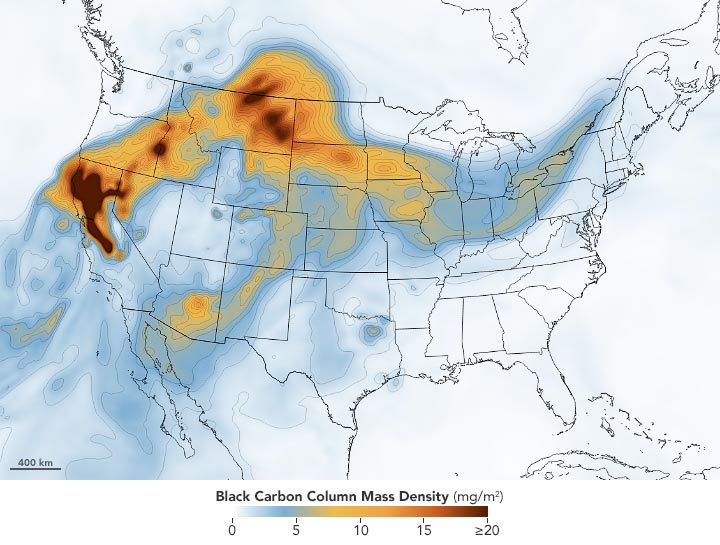
Wildfire Smoke August 25 2020 Annotated
August 25, 2020. (Click image for high-resolution view.)
Wildfire smoke has significantly degraded air quality across Northern California and is spreading toward central and eastern parts of North America. Citizens reported hazy skies in North Dakota, Indiana, and even Virginia. Wildfire smoke is particularly harmful because it contains fine particulate matter that can cause breathing and lung problems, particularly for people with asthma.
The map above shows the concentration of black carbon particulates—commonly called soot—on August 25. Black carbon can harm humans and other animals by entering the lungs and bloodstream; it also plays a role in global warming. The black carbon map comes from the GEOS forward processing (GEOS-FP) model, which assimilates data from satellite, aircraft, and ground-based observing systems. In this case, GEOS-FP assimilated fire and aerosol observations with meteorological data on air temperature, moisture, and winds to project the behavior and extent of the plume.
NASA Earth Observatory images by Lauren Dauphin and Joshua Stevens, using MODIS data from NASA EOSDIS/LANCE and GIBS/Worldview and GEOS-5 data from the Global Modeling and Assimilation Office at NASA GSFC.

UCLA Scientists Test Decontamination Methods for N95 Respirators so They Can Be Used Again

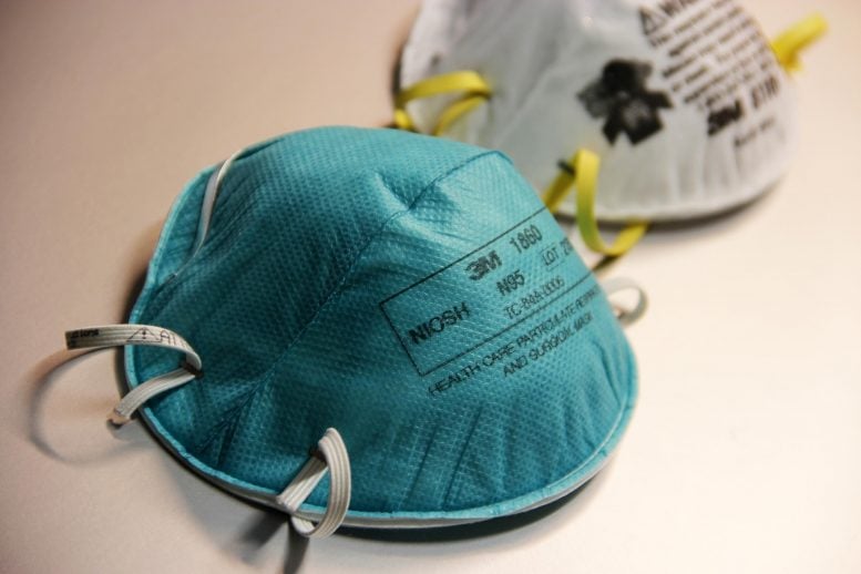
Two N95 Respirators
N95 respirators, which are widely worn by health care workers treating patients with COVID-19 and are designed to be used only once, can be decontaminated effectively and used up to three times, according to research by UCLA scientists and colleagues. Credit: CDC/Debora Cartagena
Scientists hope new methods can mitigate the chronic shortage of personal protective equipment.
N95 respirators, which are widely worn by health care workers treating patients with COVID-19 and are designed to be used only once, can be decontaminated effectively and used up to three times, according to research by UCLA scientists and colleagues.
An early-release version of their study has been published online, with the full study to appear in September in the journal Emerging Infectious Diseases.
N95 respirators reduce exposure to airborne infectious agents, including SARS-CoV-2, the virus that causes COVID-19, and are one of the key pieces of personal protective equipment used by clinical workers in preventing the spread of the virus. Critical shortages of these masks have driven efforts to find new decontamination methods that can extend their use.
“Although N95 respirators are designed for just one use before disposal, in times of shortage, N95 respirators can be decontaminated and reused up to three times,” said James Lloyd-Smith, a co-author of the study and a UCLA professor of ecology and evolutionary biology. “But the integrity of the respirator’s fit and seal must be maintained.”
In a controlled laboratory setting, the researchers tested several decontamination methods on small sections of N95 filter fabric that had been exposed to SARS-CoV-2. The methods included vaporized hydrogen peroxide, dry heat at 70 degrees Celsius (158 degrees Fahrenheit), ultraviolet light and a 70% ethanol spray. All four methods eliminated detectable viable virus traces from the N95 fabric test samples.
The investigators then treated fully intact, clean respirators with the same decontamination methods to test their reuse durability. Employees with the National Institutes of Health’s Rocky Mountain Laboratories in Montana volunteered to wear the masks for two hours to determine if they maintained a proper fit and seal over the face. The scientists decontaminated each mask three times, using the same procedure with each.
The masks treated with vaporized hydrogen peroxide experienced no failures, suggesting they potentially could be reused three times, Lloyd-Smith said. Those treated with ultraviolet light and dry heat began showing fit and seal problems after three decontaminations, suggesting these respirators potentially could be reused twice.
The study authors concluded that vaporized hydrogen peroxide was the most effective method because no traces of the virus could be detected after only a 10-minute treatment. They found that ultraviolet light and dry heat are also acceptable decontamination procedures, as long as the methods are applied for at least 60 minutes.
The ethanol spray, the scientists discovered, damaged the integrity of the respirator’s fit and seal after two sessions, and they do not recommend it for decontaminating N95 respirators.
The researchers stressed that anyone decontaminating an N95 respirator should closely check the fit and seal over the face before each reuse.
Reference: “Effectiveness of N95 Respirator Decontamination and Reuse against SARS-CoV-2 Virus” by Robert J. Fischer, Dylan H. Morris, Neeltje van Doremalen, Shanda Sarchette, M. Jeremiah Matson, Trenton Bushmaker, Claude Kwe Yinda, Stephanie N. Seifert, Amandine Gamble, Brandi N. Williamson, Seth D. Judson, Emmie de Wit, James O. Lloyd-Smith and Vincent J. Munster, 3 June 2020, Emerging Infectious Diseases.
DOI: 10.3201/eid2609.201524
Co-authors of the study include Amandine Gamble, a UCLA postdoctoral researcher in Lloyd-Smith’s laboratory, as well as researchers with Rocky Mountain Laboratories, part of the NIH’s National Institute of Allergy and Infectious Diseases.
Funding sources included the National Institute of Allergy and Infectious Diseases, the Defense Advanced Research Projects Agency, and the National Science Foundation.
In a widely cited study, Lloyd-Smith and colleagues reported in March that the virus that causes COVID-19 remains for several hours to days on surfaces and in aerosols.