Tuesday, October 11, 2022

ON THE ROAD TO HOMUNCULUS

Brain-like organoids grown in a dish provide window into autism


The structures are reminiscent of one wrinkle of a human brain at 15 to 19 weeks post-conception.

Peer-Reviewed Publication

UNIVERSITY OF UTAH HEALTH

Lab-grown organoids reveal insights into the brain 

IMAGE: SESAME SEED-SIZED BRAIN-LIKE ORGANOIDS ARE GROWN IN THE LAB FROM HUMAN CELLS. THEY ARE PROVIDING INSIGHTS INTO THE BRAIN AND UNCOVERING DIFFERENCES THAT MAY CONTRIBUTE TO AUTISM IN SOME PEOPLE. view more 

CREDIT: TREVOR TANNER

(Salt Lake City) - Whatever you do, don’t call them “mini-brains,” say University of Utah Health scientists. Regardless, the seed-sized organoids—which are grown in the lab from human cells—provide insights into the brain and uncover differences that may contribute to autism in some people.

“We used to think it would be too difficult to model the organization of cells in the brain,” says Alex Shcheglovitov, PhD, assistant professor of neurobiology at U of U Health. “But these organoids self-organize. Within a few months, we see layers of cells that are reminiscent of the cerebral cortex in the human brain.”

The research describing the organoids and their potential for understanding neural diseases publishes in Nature Communications on Oct 6 with Shcheglovitov as senior author and Yueqi Wang, PhD, a former graduate student in his lab, as lead author. They carried out the research with postdoctoral scientist Simone Chiola, PhD, and other collaborators at the University of Utah, Harvard University, University of Milan, and Montana State University.

Investigating autism

Having the ability to model aspects of the brain in this way gives scientists a glimpse into the inner workings of a living organ that is otherwise nearly impossible to access. And since the organoids grow in a dish, they can be tested experimentally in ways that a brain cannot.

Shcheglovitov’s team used an innovative process to investigate effects of a genetic abnormality associated with autism spectrum disorder and human brain development. They found that organoids engineered to have lower levels of the gene, called SHANK3, had distinct features.

Even though the autism organoid model appeared normal, some cells did not function properly:

  • Neurons were hyperactive, firing more often in response to stimuli,
  • Other signs indicated neurons may not efficiently pass along signals to other neurons,
  • Specific molecular pathways that cause cells to adhere to one another were disrupted.

These findings are helping to uncover the cellular and molecular causes of symptoms associated with autism, the authors say. They also demonstrate that the lab-grown organoids will be valuable for gaining a better understanding of the brain, how it develops, and what goes wrong during disease.

“One goal is to use brain organoids to test drugs or other interventions to reverse or treat disorders,” says Jan Kubanek, PhD, a co-author on the study and an assistant professor of biomedical engineering at the U.

Building a better brain model

Scientists have long searched for suitable models for the human brain. Lab-grown organoids are not new, but previous versions did not develop in a reproduceable way, making experiments difficult to interpret.

To create an improved model, Shcheglovitov’s team took cues from how the brain develops normally. The researchers prompted human stem cells to become neuroepithelial cells, a specific stem cell type that forms self-organized structures, called neural rosettes, in a dish. Over the course of months, these structures coalesced into spheres and increased in size and complexity at a rate similar to the developing brain in a growing fetus.

After five months in the lab, the organoids were reminiscent of “one wrinkle of a human brain” at 15 to 19 weeks post-conception, Shcheglovitov says. The structures contained an array of neural and other cell types found in the cerebral cortex, the outermost layer of the brain involved in language, emotion, reasoning, and other high-level mental processes.

Like a human embryo, organoids self-organized in a predictable fashion, forming neural networks that pulsated with oscillatory electrical rhythms and generated diverse electrical signals characteristic of a variety of different kinds of mature brain cells.

“These organoids had patterns of electrophysiological activity that resembled actual activity in the brain. I didn’t expect that,” Kubanek says. “This new approach models most major cell types and in functionally meaningful ways.”

Shcheglovitov explains that these organoids, which more reliably reflect intricate structures in the cortex, will allow scientists to study how specific types of cells in the brain arise and work together to perform more complex functions.

“We’re beginning to understand how complex neural structures in the human brain arise from simple progenitors,” Wang says. “And we’re able to measure disease-related phenotypes using 3D organoids that are derived from stem cells containing genetic mutations.”

He adds that using the organoids, researchers will be able to better investigate what happens at the earliest stages of neurological conditions, before symptoms develop.


CAPTION

Single neural rosette-derived organoids develop multiple brain cell types and have an organization and neural activity never seen before in models of this kind.

CREDIT

Trevor Tanner

Single neural rosette-derived organoids model aspects of the brain (IMAGE)

UNIVERSITY OF UTAH HEALTH

Visit UBrain browser to visualize the cells and electrical responses detected in organoids.

The research published as “Modeling human telencephalic development and autism-associated SHANK3 deficiency using organoids generated from single neural rosettes.

Support for the work came from the National Institutes of Health, Brain Research Foundation, Brain and Behavior Research Foundation, Whitehall Foundation, University of Utah Neuroscience Initiative, and University of Utah Genome Project Initiative.

About University of Utah Health

University of Utah Health  provides leading-edge and compassionate care for a referral area that encompasses Idaho, Wyoming, Montana, and much of Nevada. A hub for health sciences research and education in the region, U of U Health has a $458 million research enterprise and trains the majority of Utah’s physicians and health care providers at its Colleges of Health, Nursing, and Pharmacy and Schools of Dentistry and Medicine. With more than 20,000 employees, the system includes 12 community clinics and five hospitals. U of U Health is recognized nationally as a transformative health care system and provider of world-class care.


Creating a mouse embryo from stem cells to learn more about the mammalian development process
Creating a mouse embryo from stem cells to learn more about the mammalian development process
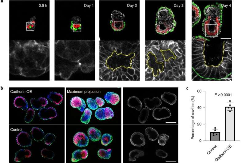
Correct self-organization is necessary for proper morphogenesis. a, Time course of the
 assembly of ETX embryos stained to reveal E-cadherin (monochrome), 
Oct4 (red) and Gata4 (green). The bottom row of images are magnifications of the images
 above and show E-cadherin staining around a nascent cavity, as indicated by the dashed
 yellow lines. The dashed green line indicates the boundary between the ES and XEN 
compartment. Scale bar, 5 μm. b, Representative images showing Oct4 (red), 
Gata4 (green), E-cadherin (monochrome) and DAPI (gray) staining in day 4 cadherin
 OE ETX structures formed by combining E-cadherin OE ES cells with P-cadherin OE 
TS cells and wild-type XEN cells. ETX structures formed by combining wild-type cells 
were used as a control. Scale bars, 100 μm. c, Comparison and quantification of joined 
cavity formation in cadherin OE and control ETX structures. n = 361 (control group) and
 n = 253 (cadherin OE group). N = 5 for each condition. The data are presented as means 
± s.d. Statistical significance was determined by unpaired two-tailed Student’s t-test. d, 
Representative image showing Oct4 (red), Gata4 (green), laminin (monochrome) and 
DAPI (blue) staining in day 4 cadherin OE ETX structures formed by combining
 E-cadherin OE ES cells with P-cadherin OE TS cells and wild-type XEN cells.
 ETX structures formed by combining wild-type cells were used as a control. Scale bars, 
100 μm. e, Quantification of the structures that contained continuous or discontinuous 
laminin. n = 40 ETX structures per condition. N = 3. The data are presented as means 
± s.d. Statistical significance was determined by unpaired two-tailed Student’s t-test. f,
 Self-organization principles in stem cell-derived ETX embryos. Differential expression of
 E-, K- and P-cadherins enables the sorting of ES (epiblast-like), XEN (VE-like) and 
TS (TE-like) stem cells. Wild-type ES cells with low E-cadherin expression and wild-type 
TS cells with low P-cadherin expression exhibited detrimental global sorting efficiency. 
This could be overcome by overexpressing E-cadherin in ES cells and P-cadherin in TS
 cells to increase the efficiency of ETX embryo formation. Proper morphogenesis, including
 cavity formation, basement membrane formation (purple) and symmetry breaking can only
 be observed in well-sorted structures. Numerical data are available as source data. 
Credit: Nature Cell Biology (2022). DOI: 10.1038/s41556-022-00984-y

A team of researchers at the California Institute of Technology, working with one colleague from The Francis Crick Institute and another from the University of Cambridge, both in the U.K., has developed a way to grow mouse embryos without using mouse eggs or sperm to learn more about early mammalian development. In their paper published in the journal Nature Cell Biology, the group describes using several types of stem cells to grow mouse embryos.

Prior research has shown that mammalian embryos differentiate into different types of cell masses as they develop. Researchers have also found that  are involved in the processes but the mechanisms responsible are still unknown. In this new effort, the researchers used three different kinds of stem cells to grow a mouse embryo that matured to the point of having a  and the beginnings of a brain.

To create such embryos, the researchers first studied communications between stem cell groups in naturally developing . They learned to recognize the elements that went into such communications and the means by which it was carried out. In essence, they "deciphered the code." They then isolated three main types of stem cells that made up the cell masses in early embryo development: pluripotent, which eventually grow to become body tissue, and two other types that grew to become the amnionic sac and placenta. They also noted the quantities of each type of stem cell.

The next step was to attempt to create a mouse embryo from scratch using the three types of stem cells in a lab setting. With careful tending, the researchers grew an embryo that matured enough to allow for study of its development.

To test further, the researchers repeated the procedure but added genetically engineered cells to see how it impacted maturation of the embryo. They found they could replicate some of the same brain development issues that have been seen in . They suggest their work could also help explain what goes wrong when mice (or people) miscarry.

Stem cell biologists create new human cell type for research
More information: Min Bao et al, Stem cell-derived synthetic embryos self-assemble by exploiting cadherin codes and cortical tension, Nature Cell Biology (2022). DOI: 10.1038/s41556-022-00984-y
Journal information: Nature Cell Biology 
© 2022 Science X Network

Light-based therapy weakens antibiotic-resistant bacteria

Light-based therapy weakens antibiotic-resistant bacteria
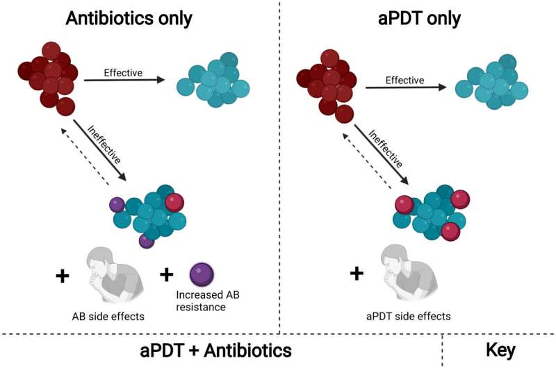
Schematic representation of single and combined antibiotic and aPDT treatment options for
 infections. Antibiotics serve an important role in medicine, successfully treating many 
common bacterial infections. However, their use leads to increased resistance, 
necessitating more dangerous antibiotic classes and higher doses. Likewise, aPDT can 
successfully eliminate bacteria, regardless of antibiotic resistance, but is not always
 applicable at large doses due to drug and light side effects. These two treatments used
 concurrently have been found to significantly lower average antibiotic resistance and 
broaden the resistance distribution, permitting the use of smaller doses of each for a more
 effective treatment. 
Credit: Proceedings of the National Academy of Sciences (2022).
DOI: 10.1073/pnas.2208378119

Antibiotics are standard treatments for fighting dangerous bacterial infections. Yet the number of bacteria developing a resistance to antibiotics is increasing. Researchers from Texas A&M University and the University of São Paulo are overcoming this resistance with light.

The researchers tailored antimicrobial  (aPDT)—a chemical reaction triggered by visible light—for use on  strains. Results showed the treatment weakened  to where low doses of current antibiotics could effectively eliminate them.

"Using aPDT in combination with antibiotics creates a synergy of interaction working together for a solution," said Vladislav Yakovlev, University Professor in the Department of Biomedical Engineering at Texas A&M and co-director of the project. "It's a step in the right direction against resistant bacteria."

The research results were published in Proceedings of the National Academy of Sciences (PNAS).

Ultraviolet light was first used to sterilize bacteria over 100 years ago. The treatment was based on the work of Niels Finsen, who won the Nobel Prize in Physiology in 1903 for using filtered sunlight—the higher frequency or ultraviolet spectrum—as a cure for skin tuberculosis. Phototherapy advances faded in popularity a few decades later when antibiotics became the weapon of choice against bacteria.

Antibiotic-resistant bacteria showed up soon after antibiotics were first used. If antibiotic treatment stops before bacteria are fully killed, the remaining bacteria develop a resistance or immunity to the antibiotic. That immunity transfers to every new bacteria cell, so more potent antibiotics or new treatment methods are needed to overcome the growing resistance.

"Photodynamic therapy was a forgotten tool," Yakovlev said. "Yet, bacteria cannot overcome it. There is no resistance."

Some human cancer cell therapies already use aPDT to prevent the growth of abnormal cells, but treating resistant bacteria with it is still a novel approach.

The researchers began their work by choosing the bacteria and the three main parts of aPDT needed to combat it: molecular oxygen, light, and a photosensitizer—something that creates a reaction between oxygen and light. An already FDA-approved dye called methylene blue served as the photosensitizer. The light sources were specially constructed panels of 25 LEDs in reflective cones built by the Technical Support Laboratory of the São Carlos Institute of Physics. Methicillin-resistant Staphylococcus aureus served as the bacteria, and the researchers grew cultures with the blue dye in them to ensure the photosensitizer alone would not affect the bacteria.

Most of the lab work occurred in the Texas A&M Health Science Center under Paul de Figueiredo, professor in the Department of Microbial Pathogenesis and Immunology in the College of Medicine.

At first, the team used aPDT by itself at various light strengths, durations, and in a specific series of follow-up treatments to log the bacteria's response. The idea was to find the lowest dose and shortest series that could weaken the bacterial membranes and other resistance mechanisms. Cell recoveries and reproductions revealed how many generations it took before antibiotic resistance returned. Next, the researchers added measured levels and combinations of antibiotics at different time intervals after aPDT treatments to note the weakened bacteria's responses.

"The use of antibiotics with aPDT is a unique idea," Yakovlev said. "We can use lower doses of both to achieve our goal in contrast to using one or the other at higher doses that could have side effects."

The goal is to shorten the treatment time and reduce the dosage to the lowest levels needed.

Getting  down to one doctor visit is especially important to Vanderlei Bagnato, professor in the Department of Physics and Materials Science at São Paulo and co-director of the project. He is trying to improve recovery odds for populations in remote areas of Brazil where patients might only see a doctor once per illness, without any chance for follow-up care.

The U.S. Department of Defense is following the project closely because battlefield wound infections also happen in remote locations and must be dealt with quickly.

So far, the results are positive. The resistant bacteria, weakened by aPDT treatments, were killed with far lower doses of current antibiotics. As a benefit, these therapies reduced the need for battling resistant bacteria with more potent and expensive  that take years to produce. Future work for the project will involve more timing and dosage investigations and tests on other  strains to see if the effectiveness is universal.

"Imagine the real-life applications," Yakovlev said. "You visit a doctor, who uses an ointment and shines a light on the infected area, and then you're done. It would be a quick and harmless treatment as needed."New drug candidate fights off more than 300 drug-resistant bacteria

More information: Jace A. Willis et al, Breaking down antibiotic resistance in methicillin-resistant Staphylococcus aureus : Combining antimicrobial photodynamic and antibiotic treatments, Proceedings of the National Academy of Sciences (2022). DOI: 10.1073/pnas.2208378119
Provided by Texas A&M University 

 EVERYONE'S FAVORITE 

How tardigrades survive freezing temperatures

How tardigrades survive freezing temperatures
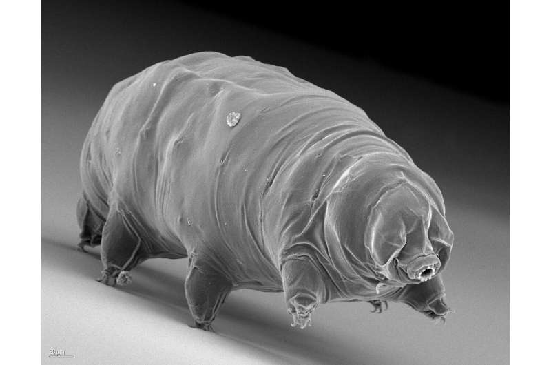
It is only under the microscope that the similarity of its namesake becomes apparent: the 
plump, round physique and the short legs are reminiscent of those of a bear. 
Credit: Ralph Schill

Tardigrades are excellent at adapting to harsh environmental conditions. Back in 2019, Ralph Schill, a professor at the Institute of Biomaterials and Biomolecular Systems at the University of Stuttgart, proved that anhydrobiotic (dried) tardigrades can survive undamaged for many years without absorbing water. Whether they age faster or slower in a frozen state, or whether aging even comes to a halt, was previously unclear. But the mystery has now been solved: Frozen tardigrades do not age.

Tardigrades, also called water bears, belong to the family of nematodes. Their gait is reminiscent of that of a bear, but that is the only similarity. The tardigrades, which are barely one millimeter in size, have managed to adapt perfectly to rapidly changing  over the course of evolution and can dry out in  and freeze in cold conditions. "They don't die, they fall into a ," explains Schill.

The Sleeping Beauty hypothesis

For a cell organism, freezing or drying out cause different kinds of stress. But tardigrades can survive both heat and cold equally unscathed. They no longer show any obvious signs of life. And this raises the question of what happens to the animals' internal clock and whether they age in this resting state.

For dried tardigrades, which wait many years in their  for the next rain, Ralph Schill and his team answered the question of aging several years ago. In a fairytale by the Grimm brothers, the princess falls into a deep sleep. When a prince kisses her 100 years later, she awakens and still looks as young and beautiful as before. It is the same with tardigrades in a dried state and therefore this is also called the "Sleeping Beauty" hypothesis ("Sleeping Beauty" model).

"During inactive periods, the internal clock stops and only resumes running once the organism is reactivated," explains Schill. "So, tardigrades, which usually only live for a few months without periods of rest, can live for many years or even decades."

Until now, it was still unclear whether this also applies to frozen animals. Do they age faster or slower than the dried animals, or does aging also come to a halt?

How tardigrades survive freezing temperatures
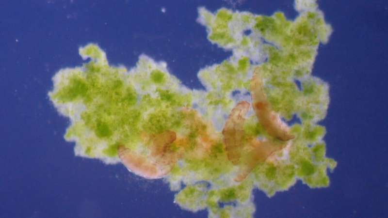
Live images taken under the microscope help Schill and his colleagues to investigate the 
(in)activity of the tardigrades. Credit: Ralph Schill

The aging process stops even when frozen

To explore this, Schill and his team conducted several experiments in which they froze a total of more than 500 tardigrades at -30 °C, thawed them out again, counted them, fed them and froze them again. This was repeated until all the animals died. At the same time, control groups were kept at constant room temperature. Excluding the time in frozen condition, the comparison with the control groups showed an almost identical lifetime. "So even in ice,  stop their internal clocks like Sleeping Beauty," concludes Schill.

Schill and his colleagues published their findings and approach in the Journal of Zoology.

Tardigrades survive impacts of up to 825 meters per second
More information: J. Sieger et al, Reduced ageing in the frozen state in the tardigrade Milnesium inceptum (Eutardigrada: Apochela), Journal of Zoology (2022). DOI: 10.1111/jzo.13018
Journal information: Journal of Zoology 
Provided by University of Stuttgart 

InSight Mars lander waits out dust storm

InSight waits out dust storm
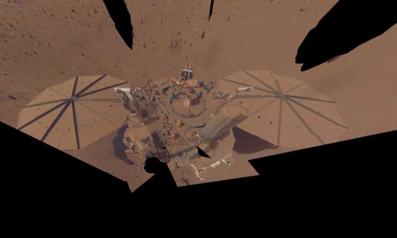
NASA’s InSight Mars lander took this final selfie on April 24, 2022, the 1,211th Martian
 day, or sol, of the mission. The lander’s solar panels have become covered with dust since
 the lander touched down on Mars in November 2018, which has led to a gradual decline in
 its power level. Credit: NASA/JPL-Caltech

NASA's InSight mission, which is expected to end in the near future, saw a recent drop in power generated by its solar panels as a continent-size dust storm swirls over Mars' southern hemisphere. First observed on Sept. 21, 2022, by NASA's Mars Reconnaissance Orbiter (MRO), the storm is roughly 2,175 miles (3,500 kilometers) from InSight and initially had little impact on the lander.

The  carefully monitors the lander's power level, which has been steadily declining as dust accumulates on its . By Monday, Oct. 3, the  had grown large enough and was lofting so much dust that the thickness of the dusty haze in the Martian atmosphere had increased by nearly 40% around InSight. With less sunlight reaching the lander's panels, its energy fell from 425 watt-hours per Martian day, or sol, to just 275 watt-hours per sol.

InSight's seismometer has been operating for about 24 hours every other Martian day. But the drop in  does not leave enough energy to completely charge the batteries every sol. At the current rate of discharge, the lander would be able to operate only for several weeks. So to conserve energy, the mission will turn off InSight's seismometer for the next two weeks.

"We were at about the bottom rung of our ladder when it comes to power. Now we're on the ground floor," said InSight's project manager, Chuck Scott of NASA's Jet Propulsion Laboratory in Southern California. "If we can ride this out, we can keep operating into winter—but I'd worry about the next storm that comes along."

The team had estimated that InSight's mission would end sometime between late October of this year and January 2023, based on predictions of how much the dust on its  will reduce its power generation. The lander has long-since surpassed its primary mission and is now close to the end of its extended mission, conducting "bonus science" by measuring marsquakes, which reveal details about the deep interior of the Red Planet.

InSight waits out dust storm
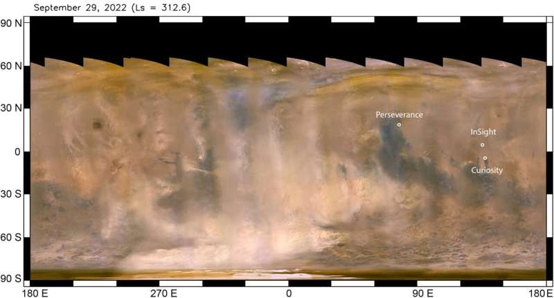
The beige clouds seen in this global map of Mars are a continent-size dust storm captured
 on Sept. 29, 2022, by the Mars Climate Imager camera aboard NASA’s Mars 
Reconnaissance Orbiter. NASA’s Perseverance, Curiosity, and InSight missions are labeled,
 showing the vast distances between them. Credit: NASA/JPL-Caltech/MSSS

Studying Martian Storms

There are signs that this large, regional storm has peaked and entered its decay phase: MRO's Mars Climate Sounder instrument, which measures the heating caused by dust absorbing sunlight, sees the storm's growth slowing down. And the dust-raising clouds observed in pictures from the orbiter's Mars Color Imager camera, which creates daily global maps of the Red Planet and was the first instrument to spot the storm, are not expanding as rapidly as before.

This regional storm isn't a surprise: It's the third storm of its kind that's been seen this year. In fact, Mars  occur at all times of the Martian year, although more of them—and bigger ones—occur during northern fall and winter, which is coming to an end.

Mars dust storms aren't as violent or dramatic as Hollywood portrays them. While winds can blow up to 60 miles per hour (97 kilometers per hour), the Martian air is thin enough that it has just a fraction of the strength of storms on Earth. Mostly, the storms are messy: They toss billowing dust high into the atmosphere, which slowly drops back down, sometimes taking weeks.

On rare occasions, scientists have seen dust storms grow into planet-encircling dust events, which cover almost all of Mars. One of these planet-size dust storms brought NASA's solar-powered Opportunity rover to an end in 2018.

Because they're nuclear-powered, NASA's Curiosity and Perseverance rovers have nothing to worry about in terms a  storm affecting their energy. But the solar-powered Ingenuity helicopter has noticed the overall increase in background haze.

Besides monitoring storms for the safety of NASA missions on the Martian surface, MRO has spent 17 years collecting invaluable data about how and why these storms form. "We're trying to capture the patterns of these storms so we can better predict when they're about to happen," Zurek said. "We learn more about Mars' atmosphere with each one we observe."

NASA's InSight enters safe mode during regional Mars dust storm