Tuesday, July 18, 2023


UMD researchers uncover privacy risks in cellphones purchased at police auctions


Their two-year study found that 27% of the phones they successfully bid on contained personal data

Reports and Proceedings

UNIVERSITY OF MARYLAND

UMD Researchers Uncover Privacy Risks in Cellphones Purchased at Police Auctions 

IMAGE: (FROM LEFT) UNIVERSITY OF MARYLAND COMPUTER SCIENCE PH.D. STUDENTS RICHARD ROBERTS AND JULIO POVEDA ARE SHOWN WITH THEIR ADVISER, ASSOCIATE PROFESSOR DAVE LEVIN. THE TRIO RECENTLY CONCLUDED A STUDY THAT UNCOVERED SIGNIFICANT PRIVACY CONCERNS WITH CELLPHONES PURCHASED FROM POLICE PROPERTY ROOM AUCTIONS view more 

CREDIT: UNIVERSITY OF MARYLAND


Law enforcement agencies nationwide regularly sell items that are seized in criminal investigations or are unclaimed from lost-and-found inventories. Many of these items—vehicles, jewelry, watches and electronic devices like cellphones—end up at online auction houses.

People looking for a bargain can bid on cellphones in bulk, snatching up dozens at rock bottom prices for parts or other uses. This ultimately provides revenue for the police agencies, making for a good deal for everyone involved. Or is it?

A recent study by University of Maryland security experts found that many of the phones sold at police property auction houses are not properly wiped of personal data. The study, conducted over two years with cellphones bought from the largest police auction house in the U.S., uncovered troves of personal information from previous owners that was easily accessible.

Of the 228 phones that the UMD team successfully bid on, 61 (27%) contained personal data like social security numbers, credit card and banking information, passport data, pictures of driver’s licenses, and more.

“We were actually surprised at the level of personal information we found, and the ease by which we could access it,” said Dave Levin, an associate professor of computer science who led the UMD team.

Levin, a core faculty member in the Maryland Cybersecurity Center, first became interested in this topic through a casual conversation with a colleague. After determining there was a security breakdown—whether through police not wiping the phones, or auction houses not taking proper safeguards before shipping items to the highest bidder—Levin and several of his graduate students set out to explore the scale of the problem

The first step was to work closely with the university’s legal counsel and institutional research review board to determine the appropriate protocols needed to view any personal data.

“There were stringent guidelines in place—how each phone we received was catalogued, the processes we used to access the phones, and most importantly, what we would be legally required to do if we found any evidence of child abuse,” said Julio Poveda, a second-year computer science Ph.D. student who was part of the research team.

The UMD team did not come across any evidence of child abuse, but did uncover other information that was unsuitable for public dissemination, such as depictions of adult nudity and drug use.

Some of the phones they accessed had been used in criminal activities like identity theft, a discovery Levin found particularly troubling.

“It’s as if people that were victims of identity theft were being ‘re-victimized’ by having their personal information available again for anyone to see,” he explained.

The UMD team determined that some of the phones had been used by sex workers, with text messages between the workers and their clients still intact.

“It's important to remember that your phone does not just have your data, it has data from anyone who has communicated with you,” said Richard Roberts, a sixth-year computer science Ph.D. student and lead author of the study.

Roberts, who presented the team’s academic work at a major security conference earlier this year, said that out of the 61 phones the researchers accessed, they determined that there had been some form of digital contact with more than 7,000 people.

Levin, Poveda and Roberts are all security experts, but decided against using using any type of sophisticated digital forensics for their study. “We wanted to attempt to gain access to any cellphone data using techniques that someone on the street might use,” Roberts said.

The researchers were shocked at how easy it was. One of the phones arrived with a sticky note attached with the phone’s passcode in plain view, a leftover from the originating police agency that had already legally hacked the phone. Multiple other phones had PINs or passcode patterns that were easy to guess.

“Sadly, passcodes like 1-2-3-4 are still in common use today,” Levin said.

Last October, the researchers reached out to the auction house where they purchased the phones. The company—PropertyRoom.com, which bills itself as the largest police auction house in the U.S. working with more than 4,400 law enforcement agencies—promised to investigate the problem. Shortly after that, the company stopped selling bulk lots of phones altogether for a short period, then started again, prompting the researchers to purchase another batch.

“We found that PropertyRoom had started wiping the phones but failed to wipe the phones’ [Secure Digital] cards, which in several cases had partial backups of the phones’ contents,” Levin said.

After pinging the company again to inform it of this oversight, the UMD researchers received no further response.

A subsequent investigative report by a local television station prodded the company to publish a message on its website stating it was aware of the security concerns and was taking corrective measures.

From a security standpoint, Levin said, police agencies should avoid auctioning used cellphones. “Just destroy them,” he said. “[The police agencies] don’t get that much money in return, and the potential damage far outweighs any financial incentives.”

He also suggested that people take better precautions in the event their phone is lost or stolen and ends up being resold.

“Use your phone under the assumption that somebody else might later become its legal owner,” Levin said. “Set a passcode that is hard to guess, minimize the private information that’s easy to access, and remotely wipe your phone if it is lost or stolen. Otherwise, our study shows just how easy it is for someone to gain an incredible amount of access to your private information.”
 

Innovative infection prevention program reduces surgical site infections, results in hospital days reduced and $500,000 savings


Could serve as model for improving outcomes

Reports and Proceedings

AMERICAN SOCIETY OF ANESTHESIOLOGISTS


Chicago — An innovative anesthesiologist-led infection prevention program helped reduce the number of surgical site infections (SSIs) in colorectal patients by 50%, the number of days in the hospital by 46%, and led to significant cost savings over a two-year period, according to research presented at the virtual American Society of Anesthesiologists’ Anesthesia Quality and Patient Safety Meeting.

“With the skyrocketing cost of medical care for patients and health care institutions, one area physicians can focus on is reducing SSIs,” said Austin Street, M.D., study author and anesthesiologist at UT Southwestern Medical Center, Dallas. “Many SSIs are preventable through well-designed, evidence-based interventions. We were very happy to reduce the SSIs in our colorectal patients by half, which also led to decreases in hospital bed days, saving the hospital and patients money, as well as freeing up beds for other patients and surgeries.”

Largely avoidable with proper infection control measures, SSIs occur either during or up to 30 days after a surgical procedure. SSIs often need to be treated with additional antibiotics, and may require interventional procedures or even re-operation. SSIs can lead to major complications, including death, as well as significantly increase the cost of care. The cost of a patient’s care increases by $20,000, on average, if they develop an SSI. According to the Centers for Disease Control and Prevention, the annual cost of SSIs to hospitals in the U.S. ranges between $3.2 billion to $10 billion a year. The SSI incidence rate in colorectal surgery is higher than many other procedures.

Prior to the intervention, the infection ratio at UT Southwestern had increased from .74 in 2018 to 3.08 in 2020, putting the program in the bottom quartile for infection rates in the country. The new infection prevention initiative leveraged the strength of the hospital’s Enhanced Recovery After Surgery (ERAS) program. An ERAS pathway is an evidence-based protocol that standardizes care to minimize surgical stress and postoperative pain, reduce complications, improve outcomes, decrease hospital length of stay and expedite recovery following elective procedures. Under the umbrella of the ERAS program, UT Southwestern’s infection prevention initiative implemented a number of interventions, each targeted at evidence-based causes of SSIs, including:

  • Giving oral antibiotics with the patient’s mechanical bowel preparation
  • Identifying the best antibiotic to use, as well as optimal timing and redosing for colorectal surgery, with the guidance of UT Southwestern’s antibiotic stewardship committee
  • Using chlorhexidine baths, a cleaning product that kills germs, prior to the surgery and wipes to the abdomen immediately prior to the operating room to decrease bacteria on the skin
  • Improving access to critical medications by storing the antibiotics directly in each operating room’s “pyxis” machines, which hold and distribute the anesthetic drugs
  • Requiring the surgical team and their assistants (scrub techs and residents) to change their gowns and gloves when the surgery was completed and they were about to close the wound, assuring no contamination from the surgical site got into the sterile areas of the wound
  • Actively warming patients both prior to and during the surgery, which has been shown to decrease the risk of wound infections
  • Increasing patient mobility as soon as possible after surgery, for example sitting up in a chair the day of surgery and walking in the hallways up to three times as soon as possible, which decreases the risk of infection

By implementing these infection control strategies, UT Southwestern met their goal of reducing colorectal SSIs by 50%. Additionally, the hospital saved an estimated $540,000 in total costs in 2021 and 2022, compared to 2020, and hospital bed days were reduced by 578 days (46%).

This program may serve as a useful model for other academic or major medical centers seeking to improve their SSI outcomes.

THE AMERICAN SOCIETY OF ANESTHESIOLOGISTS

Founded in 1905, the American Society of Anesthesiologists (ASA) is an educational, research and scientific society with more than 56,000 members organized to raise and maintain the standards of the medical practice of anesthesiology. ASA is committed to ensuring physician anesthesiologists evaluate and supervise the medical care of patients before, during and after surgery to provide the highest quality and safest care every patient deserves.

For more information on the field of anesthesiology, visit the American Society of Anesthesiologists online at asahq.org. To learn more about the role physician anesthesiologists play in ensuring patient safety, visit asahq.org/MadeforThisMoment. Like ASA on Facebook, follow ASALifeline on Twitter.

###

Merck Prize boosts work on automated air sensor for pandemic pathogens


Emory University chemist Khalid Salaita leads a visionary project


Grant and Award Announcement

EMORY UNIVERSITY

Khalid Salaita 

IMAGE: KHALID SALAITA, PROFESSOR OF CHEMISTRY AT EMORY UNIVERSITY, RECEIVED THE 2023 FUTURE INSIGHT PRIZE FROM MERCK KGAA, DARMSTADT, GERMANY. view more 

CREDIT: KAY HINTON, EMORY UNIVERSITY


Merck KGaA, Darmstadt, Germany, awarded its 2023 Future Insight Prize to Khalid Salaita, professor of chemistry at Emory University. The award comes with $540,000 to fund the next phase of research into an air sensor that can continuously monitor indoor spaces for pathogens that can cause pandemics.

“I’m extremely thankful to receive the Future Insight Prize as this enables us to continue our path toward an early-warning system for emerging threats,” Sailta says. “Our research sets the stage for fully automated detection of airborne pathogens without human intervention or sample processing.”

The Merck Future Insight Prize recognizes groundbreaking ideas to solve some of the world’s most pressing challenges in health, nutrition and energy. The Salaita lab’s sensor, a rolling micro-motor called “Rolosense,” holds the potential to help mitigate, or even prevent, a pandemic.

“The importance of being prepared is a key lesson from the COVID-19 pandemic,” says BelĂ©n Garijo, chair of the executive board and CEO of Merck, a leading science and technology company. “There are many promising collaborations to build an inclusive framework for pandemic preparedness, but we still lack an effective warning system to detect potential threats before it is too late. The pioneering work of Khalid Salaita could help fill this urgent gap in our global defenses.”

Salaita’s lab has already shown that a prototype of the sensor can detect the five variants of COVID-19 that it has tested, along with influenza type A. Theoretically, Rolosense can be programmed to simultaneously screen for a wide group of viral pathogens within a breath sample from an individual or within ambient indoor air. 

Within the next five years, the researchers hope to have viable products available to provide convenient, non-invasive, rapid ways to detect airborne viral pathogens. These products may include testing kits for the home and healthcare clinics that screen for an array of viruses within a single test, delivering a result within minutes.

“Our ultimate goal is to develop automated viral air sensors that function similar to smoke detectors,” Salaita says. “These sensors could be located in busy locations like airports, hospitals and schools to continuously monitor aerosolized particles for viruses.” 

Salaita is also on the faculty of the Wallace H. Coulter Department of Biomedical Engineering, a joint program of Georgia Tech and Emory.

Curiosity got the ball rolling on the project around a decade ago. 

“We wondered if we could convert chemical energy into mechanical work and make something move,” Salaita recalls. “Ultimately, our goal was to mimic life at the nanoscale. We wanted to make artificial, miniscule motors that match the sophistication and functionality of proteins that move cargo around in cells and perform other functions.”

As a graduate student in the Salaita lab, Kevin Yehl had the idea of constructing a DNA-based motor using a micron-sized glass sphere as its “chassis.” Hundreds of DNA strands, or “legs,” were allowed to bind to the sphere. They were then placed on a glass slide coated with the fuel: RNA.

The result was the invention of the first rolling DNA-based motor in 2015. Dubbed the “Rolosense,” the motor was 1,000 times faster than any other synthetic DNA motor. It was so fast that a simple smart phone microscope could capture its motion through video. 

Its speed and stability gave the Rolosense potential for real-world applications, such as a tool for medical diagnostics. 

Emory graduate students continued to work on refinements of the technologies through the years. Alisina Bazrafshan, who has since graduated and is now a scientist at Illumina, a DNA sequencing company, enhanced the speed and persistence of the DNA-based motors. 

When Selma Piranej joined the Salaita lab as a PhD candidate in 2018, she began working on a project to build computer programming logic into the Rolosense. She tapped a well-known reaction in bioengineering to perform the computation and then paired it with the motion of the Rolosense. The computer readout becomes simply “motion” or “no motion.”

These two logic gates of “motion” or “no motion” can be strung together to build more complicated operations, mimicking how regular computer programs build on the logic gates of “zero” or “one.” 

Piranej took the project even further by finding a way to pack many different computer operations together and still easily read the output. She simply varied the size and materials of the microscopic spheres that form the chassis for the DNA-based rolling motors. For instance, the spheres can range from three to five microns in diameter and be made of either silica or polystyrene. Each alteration provides slightly different optical properties that can be distinguished through a cell phone microscope.

When the pandemic hit in 2020, the chemists began focusing on using the Rolosense technology to develop an indoor air sensor to detect the SARS-CoV-2 virus, the infectious agent of COVID-19. 

The Salaita lab received a $883,000 grant for the project from the National Institutes of Health RADx Radical initiative, which aims to support new, non-traditional approaches for rapid-detection devices that address current gaps in testing for the presence of SARS-CoV-2, as well as potential future pandemic viruses.

Co-investigators on that grant included Gregory Melikian, a professor at Emory School of Medicine, in the Department of Pediatrics’ Division of Infectious Disease; and Yonggang Ke, assistant professor at Emory’s School of Medicine and the Coulter Department of Biomedical Engineering.

An additional key collaborator is Primordia Biosystems, a company that specializes in building microfluidic chips that can sample virus-containing aerosols in the air.

Piranej continued to work on the project along with fellow Emory PhD students. She searched through the scientific literature to find an aptamer, a piece of DNA that would bind to a universal spike protein on SARS-CoV-2, sticking to it like Velcro. 

Experiments showed that this bind stalls the Rolosense motor, giving the “no motion” readout that signals the presence of SARS-CoV-2. Variants of SARS-CoV-2 share the same spike protein, so the Rolosense is able to detect a range of them. 

By adding different aptamers, or binding agents, the researchers have shown they can detect other viruses as well, including influenza type A. Millions of nano-motors are deployed at once via the technology. The motors can be individually programmed so that they each respond only to one specific virus. That means multiple viruses could be simultaneously screened for within one test sample.

“Unlike conventional tests, we don’t have to treat a viral sample in any way to get a result,” Salaita says. “We can do the detection directly from a nasal swab, saliva sample or breath condensate. We don’t have to do any kind of amplification process to enhance the signal. That’s a huge advantage in terms of making the assay more assessable while also preserving ultrasensitive detection.”

The Merck award will support the researchers as they further refine and test the technology.

“Some of the world’s leading experts at testing and validating new COVID diagnostics happen to be on the Emory campus,” Salaita notes. His team will be drawing from this expertise, along with thousands of samples from human COVID-19 infections available in an NIH RADx Radical Diagnostic Core Resources center located at Emory School of Medicine. The samples are used to benchmark and validate the efficacy of a viral assay.

A key challenge in terms of a Rolosense product for home testing or use in a physician’s office is integration of the breath-collection tube with the readout device. 

The development of a viral sensor to continuously monitor indoor air also must overcome many chemistry and engineering challenges. 

“We’ve shown that our nano-motors can run for at least 24 hours, but we need them to run for days or weeks at a time in an automated system,” Salaita says. “We also have to engineer methods to collect air samples while filtering out enzymes in the atmosphere that chop up DNA. We need to circumvent these enzymes so that they don’t destroy the DNA nano-motors.”

While more development and clinical evaluation is needed, Salaita remains confident that the Rolosense will one day become a useful tool for public health.

“One thing is for certain,” he says. “There is a need for viral-detecting devices for public indoor air spaces as we enter an era when pandemics will likely become more common.”

SPACE

Astronomers explore the chromosphere of peculiar white dwarfs

Observations explore the chromosphere of peculiar white dwarfs
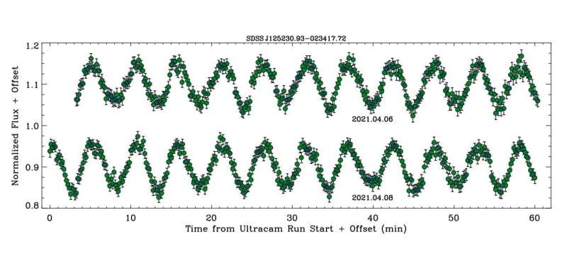
Approximately 1 h of ULTRACAM g-band light curves for SDSS J1252, each taken on a
 different night. Credit: Farihi et al, 2023

Using the 3.6-m New Technology Telescope (NTT) at the La Silla Observatory in Chile, astronomers have observed three peculiar white dwarfs of the DAHe subtype. In their results, they found dipolar chromospheres in two of these objects. The findings were reported in a paper published July 5 on the preprint server arXiv.

White dwarfs (WDs) are stellar cores left behind after a star has exhausted its nuclear fuel. Due to their high gravity, they are known to have atmospheres of either pure hydrogen or pure helium. However, a small fraction of WDs shows traces of heavier elements.

DAHe (D: degenerate, A: Balmer lines strongest, H: magnetic line splitting, e: emission) is a relatively new and small class of magnetic white dwarfs that showcase Zeeman-split Balmer emission lines. To date, only a few dozen DAHe WDs are known. The first of them was GD 356—an isolated white dwarf discovered nearly 40 years ago.

A team of astronomers led by Jay Farihi of the University College London, U.K., decided to investigate three objects of this rare class, in order to better understand the nature of the entire population. For this purpose, they employed ULTRACAM—a frame-transfer CCD imaging camera mounted on the NTT telescope. The study was complemented by data from NASA's Transiting Exoplanet Survey Satellite (TESS).

"This study focuses on light curves and the resulting periodicities of three DAHe white dwarfs, using both ground- and space-based photometric monitoring," the researchers wrote.

The three observed DAHe WDs were: SDSS J125230.93−023417.7 (or SDSS J1252 for short), LP 705-64 and WD J143019.29−562358.3 (WD J1430). It turned out that the folded ULTRACAM light curves of SDSS J1252 and LP 705-64 exhibit alternating minima that are indicative of two distinct star spots 180 degrees out-of-phase during rotation. For WD J1430, the light curves reveal a single maximum and minimum.

The astronomers found that the amplitudes of the multi-band photometric variability reported for all the three DAHe  are all several times larger than that in GD 356. They noted that all the known DAHe stars have light curve amplitudes that increase toward the blue in correlated ratios, which points to cool spots that produce higher contrasts at .

According to the authors of the paper, their findings suggest that some magnetic WDs create intrinsic chromospheres as they cool, and that no external source is responsible for the observed temperature inversion.

"Given the lack of additional periodic signals and the compelling evidence of DAHe white dwarf clustering in the HR diagram (Walters et al, 2021; Reding et al, 2023; Manser et al, 2023), an intrinsic mechanism is the most likely source for the spotted regions and chromospheric activity," the researchers concluded.

More information: J. Farihi et al, Discovery of Dipolar Chromospheres in Two White Dwarfs, arXiv (2023). DOI: 10.48550/arxiv.2307.02543


Journal information: arXiv 


© 2023 Science X Network


Astronomers discover eight new cataclysmic variables


 Two White Dwarfs


SLEEPY


XRISM mission to study ‘rainbow’ of X-rays


Business Announcement

NASA/GODDARD SPACE FLIGHT CENTER

XRISM Spacecraft 

IMAGE: XRISM, SHOWN IN THIS ARTIST’S CONCEPT, IS AN X-RAY MISSION THAT WILL STUDY SOME OF THE MOST ENERGETIC OBJECTS IN THE UNIVERSE. view more 

CREDIT: NASA'S GODDARD SPACE FLIGHT CENTER CONCEPTUAL IMAGE LAB


A new satellite called XRISM (X-ray Imaging and Spectroscopy Mission, pronounced “crism”) aims to pry apart high-energy light into the equivalent of an X-ray rainbow. The mission, led by JAXA (Japan Aerospace Exploration Agency), will do this using an instrument called Resolve.

XRISM is scheduled to launch from Japan’s Tanegashima Space Center on Aug. 25, 2023 (Aug. 26 in Japan).

“Resolve will give us a new look into some of the universe’s most energetic objects, including black holes, clusters of galaxies, and the aftermath of stellar explosions,” said Richard Kelley, NASA’s XRISM principal investigator at NASA’s Goddard Space Flight Center in Greenbelt, Maryland. “We’ll learn more about how they behave and what they’re made of using the data the mission collects after launch.”

Resolve is an X-ray microcalorimeter spectrometer instrument collaboration between NASA and JAXA. It measures tiny temperature changes created when an X-ray hits its 6-by-6-pixel detector. To measure that minuscule increase and determine the X-ray’s energy, the detector needs to cool down to around minus 460 Fahrenheit (minus 270 Celsius), just a fraction of a degree above absolute zero.

The instrument reaches its operating temperature after a multistage mechanical cooling process inside a refrigerator-sized container of liquid helium.

By collecting thousands or even millions of X-rays from a cosmic source, Resolve can measure high-resolution spectra of the object. Spectra are measurements of light’s intensity over a range of energies. Prisms spread visible light into its different energies, which we know better as the colors of the rainbow. Scientists used prisms in early spectrometers to look for spectral lines, which occur when atoms or molecules absorb or emit energy.

Now astronomers use spectrometers, tuned to all kinds of light, to learn about cosmic objects’ physical states, motions, and compositions. Resolve will do spectroscopy for X-rays with energies ranging from 400 to 12,000 electron volts by measuring the energies of individual X-rays to form a spectrum. (For comparison, visible light energies range from about 2 to 3 electron volts.)

“The spectra XRISM collects will be the most detailed we’ve ever seen for some of the phenomena we’ll observe,” said Brian Williams, NASA’s XRISM project scientist at Goddard. “The mission will provide us with insights into some of the most difficult places to study, like the internal structures of neutron stars and near-light-speed particle jets powered by black holes in active galaxies.”

The mission’s other instrument, developed by JAXA, is called Xtend. It will give XRISM one of the largest fields of view of any X-ray imaging satellite flown to date, observing an area about 60% larger than the average apparent size of the full Moon.

Resolve and Xtend rely on two identical X-ray Mirror Assemblies developed at Goddard.

XRISM is a collaborative mission between JAXA and NASA, with participation by ESA (European Space Agency). NASA’s contribution includes science participation from the Canadian Space Agency.


SwRI team identifies giant swirling waves at the edge of Jupiter’s magnetosphere


Waves produced by Kelvin-Helmholtz instabilities transfer energy in the solar system

Peer-Reviewed Publication

SOUTHWEST RESEARCH INSTITUTE

KHI at Jupiter 

IMAGE: AN SWRI-LED TEAM IDENTIFIED INTERMITTENT EVIDENCE OF KELVIN-HELMHOLTZ INSTABILITIES, GIANT SWIRLING WAVES, AT THE BOUNDARY BETWEEN JUPITER’S MAGNETOSPHERE AND THE SOLAR WIND THAT FILLS INTERPLANETARY SPACE, MODELED HERE BY UNIVERSITY CORPORATION FOR ATMOSPHERIC RESEARCH SCIENTISTS IN A 2017 GRL PAPER. view more 

CREDIT: UCAR/ZHANG, ET.AL.


SAN ANTONIO — July 17, 2023 —A team led by Southwest Research Institute (SwRI) and The University of Texas at San Antonio (UTSA) has found that NASA’s Juno spacecraft orbiting Jupiter frequently encounters giant swirling waves at the boundary between the solar wind and Jupiter’s magnetosphere. The waves are an important process for transferring energy and mass from the solar wind, a stream of charged particles emitted by the Sun, to planetary space environments.

Jake Montgomery, a doctoral student in the joint space physics program between UTSA and SwRI, noted that these phenomena occur when a large difference in velocity forms across the boundary between two regions in space. This can create a swirling wave, or vortex, at the interface that separates a planet’s magnetic field and the solar wind, known as the magnetopause. These Kelvin-Helmholtz waves are not visible to the naked eye but can be detected through instrument observations of plasma and magnetic fields in space. Plasma — a fundamental state of matter made up of charged particles, ions and electrons — is ubiquitous across the universe.

“Kelvin-Helmholtz instabilities are a fundamental physical process that occurs when solar and stellar winds interact with planetary magnetic fields across our solar system and throughout the universe,” Montgomery said. “Juno observed these waves during many of its orbits, providing conclusive evidence that Kelvin-Helmholtz instabilities play an active role in the interaction between the solar wind and Jupiter.”

Montgomery is the lead author of a study published in Geophysical Research Letters that uses data from multiple Juno instruments, including its magnetometer and the SwRI-built Jovian Auroral Distributions Experiment (JADE).

“Juno’s extensive time near Jupiter’s magnetopause has enabled detailed observations of phenomena such as Kelvin-Helmholtz instabilities in this region,” said Dr. Robert Ebert, a staff scientist at SwRI who also serves as an adjoint professor at UTSA. “This solar wind interaction is important as it can transport plasma and energy across the magnetopause, into Jupiter’s magnetosphere, driving activity within that system.”

The paper “Investigating the Occurrence of Kelvin-Helmholtz Instabilities at Jupiter’s Dawn Magnetopause” appears in Geophysical Research Letters and can be accessed at https://doi.org/10.1029/2023GL102921.

For more information, visit https://www.swri.org/planetary-science.


How fish evolved their bony, scaly armor

How fish evolved their bony, scaly armor
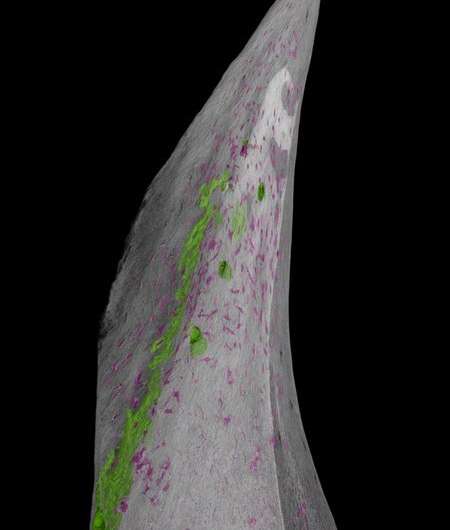
A reconstruction of a single sturgeon scute, close up. Bone-forming cells are marked in 
magenta. Credit: J. Stundl

About 350 million years ago, your evolutionary ancestors—and the ancestors of all modern vertebrates—were merely soft-bodied animals living in the oceans. In order to survive and evolve to become what we are today, these animals needed to gain some protection and advantage over the ocean's predators, which were then dominated by crustaceans.

The evolution of dermal armor, like the sharp spines found on an armored catfish or the bony diamond-shaped scales, called scutes, covering a sturgeon, was a successful strategy. Thousands of species of fish utilized varying patterns of dermal armor, composed of bone and/or a substance called dentine, an important component of modern human teeth. Protective coatings like these helped vertebrates survive and evolve further into new animals and ultimately humans.

But where did this armor come from? How did our ancient underwater ancestors evolve to grow this protective coat?

Now, using sturgeon fish, a new study finds that a specific population of stem cells, called trunk , are responsible for the development of bony scutes in fish. The work was conducted by Jan Stundl, now a Marie Sklodowska-Curie postdoctoral scholar in the laboratory of Marianne Bronner, the Edward B. Lewis Professor of Biology and director of the Beckman Institute at Caltech. A paper describing the research appears in the journal Proceedings of the National Academy of Sciences on July 17.

The Bronner laboratory has long been interested in studying neural crest cells. Found in all vertebrates including fish, chickens, and ourselves, these cells become specialized based on whether they arise from the head (cranial) or spinal cord (trunk) regions. Both cranial and trunk neural crest cells migrate from their starting points throughout the animal's developing body, giving rise to the cells that make up the jaws, heart, and other important structures.

After a 2017 study from the University of Cambridge showed that trunk neural crest cells give rise to dentine-based dermal armor in a type of fish called the little skate, Stundl and his colleagues hypothesized that the same population of cells might also give rise to bone-based armor in vertebrates broadly.

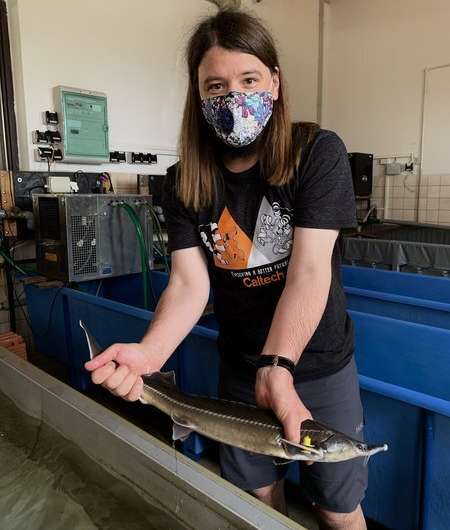
To study this, Stundl and the team turned to the sturgeon fish, specifically the sterlet sturgeon (Acipenser ruthenus). Modern sturgeons, best known for their production of the world's most expensive caviar, still have many of the same characteristics as their ancestors from millions of years ago. This makes them prime candidates for .

How fish evolved their bony, scaly armor
Jan Stundl holds a sturgeon fish in the laboratory. Credit: J. Stundl

Using sturgeon embryos grown at the Research Institute of Fish Culture and Hydrobiology in the Czech Republic, Stundl and his team used fluorescent dye to track how the fish's trunk neural crest cells migrated throughout its developing body. Sturgeons begin to develop their bony scutes after a couple of weeks, so the researchers kept the growing fish in a darkened lab in order to not disturb the  with light.

The team found fluorescently labeled trunk neural crest cells in the exact locations where the sturgeon's bony scutes were forming. They then used a different technique to highlight the fish's osteoblasts, a type of cell that forms bone. Genetic signatures associated with osteoblast differentiation were found in the fluorescent cells in the fish's developing scutes, providing strong evidence that the trunk neural crest cells do in fact give rise to bone-forming cells.

Combined with the 2017 findings about neural crest cells' role in forming dentine-based armor, the work shows that trunk neural crest cells are indeed responsible for giving rise to the bony dermal armor that enabled the evolutionary success of vertebrate .

"Working with non-model organisms is tricky; the tools that exist in standard lab organisms like mouse or zebrafish either do not work or need to be significantly adapted," says Stundl. "Despite these challenges, information from non-model organisms like  allows us to answer fundamental evolutionary developmental biology questions in a rigorous manner."

"By studying many animals on the tree of life, we can infer what evolutionary events have taken place," says Bronner. "This is particularly powerful if we can approach evolutionary questions from a developmental biology perspective, since many changes that led to diverse cell types occurred via small alterations in embryonic development."

The paper is titled "Ancient vertebrate dermal armor evolved from trunk neural ."

More information: Jan Stundl et al, Ancient vertebrate dermal armor evolved from trunk neural crest, Proceedings of the National Academy of Sciences (2023). DOI: 10.1073/pnas.2221120120