Thursday, November 11, 2021

Crown broke 1850 land treaties with First Nations, Ontario Court of Appeal rules

SEAN FINEJUSTICE WRITER
PUBLISHED NOVEMBER 8, 2021

The Ontario Court of Appeal ruled Friday that the minuscule continuing payments violated the 1850 land treaties’ promise to share the resource wealth from the territory.
COLIN PERKEL/THE CANADIAN PRESS

Ontario’s highest court has ruled that the Crown violated the terms of treaties from 1850 by capping annual payments at a few dollars per person to Indigenous peoples who ceded a vast area of the northern part of the province.

The Ontario Court of Appeal ordered a yet-to-be determined amount of compensation that could be in the billions of dollars, and that could come from Ontario, the federal government, or both.

Under the terms of the treaties, the Crown has been paying 23 First Nations just $4 per year for each of their members, in exchange for resource-rich territory the size of France. The land is located on the north shores of Lake Huron and Lake Superior, and it stretches as far north as Hudson Bay. The annual payment has not increased since 1875. In 1850, an initial lump sum of a few thousand dollars was also paid to the communities involved.

The Ontario Court of Appeal ruled 5-0 on Friday that the minuscule continuing payments violated the treaties’ promise to share the resource wealth from the territory, and sent the case back to the trial judge for the next phase: determining how the sharing should be done, and how much money it would mean for the 40,000 Anishinaabe descendants of the First Nations that ceded the territory. Which government is responsible for paying out the money is also to be determined in the next phase.

The appeal court recommended negotiations among the parties. “True reconciliation will not be achieved in the courtroom,” it said.

No place like home: On claiming land that is not our own

To understand why the land remains Indigenous, look to history

The Ontario Ministry of the Attorney General said its counsel are reviewing the decision, and that it would be inappropriate to comment because the matter is within the appeal period and before the courts.

The federal government has said it will negotiate if Ontario comes to the table. The First Nations involved have also agreed to negotiate. The court did not put a figure on the amount of money at stake, but Harley Schachter, a lawyer who represented the Red Rock and Whitesand First Nations in the case, said he expects it will be in the billions, and will address inequities in living standards.

The wealth to be shared includes government revenues connected to the use, sale or licensing of land and water in the treaties’ territory, including mineral and lumber revenues. That wealth includes both future and past revenues.

The trial judge, Ontario Superior Court Justice Patricia Hennessy, determined that the parties to the treaties had a common intention to share revenues, over and above the individual annuities, by means of collective payments to the chiefs and their tribes.

“This is not just money that goes out the window,” Mr. Schachter said in an interview. “This is an investment in a people, and everybody wins. It’s not a zero-sum game. If reconciliation is achieved, all of Canada’s the winner.

He added that he hopes Ontario will not ask the Supreme Court of Canada to hear an appeal. It has 60 days to file such a request.

“I’m hoping the Premier and cabinet will step in and say, ‘Enough’s enough. We have the business of reconciliation to attend to.’”

Catherine Boies Parker, a lawyer representing Anishinaabe beneficiaries of the treaties, also urged Ontario not to appeal.

“It is time to stop looking to the courts for assistance in avoiding your constitutional obligations,” she said, adding that there is no reasonable prospect the Supreme Court would disagree that governments had failed to share the wealth. The courts, she said, have authority to order compensation for those who were wronged.

Ontario argued the Crown has “unfettered discretion” over whether to increase the annual payments, meaning the courts could not review any government decisions related to the treaties’ compensation. In June, 2019, Justice Hennessy ruled that the Crown had acted dishonourably, and that the courts could not only review the payments but supervise to make sure governments pay compensation for treaty violations. Ontario challenged her ruling at the Court of Appeal. The federal government, which had taken different positions on some issues than Ontario, did not.

Justice Hennessy heard from historians, chiefs, elders and experts in Anishinaabe law as she analyzed the history of the treaties and the contemporaneous understanding on both sides. The Anishinaabe believed in reciprocity and renewal of relationships with the British, for whom they had fought in the War of 1812. (Some of the warriors helped negotiate the 1850 treaties.) Their wampum belts had images of two figures holding hands as part of two links in a chain.

The appeal court highlighted poor living conditions for Indigenous peoples in Northern Ontario, which it linked to the failure to share.

“The ‘share’ promised is to be determined not only based on the extent of Crown revenues but also with reference to the relative wealth and needs of the different communities,” the court said. “Obviously, the Anishinaabe would not have expected their communities to suffer a range of deprivations, including substandard housing and boil water advisories, while non-Indigenous communities thrived. Nor was it likely, based on the Anishinaabe principles discussed by the trial judge, that the Anishinaabe would have wished to enjoy great personal wealth while their fellow Canadians suffered deprivation.”

“Discretion is near and dear to the Crown, so I could see that being a motivation for Ontario to appeal,” said Thomas Slade, a lawyer for the Blood Tribe, which intervened in the case. “But the timing couldn’t be worse in terms of the calls on governments to stop fighting First Nations in court and to pursue meaningful reconciliation.”

Last month, the federal government asked the Supreme Court to hear an appeal of a child-welfare ruling involving Indigenous peoples, in which billions of dollars are at stake. Ottawa had faced pressure from the Canadian Bar Association and Indigenous groups not to appeal. The government said that it would hold settlement talks during a pause in litigation.


‘Incredible moment in . . . history’

Anishinabek leaders applaud ruling on Robinson Huron Treaty, seek negotiations with the province


Author of the article: PJ Wilson
Publishing date: Nov 09, 2021 
Mike Restoule, left, chair of the Robinson Huron Treaty Litigation Fund, Chief Duke Peltier of Wiikwemkoong, Chief Dean Sayers, Batchewana First Nation and David Nahwegahbow, co-lead counsel for the Robinson Huron Treaty Litigation Fund, address the media, Tuesday, on a recent court decision. Screen capture

The federal government has indicated it is ready to negotiate a settlement in the Robinson Huron Treaty annuities case.

Now, Chief Duke Peltier says, it is “time for the leaders of Ontario to embrace the reality, embrace the faces, embrace our history and also to embrace the rule of law relating to the Robinson Huron Treaty” and meet the leaders of the Anishinabek Nation and work on a settlement.

Speaking at a news conference Tuesday morning, Peltier, Chief Dean Sayers of the Batchewana First Nation, chair of the Robinson Huron Treaty Litigation Fund Mike Restoule and David Nahwegahbow, co-lead counsel for the litigation, said it is past time to reach a settlement.

The treaty, signed between the Crown and the Anishinabek in 1850, specified that both settlers and First Nations people were to “benefit from the resources” of the land, to “share the wealth of the territory,” Peltier said, and Ontario courts have ruled that the province must implement the augmentation clause, which ensured any increase in the revenue from lands within Robinson-Huron Treaty territory would be evenly distributed to the treaty signatories.


The annuity, initially set at $1.60 a year, was increased to $4 a year in 1874. It has remained at that level since.

“We have been successful in gaining the support of the courts in the first two appeals” filed by the province, the most recent decision being handed down by the Ontario Court of Appeal Friday.

“We unanimously reject the majority of the arguments raised on appeal,” the appeal court said in its 300-page plus ruling. “We dismiss Ontario’s appeal from the Stage Two proceedings in its entirety.”

“We believe we should immediately begin meaningful negotiations to implement the decision of Ontario’s courts of law,” Peltier said.

Nipissing MPP Vic Fedeli received a petition organized by supporters of the litigation fund Friday, calling for an end to the province’s appeals on the issue. A third stage of appeals is scheduled to be heard by the court in September, 2022.

Friday’s decision, Sayers said, is “an incredible moment in the history of our lands here in Canada,” and all Ontarians are “witnessing beautiful history unfold.”

He said the Anishinabek people are “reclaiming our inheritances.”

The province, Sayers said, “must come equipped to the table” to negotiate a just and equitable agreement.

“True reconciliation will not be achieved in the courtroom,” he said.

Sayers also pointed out that the Anishinabek people have received “overwhelming support from civic leaders” in the Robinson Huron Treaty region, which covers a significant portion of Northern Ontario from Sault Ste. Marie to North Bay and north to Kirkland Lake.

“Ontario is hanging on to an untenable position,” Sayers said, and he has met with Ontario Premier Doug Ford in an attempt to end the court cases.

He said Ford has agreed to meet with the Anishinabek leaders again.

“We truly want to go down the road of partnership” with the province, Sayers said.

The province has not yet responded to the court ruling or to the petition.

“We have won every step of the process to this point,” Restoule said, vowing to continue with court cases in the future, or to sit down and negotiate with the province.

“If litigation is what Ontario wants, we will go there.”
CAPITALI$M IS CRISIS OF OVERPRODUCTION
AUSTRALIA
Thousands of avocados dumped in South West due to massive oversupply


Monday, November 8th, 2021
By Monique Welhan
Thousands of avocados have been dumped at the Manjimup tip.
 PIC: Save our Donnelly River/Facebook

Avocado growers in the South West have been forced to dump their fruit due to a huge oversupply and a lack of workers.

A photo posted to Facebook by Save our Donnelly River shows thousands of avocados rotting at the Manjimup tip.

The bumper crop season paired with labour shortages due to the COVID-19 pandemic means avocados with minor blemishes that would make it onto supermarket shelves in previous years are now being dumped.

The oversupply is bringing down the prices of avocados with a 5.5kg tray now selling for just $18 compared to $50 last year.

According to ABC News, 8.2 million trays of avocados are expected to be picked in WA this season — a 233 per cent increase on last year's crop.

BORN AGAIN COVID SPREADERS
'We are kind of a hot zone': Amherst Nova Scotia residents worry after spike in COVID-19 cases after faith-based event
Heidi Petracek
CTV News Atlantic Reporter
Follow | Contact
Published Nov. 9, 2021 

AMHERST, N.S. -

COVID-19 testing has popped up at the Amherst Fire Department, bringing out residents concerned about case counts.

"With the amount of cases, we are kind of a hot zone, so its better to be safe than sorry," said resident Stephanie Chapell.

Fellow resident Joseph Murphy says he's worried.

"It does worry me a lot about them opening up the border, too," he said.

The area has seen a recent increase in cases, with 53 active as of Tuesday.

There is also growing concern over an outbreak at East Cumberland Lodge, a long-term care home in nearby Pugwash, N.S.

There is community spread, says public health, as a result of a faith gathering in the area last month, although it won't confirm where.

"It was a multi-day gathering and there was approximately 100 people that attended the gathering," Dr. Shelley Deeks, Nova Scotia's deputy chief medical officer said on Monday.

One Amherst church did hold such an event in late October.

The pastor for Gospel Light Baptist Church would not speak to CTV News on camera, but Pastor Robert Smith calls what happened "unfortunate" and told CTV the event followed the rules for faith gatherings, which do not require proof of vaccination.

He says parishioners did what 811 told them to when they learned of an exposure.

"On Friday, Dr. Strang did reach out to faith-based leaders to clarify when proof of vaccine requirements are needed," Deeks said.

Smith says 70 per cent of parishoners at the church are fully vaccinated, and the church is working with Nova Scotia Public Health.

Health officials say the outbreak at East Cumberland Lodge is linked to a faith event.

Seventeen residents at the home have tested positive, along with two employees. One resident is in hospital.

"East Cumberland Lodge was one of the members who have had excellent uptake on their vaccinations," said Michele Lowe of Nursing Homes of Nova Scotia Association, the group representing care homes in the province.

Lowe says all residents, and 96 per cent of staff at the lodge have their shots, but community spread, means heightened concern.


"This is where, as a community, we need to support people to get tested through rapid testing," Lowe said.

That is something the area's MLA says should have been happening here sooner.

"Public health and government took away asymptomatic testing from us here," said Elizabeth Smith-McCrossin.

And, she says, take-home test kits aren't widely available either.

"Fully available in HRM, but not in rural Nova Scotia," Smith-McCrossin said


Listen to honey bees shriek a warning to their hive when murder hornets approach

Scientists have compared the calls, named 'antipredator pipes', to fear screams and panic calls

Author of the article:Devika Desai
Publishing date:Nov 10, 2021
Giant 'murder' hornets attack a honey bee hive in Vietnam 
PHOTO BY HEATHER MATTILA/WELLESLEY COLLEGE

Bees have developed their own version of a fire alarm to warn their hive of predatory murder hornets, a new study has found.

Once they’ve spotted a lurking giant or murder hornet, Asian honey worker bees will immediately string along series of panic calls — termed as “antipredator pipes” — which harsher and more irregular in their frequency.

Giant hornets or even Asian hornets which are closely related to the murder hornets recently discovered in North America are known to prey on bees and often send out scouting insects to search for hives to prey on. On finding one, the scout then returns to inform its nest, which then track the hive and often slaughter the entire colony.

“Antipredator pipes share acoustic traits with alarm shrieks, fear screams and panic calls of primates, birds and meerkats”, the study explains.

These pipes, scientists explain in their study published on Wednesday in the Royal Society Open Science journal, are actually vibroacoustic signals made by raising their abdomens, buzzing their wings and flying about “frantically.”

“These sophisticated defences require timely predator detection and swift activation of a defending workforce,” the authors wrote in the study

“Vibroacoustic signals likely play an important role in organizing these responses because they are transmitted quickly between senders and receivers within nests.”

Lead researcher Heather Mattila, professor of biological sciences at Wellesley College, Massachusetts and her team studied interactions between giant hornets and Asian honey bees in Vietnam for over seven years, by placing microphones in hives belonging to local beekeepers. The study collected almost 30,000 signals made by the bees over 1,300 minutes of monitoring.

“[Bees] are constantly communicating with each other, in both good times and in bad, but antipredator signal exchange is particularly important during dire moments when rallying workers for colony defense is imperative,” researchers wrote in their study.

In some cases, scientists observed that the signals prompted the worker bees at the hive’s entrance to go into defence mode, either attacking the scouting hornet by forming a ball around it to heat it to death or spreading animal dung on the hive to repel predators — the first document use of a tool by bees.

It’s unclear the signals do in fact indicate preparation for a specific type of defence, but as a whole, scientists suggest the the bee pipes are a “rallying call for collective defense.”

Scientists also found ‘striking differences’ in the way the bees responded to two types of predators – the giant hornet which attack in numbers and a smaller hornet species which hunts solitarily. The anti-predator pipes didn’t sound as much as for the latter, suggesting that the response may have been specifically developed for the larger, more dangerous predator.

“Colony soundscapes showcase the diversity of (the bees’) alarm signalling repertoire, including a novel antipredator pipe made by workers when (giant hornet) workers were present at nest entrances,” the study states.

“This research shows how amazingly complex signals produced by Asian hive bees can be,” behavioural ecologist Gard Otis was quoted by science website EurekaAlert! .

“We feel like we have only grazed the surface of understanding their communication. There’s a lot more to be learned.”


Captured on video: Bees pipe out alarms to warn of “murder hornet” attacks

When murder hornets approach an Asian beehive, everyone can hear the screams.


JENNIFER OUELLETTE - 11/9/2021, 

Wellesley College researchers have identified a sound that Asian honeybees use to warn the hive of a "murder hornet" attack.

Asian honeybees (Apis cerana) produce a unique alarm sound to alert hive members to an attack by giant "murder hornets," according to a new paper published in the journal Royal Society Open Science. For the first time, scientists at Wellesley College have documented these so-called "anti-predator pipes," which serve as clarion calls to the hive members to initiate defensive maneuvers. You can hear a sampling in the (rather disturbing) video, embedded above, of bees under a hornet attack.

“The [antipredator] pipes share traits in common with a lot of mammalian alarm signals, so as a mammal hearing them, there's something that is instantly recognizable as communicating danger,” said co-author Heather Mattila of Wellesley College, who said the alarm signals gave her chills when she first heard them. “It feels like a universal experience.”

As I've written previously, so-called murder hornets rocketed to infamy after November 2019, when a beekeeper in Blaine, Washington, named Ted McFall, was horrified to discover thousands of tiny mutilated bodies littering the ground—an entire colony of his honeybees had been brutally decapitated. The culprit: the Asian giant hornet species Vespa mandarinia, native to Southeast Asia and parts of the Russian Far East. Somehow, these so-called "murder hornets" had found their way to the Pacific Northwest, where they now pose a dire ecological threat to North American honeybee populations.

FURTHER READING Attack of the Murder Hornets is a nature doc shot through horror/sci-fi lens

There are other species of Asian giant hornets, too. They are apex predators and sport enormous mandibles that they use to rip the heads off their prey and remove the tasty thoraxes (which include muscles that power the bee's wings for flying and movement). A single hornet can decapitate 20 bees in one minute, and just a handful can wipe out 30,000 bees in 90 minutes. The hornet has a venomous, extremely painful sting—and its stinger is long enough to puncture traditional beekeeping suits. And while Asian honeybees have evolved defenses against the murder hornet, North American honeybees have not, as the slaughter of McFall's colony clearly demonstrated.

Mattila has been studying honeybees for 25 years, fascinated by their organization and ability to communicate, and she turned her attention to Asian bees in 2013. "They have evolved in a much scarier predator landscape," she told Ars, pointing to the 22 known species of hornets worldwide for whom Asia is a particular hot zone. Many of these species rely on insects like honeybees to grow their colonies, so they are among the bees' most relentless predators. The deadliest of all are the giant hornets (aka, "murder hornets") because they coordinate in groups to attack beehives.

"As humans, I think there is something fundamentally attractive about understanding predator-prey interactions," said Mattila. "Humans are both predators and prey, depending on the situation, so we've evolved under analogous circumstances as the bees. We can recognize their plight in the face of giant hornets."

Last year, Mattila and her team documented the first example of tool use by honeybees in Vietnam. The researchers discovered that Asian bees forage for animal dung and use it to line the entrances to their hives—a practice dubbed "fecal spotting." It serves as a kind of chemical weapon to ward off giant hornets. Mattila and her team found that hornets were far less likely to land on or chew their way into hives with entrances lined with animal dung.

How Honeybees use animal poop as a chemical weapon to protect hives from giant “murder hornets."

While Mattila and her team were in Vietnam for the dung study, they noticed that noise levels in the hives increased dramatically whenever the giant hornets approached. "We could hear the bees' sounds from several feet away," she said. "So we started popping microphones into colonies so that we could eavesdrop on them." They also took extensive video recordings of activity in the apiaries of local beekeepers.

Ultimately, they collected some 30,000 signals made by the bees over 1,300 minutes and then translated those sounds into spectrograms for analysis. Bees produce a surprisingly complex array of sounds, which they perceive either as air-particle movements they detect with their antennae or as vibrations they detect via special organs in their legs. So bee signals are "vibroacoustic" and are transmitted within colonies as both airborne sounds and vibrations.

There are hisses, for example, usually made by all the bees at once as they lower their bodies and move their wings in near-synchrony, Mattila said. They hiss constantly, but more so when hornets are present, and the exact purpose of the hissing is not yet fully understood.

"Hisses in other animals are often used to intimidate a predator, but that is not likely the case with bees, mostly because they hiss a lot without predators, too," said Mattila. "One idea that has been proposed (not by us) is that hissing helps to momentarily hush the colony because bees stand still for a beat after a hiss. It might help workers perceive other sounds in the nest if most bees stop moving for a second."


Enlarge / Giant murder hornets attack a honey bee hive in Vietnam.
Heather Mattila/Wellesley College

Piping signals accounted for the vast majority (about 95 percent) of the signals in the data set, but there was considerable variability, according to the authors. So-called "stop signals" were the most common (62 percent of the piping signals in the data set). These are produced when a worker vibrates its wings or thorax, typically in response to predator attacks, when fighting over a food source, and during swarming. "Stop signals are really short, [with a] relatively even frequency, and often delivered to other workers via a head butt," said Mattila. Asian honeybees produce more stop signals when hornets appear, and they will change the properties of those signals depending on the type of hornet that is attacking.

Although they are also produced by vibration of the wings or thorax, the newly discovered antipredator pipes are distinct from stop signals. They are harsher and more irregular, with abruptly shifting frequencies. Those colonies used in the study that were attacked by giant hornets were much noisier and frenetic, said Mattila, compared to the relative quiet and calm of the control colonies.

The bees under attack would run about frantically, often congregating at the hive entrance to form defensive "bee carpets." The mode of defense depends in part on the species of attacking hornets. Bees have also been known to form "bee balls" to kill attacking hornets collectively via overheating and suffocation. In fact, the authors wrote, "We observed workers attempt to bee ball a [hornet of the Vespa soror species] immediately after she killed a nest mate that was making antipredator pipes at the [hive] entrance." By contrast, the smaller Vespis velutina hornet attacks alone, targeting individual bees as it hovers in front of hives. The bees typically respond with coordinated "body shaking" as an intimidating visual display.


Enlarge / Biologist Heather Mattila of Wellesley College examines a group of honeybees.
Wellesley College

There was also a broad categorization of piping signals known as "long pipes" that accounted for 34 percent of all the piping signals in the data set. These lack the rapid and unpredictable frequency modulation of the antipredator pipes, according to the authors. "When frequency changed over the duration of these pipes, it did so in smooth sweeps," they wrote. Nor was the long pipe signaling affected by the type of hornet attack.

“[Bees] are constantly communicating with each other, in both good times and in bad, but antipredator signal exchange is particularly important during dire moments when rallying workers for colony defense is imperative,” the authors wrote. Furthermore, when they make those signals, the bees expose a pheromone-producing gland, suggesting they may employ multiple communication strategies to grab the attention of their nest mates. A future focus of Mattila's research will be investigating the role this gland might play in organizing the bees' response to hornet attacks.

The ongoing pandemic has limited field work for the time being, but Mattila says her team has been mailing giant hornets to their labs around the world and have even brought on some new collaborators. Currently, the researchers are re-analyzing the hornet attack videos from the perspective of the hornets rather than the bees. "We're trying to figure out how they disseminate pheromones to recruit their nest mates to a group attack," she said. "These hornet pheromones are key stimuli that bees would be paying attention to—in addition to the alarm signals they produce for each other—to determine what kind of hornet is attacking them."

DOI: Royal Society Open Science, 2021. 10.1098/rsos.211215 (About DOIs).
'Cannibal' sun eruption gives departing astronauts their best aurora views yet


By Tereza Pultarova 

Astronauts get treated to mesmerizing polar lights fairly regularly, so this one must have been quite something.
Astronauts at the International Space Station enjoyed the most stunning display of aurora borealis thanks to a cannibal coronal mass ejection.o (Image credit: ESA/Thomas Pesquet)

Astronauts bidding farewell to the International Space Station enjoyed the most spectacular aurora display of their entire mission over the weekend after a massive blast of material from the sun reached our planet.

The sun has been acting out lately, waking up to its new period of activity after years of quietness. This variation is part of the sun's regular 11-year cycle, the little understood ebb and flow of sunspots and solar flares that is next expected to peak in 2025.

"We were treated to the strongest auroras of the entire mission, over North America and Canada," European Space Agency's astronaut Thomas Pesquet tweeted with a mesmerizing photo of greenish glow. "Amazing spikes higher than our orbit. Star-struck, and we flew right above the centre of the ring, rapid waves and pulses all over

The spectacle must have been quite something since astronauts do get treated to aurora displays fairly regularly. Pesquet himself has shared many images of the magnificent polar lights on his Flickr account since his arrival at the orbital outpost with SpaceX Crew-2 in April.

The latest aurora display was triggered by a series of coronal mass ejections, bursts of magnetized plasma that the sun blasted out last week within a short period of time. The second outburst, travelling a bit faster than the first one, cannibalized its predecessor on the way, resulting in a much more powerful plasma cloud than originally expected.

Auroras occur in Earth's atmosphere when magnetized plasma particles from the sun hit Earth's magnetic field, creating a temporary magnetic havoc around the planet. In addition to providing the glowing spectacle the magnetic storms can damage satellites and knock out power grids. The worst geomagnetic storm in recorded history, the so-called Carrington Event of 1859, disabled telegraph networks all over Europe and North America.

For Pesquet and his Crew-2 companions, NASA astronaut Shane Kimbrough and Megan McArthur, and Japan's Akihiko Hoshide, the latest aurora provided a memorable conclusion to their six-month orbital adventure. Crew-2 is set to return to Earth today. You can watch their departure from the orbital outpost aboard SpaceX's Dragon capsule here.

A view of the Gulf of Morbihan, France
Thomas Pesquet: The best pictures from French astronaut's space missionUpdated: 09/11/2021By James Thomas
France's Thomas Pesquet and three other astronauts have landed back on Earth after a six-month mission aboard the International Space Station (ISS).
Pesquet, representing the European Space Agency, and his crewmates landed in the Gulf of Mexico off the coast of Florida on Monday
Pesquet was known for his active social media presence while in space, regularly posting pictures of his life aboard the ISS, breathtaking views of the Earth and stars and also the occasional meme
To mark his return, we've chosen some of his most popular and striking images from his time in orbit.
A view of the Northern Lights from the International Space Station. Pesquet described this particular sighting on 6 November as “the most intense” of the whole mission.Credit: European Space Agency
A view of the space station in orbit.Credit: European Space Agency
The crew grows chilli peppers for the first time in outer space.
Credit: European Space Agency
A view of the stars from the space station.Credit: European Space Agency
A view of the Gulf of Morbihan, FranceCredit: European Space Agency
The Northern Lights.Credit: European Space Agency
The four astronauts conduct their final checks before their return to Earth.Credit: European Space Agency
A view of Berck, France, near where Pesquet went to flight school.Credit: European Space Agency
A view of Egypt by night.Credit: European Space Agency
A view of Florida. Pesquet notes that unlike other forms of travel, space travel allows you to see your destination before you depart.Credit: European Space Agency

Modified silk cloth keeps skin cooler than cotton

Credit: DOI: 10.1038/s41565-021-00987-0

A team of researchers affiliated with a host of entities in China and one in the U.S. has developed a modified textile that can keep skin cooler than materials made of cotton. In their paper published in the journal Nature Nanotechnology, the group describes their approach to developing garments that are cooler when worn in outdoor conditions.

Humans have been wearing clothes for hundreds of thousands of years, and over that time, have been refining them to suit the needs of their environments, which are mostly cold environments. In this new effort, the researchers wondered if it might be possible to create a type of material that would be cooler to wear than other  and or bare  under direct sunlight. To find out, they started with silk fabric, a material that has been used for thousands of years because of its looks and comfort.

The researchers noted that silk does a good job of reflecting sunlight in the mid-infrared range, which suggests it could be suitable as a cooling garment material. But because it is made by spiders, it contains a protein component that tends to absorb , making the material and its wearer grow hotter under .

To make the silk material UV reflective, the researchers dipped a standard piece of silk fabric into a liquid solution containing highly refractive inorganic oxide nanoparticles. These adhered to the  fabric, allowing it to become evenly saturated throughout the material. They allowed the fabric to dry and then tested it to see if the addition of the nanoparticles made the material more UV reflective. They found that under peak sunlight conditions, the temperature under the material was approximately 3.5 degrees Celsius cooler than the ambient air temperature. Next, they placed the material on a patch of simulated skin and found the skin temperature was approximately 8 degrees Celsius cooler than the same type of simulated skin without the material covering. They also found that it kept the artificial skin approximately 12.5 degrees Celsius cooler than standard cotton material. Further testing showed that the material was able to reflect approximately 95% of , preventing it from passing through to the skin underneath.Microfiber-based metafabric provides daytime radiative cooling

More information: Bin Zhu et al, Subambient daytime radiative cooling textile based on nanoprocessed silk, Nature Nanotechnology (2021). DOI: 10.1038/s41565-021-00987-0

Journal information: Nature Nanotechnology 

© 2021 Science X Network

POSTMODERN ALCHEMY

Going for gold to reduce antibiotic resistance

Going for gold to reduce antibiotic resistance
The gold nanoclusters in their "molecular envelope." The ligands in blue are the zwitterionic ones while those in red are positively charged ones. They are bound to the Au25 cluster (in brown) via thiol molecules (yellow). Credit: University of Leeds

Tiny particles of gold could be the new weapon in the fight against bacterial antibiotic resistance, according to research just published.

Scientists have been investigating the use of gold nanoclusters—each made up of about 25 atoms of gold—to target and disrupt , making them more susceptible to standard antibiotic treatments.

A report from the World Health Organization last year said, "Antibiotic resistance is rising to dangerously high levels in all parts of the world," and called for greater investment in ways to tackle the problem.

For several years, researchers have recognized the antimicrobial properties of specially-adapted gold nanoparticles, but they have struggled to find a way of getting the nanoparticles to the site of a bacterial infection without harming healthy host mammalian .

Now a study by an international team of scientists from the University of Leeds, Southern University of Science and Technology in Shenzhen and Fudan University, Shanghai, both in China, has identified a way of packaging the gold nanoclusters in a molecular envelope that makes them less toxic to healthy tissue without affecting their antibacterial properties.

Laboratory studies have shown that the approach has had a "strong effect" in terms of killing a range of bacteria, some linked to hospital acquired infections and resistant to standard antibiotic treatments.

The findings, which are based on laboratory investigations and not patient trials, have been published in the journal Chemical Science.

Forces of nature

The scientists' solution exploits electrostatic forces in nature.

Bacterial cell walls are more strongly negatively charged than the cells of mammals. Using the idea that opposite charges attract, the gold nanoclusters are wrapped in a molecule called a ligand that is positively charged. Like a carrier pigeon, it finds and delivers the nanoclusters to the wall of bacteria cells, where they disrupt the bacterial cell membrane.

The disruption to the cell membrane increases the permeability of the bacterial cell to standard antibiotic treatments, giving a new lease of life to  that are either ineffective or have waning effectiveness against resistant bacteria.

The problem, though, is the positively charged molecule wrapped around each  is also toxic to healthy host mammalian cells.

Reducing toxicity

To protect host cells, the scientists have added a second ligand to the envelope around each nanocluster. These molecules have both positive and negative charges and are called zwitterionic groups, which are also found in the lipids of cell membranes in mammals. This makes the gold nanoclusters more compatible with host mammalian cells, and easier for the gold nanoclusters to pass through the kidney and be excreted from the body.

In , the scientists investigated whether the gold nanoclusters would be effective in reducing the defenses of the bacterial cells—and make them more susceptible to antibiotic treatment.

They used a bacterial strain called methicillin resistant Staphylococcus epidermidis (MRSE), which is responsible for some hospital-acquired infections.

They tested three antibiotics—each representing a class of antibiotics—against MRSE with and without the gold nanoclusters.

In those cases where the antibiotic was used in combination with the gold nanoclusters, there was an improved antimicrobial effect. With one class of antibiotics, there was a 128-fold decrease in the amount of antibiotic needed to inhibit growth of MRSE.

Dejian Zhou, Professor at Nanochemistry at the University of Leeds and one of the supervisors of the research, said, "Despite extensive research in antibacterial nanomaterials, most of the research has only focused on boosting antibacterial potency without considering their biocompatibility, stability and ability to be excreted from the body. These are essential requirements for clinical approval. As a result, many of the promising antibacterial nanomaterials will not progress to become therapeutic agents to be used in medicine.

"By systematically tuning the ratio of the two ligands, we have identified a way of using gold nanoclusters not only to act as effective antimicrobial agents, but as a mechanism to enhance the potency of antibiotics which have become ineffective because of bacterial drug resistance.

"The research has a significance on the way we should be thinking about responding to antimicrobial resistance."

Professor Zhou hopes that the research findings will be picked up by the pharmaceutical industry. He believes combing  nanoclusters with existing antibiotics will be a faster and cheaper alternative to developing a host of new antibiotics in response to .Atomically precise noble metal nanoclusters

More information: Zeyang Pang et al, Controlling the pyridinium–zwitterionic ligand ratio on atomically precise gold nanoclusters allowing for eradicating Gram-positive drug-resistant bacteria and retaining biocompatibility, Chemical Science (2021). DOI: 10.1039/D1SC03056F

Journal information: Chemical Science 

Provided by University of Leeds 

Sponge Cells Hint On Evolution Of Nervous System

November 10, 2021

Sponges are simple multicellular organisms, though they are skillful filter feeders, filtering tens of thousands of liters of water through their bodies each day to get food. Their ability for this complicated behavior is more exceptional as they do not have a brain or a single neuron.

According to a study published in Science, sponges use a complex cell communication system to regulate their feeding and potentially annihilate invading bacteria. This is exciting research that enables us to look at sponges in a whole new way. According to Casey Dunn, an evolutionary biologist at Yale University, Connecticut, the results could benefit us in understanding the evolution of animals’ nervous systems.
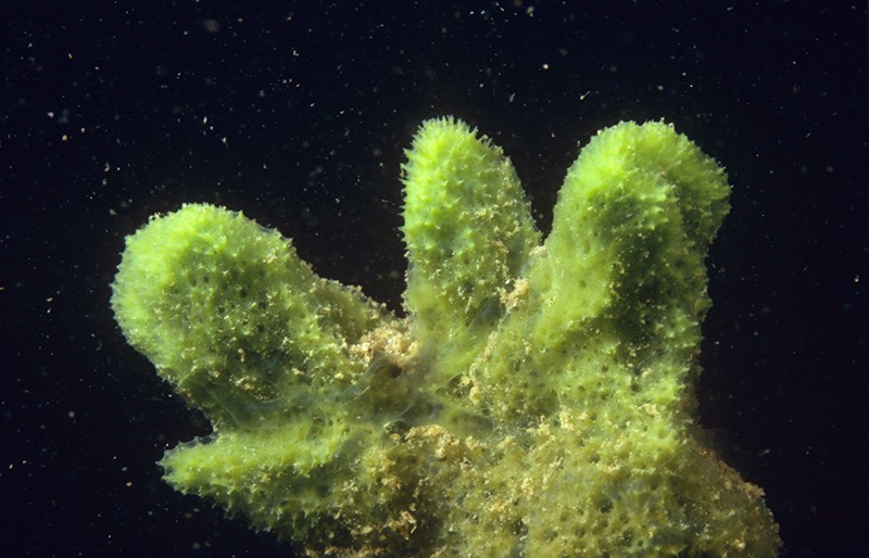
This freshwater sponge (Spongilla lacustris) may hold clues about the evolution of the nervous system. Image Credits: Willem Kolvoort/Nature Picture Library

Neurons interact with one another by transferring electrical or chemical signals via small and targeted connections termed synapses. Despite the animals’ lack of neurons, earlier research discovered that sponges have genes encoding proteins that support synapses function.

Detlev Arendt, an evolutionary biologist, EMBL, Germany, and team sequenced the RNA in several separate cells from a freshwater sponge (Spongilla lacustris) to
determine which cells expressed these genes.

They observed that the sponge has 18 distinguished cell types. Synaptic genes were active in some of them, which were grouped near the digestive cells of the sponges. This implies that some kind of cellular interaction may coordinate the animal’s filter-feeding behavior.

The team then studied one of these cell types, which they named secretory neuroid cells, using X-ray imaging and electron microscopy. The study uncovered that neuroids extend long arms to reach choanocytes, a type of cell with hair-like projections that power sponges’ water-flow systems and capture the majority of their food.


Nervous-system precursor

The team believes that these arms enable neuroids to interact with choanocytes, allowing them to halt the water-flow system and clean out any detritus or foreign microorganisms, based on the proximity of the two cell types and the expression of genes that may allow for chemical secretion. Though these neuroid cells are not nerves, and there is no evidence of the synapses that allow neurons to interact so fast.

Jacob Musser, an evolutionary biologist, EMBL, and co-author of the study, stated that this cell type could be an evolutionary precursor to an actual nervous system.

He further added that they are at an intermediary position, where they have gone from having all these independent pieces to bringing them together more extensively, yet all the interconnectivity required to form a fast synapse is not obtained.

Some experts believe that referring to these cells as nervous system precursors is a stretch. Linda Holland, an evolutionary developmental biologist, UC San Diego, stated that it’s intriguing but not conclusive. She commented that it would be challenging to determine whether the evolution of nervous systems happened from this cellular communication system or arose earlier or even several times, as suggested by some experts. According to Sally Leys, a marine biologist, the University of Alberta, Canada, several other organisms, like unicellular eukaryotes, have the same synaptic genes.

April Hill, a developmental geneticist, Bates College, Maine, believes that this research and its methods will serve as a “launchpad” for further studies into this ubiquitous sponge. She further stated that whether other sponges utilize a comparable cellular interaction system is an important unanswered mystery.

What sponges can tell us about the evolution of the brain

More than a gut reaction
Sponge neuroid cell (orange) extends arms that enwrap the feeding apparatus of a 
sponge digestive cell (green) to create a link for targeted communication. 
The image was taken using electron microscopy. Credit: Jacob Musser, Giulia Mizzon, 
Constantin Pape, Nicole Schieber / EMBL

Despite its central importance, the brain's origins have not yet been uncovered. The first animal brains appeared hundreds of millions of years ago. Today, only the most primitive animal species, such as aquatic sponges, lack brains. Paradoxically, these species may hold the key to unlock the mystery of how neurons and brains first evolved.

Individual neurons in a  communicate via synapses. These connections between  lie at the heart of brain function and are regulated by a number of different genes. Sponges do not have these synapses, but their genome still encodes many of the synaptic genes. EMBL scientists asked the question why this might be the case. Their latest findings are published today in the journal Science.

"We know that these synaptic genes are involved in neuronal function in higher . Finding them in primitive species like sponges begs the question: if these animals don't have brains, what is the role of these genes?" said Detlev Arendt, EMBL group leader and senior scientist at EMBL Heidelberg. "As simple as that sounds, answering this question was beyond our technological abilities so far."

To study the role of these synaptic genes in sponges, the Arendt lab established microfluidic and genomic technologies in the freshwater sponge Spongilla lacustris. Using these techniques, the scientists captured  from several  inside microfluidic droplets and then profiled each cell's genetic activity.

Neuroid cells (purple and red) send cellular arms to contact and communicate with specific digestive cells in the sponge digestive chamber (blue, green, yellow). Other digestive chamber cells are shown in grey. Credit: Jacob Musser, Giulia Mizzon, Constantin Pape, Nicole Schieber / EMBL

"We showed that certain cells in the sponge digestive chambers activate the synaptic genes. So even in a primitive animal lacking synapses, the synaptic genes are active in specific parts of its body," said Jacob Musser, research scientist in the Arendt group and lead author on the study.

Sponges use their digestive chambers to filter out food from the water and interact with environmental microbes. To understand what the cells expressing synaptic  do, the Arendt group joined forces with six EMBL teams as well as collaborators in Europe and worldwide. Working with EMBL's Electron Microscopy Core Facility, Yannick Schwab's team and Thomas Schneider's group operating synchrotron beamlines at EMBL Hamburg the researchers developed a new correlative imaging approach. "By combining  with X-ray imaging on a synchrotron beamline we were able to visualize the stunning behavior of these cells," Dr. Schwab explained.

The scientists captured three-dimensional snapshots of cells crawling throughout the digestive chamber to clear out bacterial invaders and sending out long arms that enwrap the feeding apparatus of specific digestive cells. This behavior creates an interface for targeted cell-cell communication, as it also happens across synapses between neuronal cells in our brains.

"Our results point to the cells regulating feeding and controlling the microbial environment as possible evolutionary precursors for the first animal brains," Dr. Musser said.Microglia pruning brain synapses captured on film for the first time

More information: Jacob Musser et al, Profiling cellular diversity in sponges informs animal cell type and nervous system evolution, Science (2021). DOI: 10.1126/science.abj2949. www.science.org/doi/10.1126/science.abj2949

Journal information: Science 

Provided by European Molecular Biology Laboratory 


Sponge Genes Hint at the Origins of Neurons and Other Cells

A new study of gene expression in sponges reveals the complex diversity of their cells as well as some possibly ancient connections between the nervous, immune and digestive systems.


A new atlas of gene expression in the sponge Spongilla has revealed surprising levels of cellular diversity in these primitive animals.
Allexxandar / Dreamstime.com


Viviane Callier

November 4, 2021

When the first sponge genomes were sequenced in the early 2000s, researchers were surprised to find that sponges not only have roughly as many genes as humans and other complex creatures but also have many of the same genes. Sponges are among the earliest branching lineages on the evolutionary tree of animal life; their simple bodies don’t even have a pattern of symmetry or a set number of parts. The presence of those genes implied that the genetic information for functions like muscle contraction and the differentiation of neurons was much more ancient than muscles or nervous systems themselves.

But what were those genes doing in an animal without neurons or muscles? Researchers could only make educated guesses and investigate expression patterns on a painstaking gene-by-gene basis.

Today, however, a new study taking advantage of rapid advances in genomic technologies has illuminated where about 26,000 genes are expressed in the freshwater sponge Spongilla. This atlas of gene expression reveals the genetic configuration of cell types throughout the sponge’s body, including some cell types never described before. It offers important hints about how cell types evolved in the first place, and it may help to settle a long, thorny debate about whether neurons evolved just once or many times. The study appears in the latest issue of Science.

This ambitious paper “leapfrogs” over previous work, according to Scott Nichols, who studies sponge evolution at the University of Denver. “What is extraordinary about it is that really fascinating hypotheses have emerged from this data set,” he said. “But I would emphasize strongly that they need to be experimentally tested.”

The most exciting hypothesis concerns cells inside the sponge’s digestive chambers. The chambers are lined with distinctive cells called choanocytes, which have a collar of fingerlike protrusions (microvilli) and a flagellum. The choanocytes beat their flagella to regulate the flow of water through the digestive chamber, all the while feeding on small particles and debris the water carries. The digestive chambers also contain mobile “neuroid” cells that were described years ago, although their identity and function were mysterious.

Using high-throughput single-cell RNA sequencing technology, Detlev Arendt’s team at the European Molecular Biology Laboratory in Heidelberg discovered that choanocytes express genes that in neurons produce the postsynaptic “scaffolding” involved in receiving and responding to neurotransmitters. They also discovered that the mobile neuroid cells express a suite of genes that are typically active in the presynaptic bulb of a neuron. This led the researchers to hypothesize that the neuroid cells might be talking to the choanocytes, and that the neuroid cells’ job might be to patrol the microbial environment in the digestive chamber and regulate the choanocytes’ feeding behaviors accordingly.


Sponges have digestive chambers lined with cells called choanocytes. Waving their flagella to propel water through the chambers, the choanocytes digest small particles in the flow.

Caterina Longo, Bari University; source: doi.org/10.1371/journal.pone.0042392.g005

When Jacob Musser, the postdoctoral fellow in Arendt’s lab who led the project, stained the sponge to look at where exactly the pre- and postsynaptic genes were being expressed, he saw that the neuroid cells expressing presynaptic genes were indeed near the choanocytes expressing postsynaptic genes. In fact, the neuroid cells reached out pseudopod arms that seemed to touch the choanocytes.

“This was obviously really tantalizing,” Musser said. “But you can’t really tell what is going on.”

To get a more detailed picture of what the cells were doing, Musser and the team used focused ion beam electron microscopy at the X-ray synchrotron facility in Hamburg to get very high-resolution 3D images of the cells, which could distinguish cellular features as small as 15 nanometers, roughly the size of many folded proteins. They saw that projections from the neuroid cells enveloped the choanocytes’ microvilli collar and flagellum, and that the neuroid cells held vesicles like those in the presynaptic bulb of a neuron. They suspect the vesicles are probably releasing glutamate, a neurotransmitter.

But tempting as it is to imagine these sponges as having primitive synapses, the researchers never observed direct, stable contacts between the neuroid cells and choanocytes. The connections between the cells instead seem to be transient. Furthermore, the DNA of sponges lacks genes for some of the key ion channels needed to create an action potential — the sharp electrical signal that stimulates the release of neurotransmitters in neurons.

Nevertheless, because sponges have always been thought to lack anything even resembling a nervous system, the suggestion that they have cellular mechanisms with a deep evolutionary relationship to neurons “is an exciting path forward to connect sponge biology to neural cell biology, to understand where neuronal signaling came from at all in animals,” Nichols said.


A colorized micrograph of the cells in a sponge digestive chamber (left) reveals the interaction of a neuroid cell (magenta) with a choanocyte (green). In a magnified detail (right), the transient contact between the two cells could be suggestive of the synaptic contact between neurons.
Quanta Magazine; source: Jacob Musser, Giulia Mizzon, Constantin Pape, Nicole Schieber / EMBL

The origin of neurons and nervous systems — and in particular, the question of whether neurons arose once or multiple times — is one of the most contentious topics in the field of evolutionary developmental biology, according to Maria Antonietta Tosches, who studies the evolution of cell types in vertebrates at Columbia University and previously trained in Arendt’s lab. The findings from this new study seem to bear on that mystery because the researchers found presynaptic gene sets expressed in neuroid cells and postsynaptic genes expressed in choanocytes. (Both sets of genes were active in other cell types as well.) That fact suggests that the genetic modules responsible for both the sending and receiving ends of cell-cell communication systems were deployed in various types of ancestral animal cells. Neurons could therefore have evolved repeatedly and independently through different applications of these gene modules, Tosches said.

In fact, many multifunctional cells in sponges express modules of genes usually associated with specialized cells in more complex animals like vertebrates. For example, sponge neuroid cells not only express some of the presynaptic machinery of neurons, but also express immune genes. (It’s possible that if neuroid cells monitor the microbial content of the digestive chambers for sponges, these immune genes assist in that role.) Sponges also have cells called pinacocytes that contract in unison like muscle cells to squeeze the animal and expunge waste or unwanted debris; pinacocytes have some sensory machinery that responds to nitric oxide, a vasodilator.

“Nitric oxide is what relaxes our smooth muscle in our blood vessels, so when our blood vessels expand, that’s nitric oxide causing that relaxation,” Musser said. “And we’ve actually shown through experiments in the paper that nitric oxide is also regulating contractions in this sponge.” Like glutamate, nitric oxide might have been part of an early signaling mechanism to coordinate primitive behaviors in the sponge, he suggests.


EVOLUTION
Scientists Debate the Origin of Cell Types in the First Animals

JULY 17, 2019


“Our data are very consistent with this notion that a large number of important functional pieces of machinery existed early in animal evolution,” Musser said. “And a lot of early animal evolution was about starting to subdivide this out to different cells. But likely these very first cell types were very multifunctional, and they had to do multiple things.” The earliest animal cells, like their close relatives the protozoans, probably had to be cellular Swiss Army knives. As multicellular animals evolved, their cells may have taken on different roles, a division of labor that may have led to more specialized cell types. But different lineages of animals may have divvied things up differently and to different degrees.

If the mixing and matching of genetic modules was a crucial theme of early animal evolution, then comparing the arrangement and expression of those modules in different species could tell us about their history — and about possible limitations on how haphazardly they can be shuffled. One researcher looking for those answers is Arnau Sebé-Pedrós, who studies cell type evolution at the Center for Genomic Regulation in Barcelona and who published the first atlases of cell types in sponges, placozoans and comb jellies in 2018.

Sebé-Pedrós thinks that the spatial configuration of the genes along the chromosomes could be revelatory because genes located together can share regulatory machinery. “I’m absolutely shocked by the degree of conservation of the gene orders in animal genomes,” he said. He suspects that the need to co-regulate sets of functionally related genes keeps them in the same chromosomal neighborhood.

Scientists are still in the early days of learning how cell types evolve and relate to one another. But as important as it is to clarify the muddy origins of animal evolution, sponge cell atlases are also making a major contribution by revealing the possibilities in animal cell biology. “It is not just important for us to understand the very origin of animals,” Sebé-Pedrós said, “but also to understand things that may be radically different from anything else that we know about other animals.”
A big discovery of a tiny critter


Water bear fossil found in 16-million-year-old amber is the third tardigrade species ever discovered

Amber specimen including tardigrades and other invertebrates.


Image by Phil Barden/NJIT

BY Juan Siliezar Harvard Staff Writer

DATE November 5, 2021

Good luck finding an animal tougher than a tardigrade.

These tiny creatures are famous for their ability to survive in the most extreme conditions, including boiling water, freezing water, and even the vacuum of space. Called water bears or moss piglets because of their appearance under a microscope, tardigrades are the smallest-known animals with legs. They have a pudgy body — no larger than a pencil point — their eight legs have several pointed claws at the end, and they have a spear-like sucker that extends from their mouth.

Tardigrades are found on all the continents (basically wherever there is water) and have survived on Earth for more than 500 million years. Despite such a long evolutionary history and global presence, the fossil record on tardigrades is thin, with only two clear examples identified as separate species ever found. But thanks to a 16-million-year-old piece of amber discovered in the Dominican Republic, scientists can now add a third — a discovery immortalized in word and song.


Magnified example of a tardigrade, with its pudgy body that is no larger than a pencil point.

Credit: Phil Barden/NJIT


The researchers from Harvard and the New Jersey Institute of Technology who made the discovery describe their finding in a new paper in the Proceedings of the Royal Society B. The ultra-rare fossil can help shed new light on these ancient animals and provide new evolutionary insight into how the 1,300 tardigrade species that exist today evolved.

The specimen is the first water bear fossil ever recovered from the Cenozoic era, which started 66 million years ago and encompasses the planet’s current geological era. The researchers believe this is the best-imaged tardigrade fossil to date. In fact, they were able to describe microscopic details on parts of the mouth and needle-like claws that are about 20 to 30 times finer than a human hair. They were also able to get an unprecedented look at the internal anatomy of its foregut, which played the key role in identifying the fossil as a new genus and species.

“We could see it had this unique foregut organization that warranted for us to formalize a new genus within this extant group of tardigrade superfamilies,” said Marc A. Mapalo, a Ph.D. candidate who does his work in the Department of Organismic and Evolutionary Biology, Harvard’s Graduate School of Arts and Sciences, and is lead author of the study. “We saw characters that are not observed in extant species, but are observed in the fossils. This helps us understand what changes in the body occurred across millions of years.”

The researchers were able to examine these distinct anatomical features with a confocal laser microscope, a piece of equipment that uses a laser instead of visible light to peer into the specimen and produce a high-quality image. Usually, it is used to see biological and molecular processes such as cell division 

.
Reconstruction of tardigrades from the Miocene of Dominican Republic. Artwork by Holly Sullivan

“Using this high-powered technique that is usually employed for studying cell biology, it was possible to obtain extremely detailed anatomical information,” said Javier Ortega-Hernández, an assistant professor in OEB and curator of invertebrate paleontology in the Museum of Comparative Zoology. “We saw the whole animal in way better detail than previously possible using conventional light microscopy.”

The researchers called the new species Paradoryphoribius chronocaribbeus. The name uses the Greek word for time, “chrono,” and refers to the Caribbean region where it was found, “caribbeus.” The new species is a relative of the modern living family of tardigrades known as Isohypsibioidea.

The other two fully described unequivocal tardigrade fossils are Milnesium swolenskyi and Beorn leggi, both known from Cretaceous-age amber in North America. This makes the new Dominican species the first water bear fossil to be found outside that region.

Co-author Phillip Barden from the New Jersey Institute of Technology introduced the fossil to Ortega-Hernández and Mapalo in 2019 after visiting the Museum of Comparative Zoology as a guest speaker. Mapalo, who was new to the lab, is a specialist in tardigrades and took the lead in analyzing the fossil using confocal microscopes in the Harvard Center for Biological Imaging.

Mapalo and Ortega-Hernández hope their success with the confocal microscope will inspire other researchers to use it to examine their own amber samples. The pair will also continue to employ the technology to study other tardigrades fossils trapped in amber.

Meanwhile, Mapalo has written a song (as one does) to celebrate number three.