And its tiny movements - less than the width of a human hair - have been caught on film for the first time.
by Joe Mellor
in News, Science
The brain actually ‘beats’… just like the heart, say scientists.
And its tiny movements – less than the width of a human hair – have been caught on film for the first time.
The breakthrough could lead to better ways of spotting concussions and other brain injuries before they become life threatening.
It also offers hope of developing cycling and motorbike crash helmets that provide better protection.
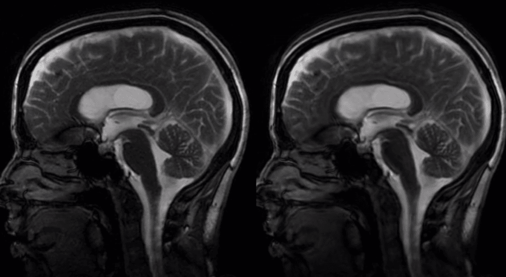
The US team used a revolutionary imaging technique to capture and magnify the movement of the brain every time the heart beats – in real time.
They say it is a promising and long awaited diagnostic tool for a host of brain disorders that will improve and hasten treatment.
These include weakened blood vessels in the brain called aneurysms that can trigger potentially fatal haemorrhages.
Understanding how the brain moves – at rest and upon impact – has been crucial to shedding light on them, but technology to clearly see it has lagged behind.
Co lead author Professor Mehmet Kurt, a biomechanical engineer at Stevens Institute of Technology, New Jersey, said: “It is proof of concept.
“We wanted to see if we could amplify the tiny movements of the brain with every heartbeat and capture that movement as it naturally occurs – so without introducing noise.
“That is important when you are trying to do what we are trying to do – detect abnormal motions in the brain to diagnose and monitor disorders.”
The brain moves minutely with each heartbeat – on the order of ten to 150 micrometres which is less than the width of a single human hair.
The movements are so small standard MRI (magnetic resonance imaging) brain scans are unable to film them in detail.
The new technique reported in Magnetic Resonance in Medicine is called phase-based amplified MRI and was originally developed while the researchers were based at the University of Stanford, California.
In the past two years they have fine tuned the method to show it can be used for diagnostic benefit.
In experiments they attached a pulsometer that monitors heart rate to the wrists of healthy subjects and coordinated the timing of the beat with images of the brain, stitching the slices together to create a smooth movement.
An algorithm, tailored to the piston-like motions of blood and spinal fluid coursing through the brain, then intelligently magnifies the brain’s motion to a more visible scale while keeping potential noise subdued.
The resulting video images, reconstructed slice by slice, retain the spatial characteristics of an MRI.
The skull and all anatomical features are displayed at actual scale. But the pulse-driven motion is amplified significantly as they animate.
Co lead senior author Dr Samantha Holdsworth, a medical physicist who is now at the University of Auckland, explained: “You can actually capture the whole head ‘nodding’ in the scanner due to the force of the blood pumping into the brain every time the heart beats.”
The researchers found the technique provided few errors and good visibility, particularly in areas of the brain that move most. These included the mid brain and spinal cord, which helps relay sensory information.
It also spots movement in areas resistant to motion such as the frontal cortex which is important for planning, reasoning and judgement.
The team applied the technique on two subjects, a control and a patient with Chiari malformation I.
The condition, present at birth, can cause many symptoms, including headaches or stiffness in the neck, due to malformations at the base of the skull and upper spinal area.
Unlike the control, video images of the patient showed significantly abnormal brain movement in at least two locations.
The researchers will continue to use the technology in clinical settings involving larger numbers of patients with known medical diagnoses of various conditions such as concussion, aneurysm and structural brain abnormalities.
Prof Kurt, who is also known for his work on concussions, added: “Better visualisation and understanding of the biomechanical properties of the brain could lead to earlier detection and monitoring of brain disorders.
“It could also help with prevention, as it could lead to the design of better helmets.”
in News, Science
The brain actually ‘beats’… just like the heart, say scientists.
And its tiny movements – less than the width of a human hair – have been caught on film for the first time.
The breakthrough could lead to better ways of spotting concussions and other brain injuries before they become life threatening.
It also offers hope of developing cycling and motorbike crash helmets that provide better protection.
The US team used a revolutionary imaging technique to capture and magnify the movement of the brain every time the heart beats – in real time.
They say it is a promising and long awaited diagnostic tool for a host of brain disorders that will improve and hasten treatment.
These include weakened blood vessels in the brain called aneurysms that can trigger potentially fatal haemorrhages.
Understanding how the brain moves – at rest and upon impact – has been crucial to shedding light on them, but technology to clearly see it has lagged behind.
Co lead author Professor Mehmet Kurt, a biomechanical engineer at Stevens Institute of Technology, New Jersey, said: “It is proof of concept.
“We wanted to see if we could amplify the tiny movements of the brain with every heartbeat and capture that movement as it naturally occurs – so without introducing noise.
“That is important when you are trying to do what we are trying to do – detect abnormal motions in the brain to diagnose and monitor disorders.”
The brain moves minutely with each heartbeat – on the order of ten to 150 micrometres which is less than the width of a single human hair.
The movements are so small standard MRI (magnetic resonance imaging) brain scans are unable to film them in detail.
The new technique reported in Magnetic Resonance in Medicine is called phase-based amplified MRI and was originally developed while the researchers were based at the University of Stanford, California.
In the past two years they have fine tuned the method to show it can be used for diagnostic benefit.
In experiments they attached a pulsometer that monitors heart rate to the wrists of healthy subjects and coordinated the timing of the beat with images of the brain, stitching the slices together to create a smooth movement.
An algorithm, tailored to the piston-like motions of blood and spinal fluid coursing through the brain, then intelligently magnifies the brain’s motion to a more visible scale while keeping potential noise subdued.
The resulting video images, reconstructed slice by slice, retain the spatial characteristics of an MRI.
The skull and all anatomical features are displayed at actual scale. But the pulse-driven motion is amplified significantly as they animate.
Co lead senior author Dr Samantha Holdsworth, a medical physicist who is now at the University of Auckland, explained: “You can actually capture the whole head ‘nodding’ in the scanner due to the force of the blood pumping into the brain every time the heart beats.”
The researchers found the technique provided few errors and good visibility, particularly in areas of the brain that move most. These included the mid brain and spinal cord, which helps relay sensory information.
It also spots movement in areas resistant to motion such as the frontal cortex which is important for planning, reasoning and judgement.
The team applied the technique on two subjects, a control and a patient with Chiari malformation I.
The condition, present at birth, can cause many symptoms, including headaches or stiffness in the neck, due to malformations at the base of the skull and upper spinal area.
Unlike the control, video images of the patient showed significantly abnormal brain movement in at least two locations.
The researchers will continue to use the technology in clinical settings involving larger numbers of patients with known medical diagnoses of various conditions such as concussion, aneurysm and structural brain abnormalities.
Prof Kurt, who is also known for his work on concussions, added: “Better visualisation and understanding of the biomechanical properties of the brain could lead to earlier detection and monitoring of brain disorders.
“It could also help with prevention, as it could lead to the design of better helmets.”
No comments:
Post a Comment