Monday, October 24, 2022

ALCHEMY

Nanomaterial from the Middle Ages
Nanomaterial from Middle Ages
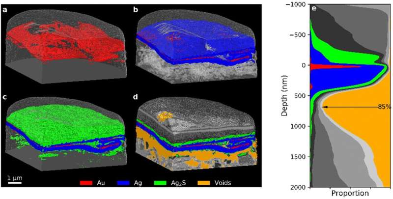
PXCT 3D images of the 35-year old Zwischgold sample showing the addition of (a) Au, (b) Ag (with transparent voids), (c) “silver corrosion products”, and (d) other segments. (e) Stack plot of the depth profile of the single-layered section of the sample, aligned to the main layer of the Au segment. Credit: Nanoscale (2022). DOI: 10.1039/D2NR03367D

To gild sculptures in the late Middle Ages, artists often applied ultra-thin gold foil supported by a silver base layer. For the first time, scientists at the Paul Scherrer Institute PSI have managed to produce nanoscale 3D images of this material, known as Zwischgold. The pictures show this was a highly sophisticated medieval production technique and demonstrate why restoring such precious gilded artifacts is so difficult.

The samples examined at the Swiss Light Source SLS using one of the most advanced microscopy methods were unusual even for the highly experienced PSI team: minute samples of materials taken from an altar and wooden statues originating from the fifteenth century. The altar is thought to have been made around 1420 in Southern Germany and stood for a long time in a mountain chapel on Alp Leiggern in the Swiss canton of Valais.

Today it is on display at the Swiss National Museum (Landesmuseum Zürich). In the middle you can see Mary cradling Baby Jesus. The material sample was taken from a fold in the Virgin Mary's robe. The tiny samples from the other two medieval structures were supplied by Basel Historical Museum.

The material was used to gild the sacred figures. It is not actually  leaf, but a special double-sided foil of gold and  where the gold can be ultra-thin because it is supported by the silver base. This material, known as Zwischgold (part-gold) was significantly cheaper than using pure gold leaf.

"Although Zwischgold was frequently used in the Middle Ages, very little was known about this material up to now," says PSI physicist Benjamin Watts: "So we wanted to investigate the samples using 3D technology which can visualize extremely fine details."

Although other microscopy techniques had been used previously to examine Zwischgold, they only provided a 2D cross-section through the material. In other words, it was only possible to view the surface of the cut segment, rather than looking inside the material. The scientists were also worried that cutting through it may have changed the structure of the sample.

The advanced microscopy imaging method used today, ptychographic tomography, provides a 3D image of Zwischgold's exact composition for the first time.

X-rays generate a diffraction pattern

The PSI scientists conducted their research using X-rays produced by the Swiss Light Source SLS. These produce tomographs displaying details in the nanoscale range—millionths of a millimeter, in other words.

"Ptychography is a fairly sophisticated method, as there is no objective lens that forms an image directly on the detector," Watts explains. Ptychography actually produces a  of the illuminated area, in other words an image with points of differing intensity.

By manipulating the sample in a precisely defined manner, it is possible to generate hundreds of overlapping diffraction patterns. "We can then combine these diffraction patterns like a sort of giant Sudoku puzzle and work out what the original image looked like," says the physicist. A set of ptychographic images taken from different directions can be combined to create a 3D tomogram.

The advantage of this method is its extremely high resolution. "We knew the thickness of the Zwischgold sample taken from Mary was of the order of hundreds of nanometers," Watts explains. "So we had to be able to reveal even tinier details."

The scientists achieved this using ptychographic tomography, as they report in their latest article in the journal Nanoscale. "The 3D images clearly show how thinly and evenly the gold layer is over the silver base layer," says Qing Wu, lead author of the publication.

The art historian and conservation scientist completed her Ph.D. at the University of Zurich, in collaboration with PSI and the Swiss National Museum. "Many people had assumed that technology in the Middle Ages was not particularly advanced," Wu comments. "On the contrary: this was not the Dark Ages, but a period when metallurgy and gilding techniques were incredibly well developed."

Secret recipe revealed

Unfortunately there are no records of how Zwischgold was produced at the time. "We reckon the artisans kept their recipe secret," says Wu. Based on nanoscale images and documents from later epochs, however, the art historian now knows the method used in the 15th century: first the gold and the silver were hammered separately to produce thin foils, whereby the gold film had to be much thinner than the silver.

Then the two metal foils were worked on together. Wu describes the process: "This required special beating tools and pouches with various inserts made of different materials into which the foils were inserted," Wu explains. This was a fairly complicated procedure that required highly skilled specialists.

"Our investigations of Zwischgold samples showed the average thickness of the gold layer to be around 30 nanometers, while gold leaf produced in the same period and region was approximately 140 nanometers thick," Wu explains. "This method saved on gold, which was much more expensive." At the same time, there was also a very strict hierarchy of materials: gold leaf was used to make the halo of one figure, for example, while Zwischgold was used for the robe.

Because this material has less of a sheen, the artists often used it to color the hair or beards of their statues. "It is incredible how someone with only hand tools was able to craft such nanoscale material," Watts says. Medieval artisans also benefited from a unique property of gold and silver crystals when pressed together: their morphology is preserved across the entire metal film. "A lucky coincidence of nature that ensures this technique works," says the physicist.

Golden surface turns black

The 3D images do bring to light one drawback of using Zwischgold, however: the silver can push through the gold layer and cover it. The silver moves surprisingly quickly—even at room temperature. Within days, a thin silver coating covers the gold completely. At the surface the silver comes into contact with water and sulfur in the air, and corrodes.

"This makes the gold surface of the Zwischgold turn black over time," Watts explains. "The only thing you can do about this is to seal the surface with a varnish so the sulfur does not attack the silver and form silver sulfide." The artisans using Zwischgold were aware of this problem from the start. They used resin, glue or other organic substances as a varnish. "But over hundreds of years this protective layer has decomposed, allowing corrosion to continue," Wu explains.

The corrosion also encourages more and more silver to migrate to the surface, creating a gap below the Zwischgold. "We were surprised how clearly this gap under the metal layer could be seen," says Watts. Especially in the sample taken from Mary's robe, the Zwischgold had clearly come away from the base layer.

"This gap can cause mechanical instability, and we expect that in some cases it is only the protective coating over the Zwischgold that is holding the metal foil in place," Wu warns. This is a massive problem for the restoration of historical artifacts, as the silver sulfide has become embedded in the varnish layer or even further down.

"If we remove the unsightly products of corrosion, the varnish layer will also fall away and we will lose everything," says Wu. She hopes it will be possible in future to develop a special material that can be used to fill the gap and keep the Zwischgold attached. "Using ptychographic tomography, we could check how well such a consolidation material would perform its task," says the art historian.Maintaining the structure of gold and silver in alloys

More information: Qing Wu et al, A modern look at a medieval bilayer metal leaf: nanotomography of Zwischgold, Nanoscale (2022). DOI: 10.1039/D2NR03367D
Journal information: Nanoscale 
Provided by Paul Scherrer Institute 


Scientists hit their creative peak early in their careers, study finds

scientist
Credit: Pixabay/CC0 Public Domain

A new study provides the best evidence to date that scientists overall are most innovative and creative early in their careers.

Findings showed that, on one important measure, the impact of biomedical scientists' published work drops by between one-half to two-thirds over the course of their careers.

"That's a huge decline in impact," said Bruce Weinberg, co-author of the study and professor of economics at The Ohio State University.

"We found that as they get older, the work of biomedical scientists was just not as innovative and impactful."

But the reasons behind this trend of declining innovativeness make the findings more nuanced and show why it is still important to support scientists later in their careers, Weinberg said.

The study was published online Oct. 7, 2022 in the Journal of Human Resources.

Researchers have been studying the relationship between age or experience with innovativeness for nearly 150 years, but no consensus has emerged. Findings, in fact, have been "all over the map," Weinberg said.

"For a topic that so many people with so many approaches have studied for so long, it is pretty remarkable that we still don't have a conclusive answer."

One advantage of this study is that the authors had a huge dataset to work with—5.6 million biomedical science articles published over a 30-year period, from 1980 to 2009, and compiled by MEDLINE. These data include detailed information on the authors.

This new study measured the innovativeness of the articles by biomedical scientists using a standard method—the number of times other scientists mention (or "cite") a study in their own work. The more times a study is cited, the more important it is thought to be.

With detailed information on the authors of each paper, the researchers in this study were able to compare how often scientists' work was cited early in their careers compared to later in their careers.

As they analyzed the data, Weinberg and his colleagues made a discovery that was a key to understanding how innovation changes over a .

They found that scientists who were the least innovative early in their careers tended to drop out of the field and quit publishing new research. It was the most productive, the most important young scholars who were continuing to produce research 20 or 30 years later

"Early in their careers, scientists show a wide range of innovativeness. But over time, we see selective attrition of the people who are less innovative," Weinberg said.

"So when you look at all biomedical scientists as a group, it doesn't look like innovation is declining over time. But the fact that the least innovative researchers are dropping out when they are relatively young disguises the fact that, for any one person, innovativeness tends to decline over their career."

Results showed that for the average researcher, a scientific article they published late in their career was cited one-half to two-thirds less often than an article published early in their careers.

But it wasn't just citation counts that suggest researchers were less innovative later in their career.

"We constructed additional metrics that captured the breadth of an article's impact based on the range of fields that cite it, whether the article is employing the best and latest ideas, citing the best and latest research, and whether the article is drawing from multiple disciplines," said Huifeng Yu, a co-author, who worked on the study as a Ph.D. student at the University at Albany, SUNY.

"These other metrics also lead to the same conclusion about declining innovativeness."

The findings showing selective attrition among less-innovative scientists can help explain why previous studies have had such conflicting results, Weinberg said.

Studies using Nobel Laureates and other eminent researchers, for whom attrition is relatively small, tend to find earlier peak ages for innovation. In contrast, studies using broader cross-sections of scientists don't normally find an early peak in creativity, because they don't account for the attrition.

Weinberg noted that  in the  may not relate only to innovativeness. Scientists who are women or from underrepresented minorities may not have had the opportunities they needed to succeed, although this study can't quantify that effect.

"Those scientists who succeeded probably did so through a combination of talent, luck, personal background and prior training," he said.

The findings suggest that organizations that fund scientists have to maintain a delicate balance between supporting youth and experience.

"Young scientists tend to be at their peak of creativity, but there is also a big mix with some being much more innovative than others. You may not be supporting the very best researchers," said Gerald Marschke, a co-author of the study and associate professor of economics at the University at Albany,

"With older, more experienced scientists, you are getting the ones who have stood the test of time, but who on average are not at their best anymore."

Creativity is not just for the young, study finds

More information: Huifeng Yu et al, Publish or Perish: Selective Attrition as a Unifying Explanation for Patterns in Innovation over the Career, Journal of Human Resources (2022). DOI: 10.3368/jhr.59.2.1219-10630R1
Journal information: Journal of Human Resources

A new approach, not currently described by the Clean Air Act, could eliminate air pollution disparities

air quality
Credit: Unsplash/CC0 Public Domain

While air quality has improved dramatically over the past 50 years thanks in part to the Clean Air Act, people of color at every income level in the United States are still exposed to higher-than-average levels of air pollution.

A team led by researchers at the University of Washington wanted to know if the Clean Air Act is capable of reducing these disparities or if a new approach would be needed. The team compared two approaches that mirror main aspects of the Clean Air Act and a third approach that is not commonly used to see if it would be better at addressing disparities across the contiguous U.S. The researchers used national emissions data to model each strategy: targeting specific emissions sources across the U.S.; requiring regions to adhere to specific concentration standards; or reducing emissions in specific communities.

While the first two approaches—based on the Clean Air Act—didn't get rid of disparities, the community-specific approach eliminated  disparities and reduced pollution exposure overall.

The team published these findings Oct. 24 in the Proceedings of the National Academy of Sciences.

"In earlier research, we wanted to know which  were responsible for these disparities, but we found that nearly all sources lead to unequal exposures. So we thought, what's it going to take? Here, we tried three approaches to see which would be the best for addressing these disparities," said senior author Julian Marshall, a UW professor of civil and environmental engineering. "The two approaches that mirror aspects of the Clean Air Act were pretty weak at addressing disparities. The third approach, targeting emissions in specific locations, is not commonly done, but is something overburdened communities have been asking for for years."

Fine particulate matter pollution, or PM2.5, is less than 2.5 micrometers in diameter—about 3% of the diameter of a human hair. PM2.5 comes from vehicle exhaust; fertilizer and other agricultural emissions; electricity generation from ; forest fires; and burning of fuels such as wood, oil, diesel, gasoline and coal. These tiny particles can lead to heart attacks, strokes,  and other diseases, and are estimated to be responsible for about 90,000 deaths each year in the U.S.

The researchers tested the three potential strategies using a tool called InMAP, which Marshall and other co-authors developed. InMAP models the chemistry and physics of PM2.5, including how it is formed in the atmosphere, how it dissipates and how wind patterns move it from one location to another. The team modeled these approaches with national emissions data from 2014 because it was the most recent data set available at the time of this study.

The researchers looked at how efficiently and effectively each approach reduced average  for all people and how well it eliminated the disparities for people of color.

While the emission source and concentration standards approaches were successful in reducing overall exposure across the country, these methods failed to address pollution disparities.

"Our optimization models what happens if we maximize the reductions in disparities. If an approach cannot address disparities even when optimized to do so, then any real-world implementation of the approach will also not address disparities," said lead author Yuzhou Wang, a doctoral student in civil and environmental engineering. "But we saw that even with less than 1% of emission reductions targeting specific locations, the pollution  that have persisted for decades were reduced to zero."

Implementing this location-specific approach would require additional work to identify which locations would be the best to target and working with the communities there to identify how to reduce emissions, the team said.

"Current regulations have improved average air pollution levels, but they have not addressed structural inequalities and often have ignored the voices and lived experiences of people in overburdened communities, including their requests to focus greater attention on sources impacting their communities," Marshall said. "These findings reflect historical experiences. Because of redlining and other racist urban planning from many decades ago, many pollution sources are more likely to be located in Black and brown communities. If we wish to address current inequalities, we need an approach that reflects and acknowledges this historical context."

Additional co-authors are Joshua Apte and Cesunica Ivey, both at the University of California, Berkeley; Jason Hill at the University of Minnesota; Regan Patterson at the University of California, Los Angeles; Allen Robinson at Carnegie Mellon University; and Christopher Tessum at the University of Illinois Urbana-Champaign.People of color hardest hit by air pollution from nearly all sources

More information: Wang, Yuzhou, Location-specific strategies for eliminating US national racial-ethnic PM2.5 exposure inequality, Proceedings of the National Academy of Sciences (2022). DOI: 10.1073/pnas.2205548119doi.org/10.1073/pnas.2205548119


A step closer to understanding why some lizards are immune to black widow spider venom

A step closer to understanding why some lizards are immune to black widow spider venom
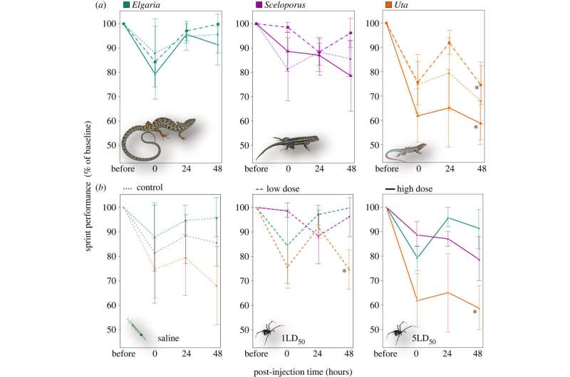
Whole-animal performance (sprint) in response to control (saline injection), low (1LD50) or
 high (5LD50) treatments of venom from the western black widow spider (Latrodectus 
hesperus). Changes in sprint speed quantified as the difference in post-injection velocity 
compared with baseline sprint speed (pre-injection), and recorded across three time points
 (0: immediately after injection; 24: 1 day after injection; 48: 2 days after injection). 
(a) Plots grouped by species: Elgaria multicarinata (southern alligator lizard); Sceloporus
 occidentalis (western fence lizard); Uta stansburiana (side-blotched lizard). 
(b) Plots grouped by treatment. Note that U. stansburiana is the only species that showed 
significant reduction in sprint speed compared with baseline speed (table 1), denoted by 
* (p < 0.05). Photos courtesy RW Hansen. Credit: Royal Society Open Science (2022).
 DOI: 10.1098/rsos.221012

A team of researchers at the University of Nevada has taken a step toward understanding how it is some lizards are able to withstand a black widow spider bite with few to no ill effects. In their paper published in the journal Royal Society Open Science, the group describes how they injected several species of lizards with black widow venom and then took a close look to see how they responded.

Prior research has shown that the black widow spider is one of the most poisonous spiders in North America. Luckily,  are not hostile and thus do not attack humans and they also produce very little venom. Because of that, very few people have ever died from a bite.

Prior research and anecdotal evidence has also shown that some  not only are not bothered by black widows but actively seek out the spiders as a . In this new effort, the researchers sought to learn more about such lizards to find out if their ability to withstand black widow bites might offer some clues for better treating humans who have been bitten.

The work by the researchers started with them going out into the field and collecting several specimens of three types of lizards: alligator, Western fence and side-blotched. The first two are known to be immune to black-widow spider bites while the third is very susceptible—they die if bitten.

The researchers then injected several of each of the three types of lizards with black widow venom and then set them down on a small racecourse and encouraged them to run. Both the alligator and Western fence were able to do so, while the side-blotched were not.

The researchers then dissected several of each of the lizards after injecting them with the venom. They noted that prior research has shown that creatures adversely impacted by black widow spider bites suffer from tissue and muscle damage. They found such damage in both the side blotched and Western fence but none in the alligator. Taking a closer look, the researchers discovered that there were no signs at all that the alligator lizards had been injected with the venom.

The researchers note that the lack of reaction to the venom by the alligator lizards suggests that they have a very strong protection mechanism. They note that some other species of animals have evolved a known mechanism to protect against venom—king snakes, for example, are immune to rattlesnake venom.

They have something in their blood that binds to the , neutralizing it. The researchers suggest it is likely the alligator lizards have something similar. They will not know what it is, however, until the genome of the lizard is sequenced.

Eating fire ants could prepare lizards for future fire ant attack
More information: Vicki L. Thill et al, Preying dangerously: black widow spider venom resistance in sympatric lizards, Royal Society Open Science (2022). DOI: 10.1098/rsos.221012
Journal information: Royal Society Open Science 
© 2022 Science X Network

Gestational exposure to flame retardant alters brain development in rats

brain
Credit: CC0 Public Domain

A new study from North Carolina State University shows that exposure in utero to the flame-retardant FireMaster 550 (FM 550), or to its individual brominated (BFR) or organophosphate ester (OPFR) components, resulted in altered brain development in newborn rats. The effects—most notably evidence of mitochondrial disruption and dysregulated choline and triglyceride levels in brain tissue—were greater in male offspring than in females.

The work adds to the body of evidence that both OPFRs and BFRs can be neurotoxic. It appears in a special issue of Neuroendocrinology. Shannah Witchey, former NC State postdoctoral researcher, is first author.

FM 550 is a flame-retardant mixture first identified a decade ago. It was developed to replace PBDEs, a class of fire retardants being phased out due to safety concerns.

"While some new flame-retardant mixtures still contain BFRs, the OPFRs are a popular substitute for PBDEs, since it is believed that OPFRs don't accumulate in the body and thus cannot be as harmful," says Heather Patisaul, associate dean for research in NC State's College of Sciences and corresponding author of the study. "Specifically, it was thought that OPFRs wouldn't impact acetylcholinesterase—a key neurotransmitter. But it looks as though OPFRs still impact choline signaling and are just as bad if not worse than PBDEs for the developing brain."

Patisaul and her colleagues performed transcriptomic and lipidomic studies on the prefrontal cortexes of newborn rats whose mothers had been exposed to FM550, or to BFR or OPFR elements individually, during gestation.

"Getting genetic information from transcriptomics is what researchers commonly do to tease out potential connections between toxicity and health effects," Patisaul says. "In this case, we also wanted to see if the lipid (or fat) composition of the brain was altered—our brains are essentially balls of fat, and lipidomics can reveal how exposure may affect the brain in its earliest developmental stages."

Both the transcriptomic and lipidomic analyses showed evidence of mitochondrial disruption, although the disruptions were more pronounced in the offspring that had been exposed to OPFRs. Mitochondria are found in almost every cell and serve as cells' energy generators, playing a vital role in cellular respiration.

The transcriptomic analysis found disruption of  genes associated with neurodegenerative disorders like Alzheimer's and ALS, while lipidomics pointed to disrupted choline and triglyceride levels in the brain.

In males exposed to OPFRs, genes associated with axon guidance and choline signaling were also dysregulated. Axon guidance is the process by which neurons make the proper connections during neural development. Choline is a precursor to the neurotransmitter acetylcholine, which affects critical aspects of neuron function and neuronal signaling.

"That so many altered genes are involved in respiration and choline, there is concern that these FRs impair basic autonomic function and cognition," Patisaul says. "So, the bottom line is that exposure to BFRs and OPFRs is disrupting both neuronal signaling and the ability of cells to properly produce and utilize energy."

The researchers also found that  were more affected than females.

"In earlier rat studies, we found that OPFR levels are higher in placentas attached to males than females," Patisaul says. "So that difference in exposure could be why we see different and more severe effects in males.

"The important message here is that the presumption that OPFRs are safer that other FRs is likely wrong. Both OPFRs and BFRs can disrupt cortical development and function. And the fact that these chemicals are detectable in the placenta means they aren't breaking down quickly enough to do no damage."Placental accumulation of flame retardant chemical alters serotonin production in rats

More information: Shannah K. Witchey et al, Impacts of Gestational FireMaster 550 (FM 550) Exposure on the Neonatal Cortex are Sex Specific and Largely Attributable to the Organophosphate Esters, Neuroendocrinology (2022). DOI: 10.1159/000526959


Fossil bird's skull reconstruction reveals a brain made for smelling and eyes made for daylight

Fossil bird's skull reconstruction reveals a brain made for smelling and eyes made for daylight
Artistic reconstruction of Jeholornis in life. Credit: Michael Rothman

Jeholornis was a raven-sized bird that lived 120 million years ago, among the earliest examples of dinosaurs evolving into birds, in what's now China. The fossils that have been found are finely preserved but smashed flat, the result of layers of sediment being deposited over the years. That means that no one's been able to get a good look at Jeholornis's head. But in a new study, researchers digitally reconstructed a Jeholornis skull, revealing details about its eyes and brain that shed light on its vision and sense of smell.

"Jeholornis is my favorite Cretaceous bird, it has a lot of unusual, primitive traits, and it helps shed light on the bigger story of how different birds evolved," says Jingmai O'Connor, associate curator of fossil reptiles at the Field Museum and one of the authors of the paper describing the discovery in the Zoological Journal of the Linnean Society. "This study is the first time we're really getting at what this bird's  looked like, what its brain must have been like, which is really exciting."

The study's first author, Han Hu, went through roughly 100 fossils at China's Shandong Tianyu Museum of Nature and selected the one with the best-preserved skull—still a little flattened, but intact. "It is very difficult to find the right skull among around 100 fossils, since we won't know if one skull will provide us the information we want before the scanning, and due to the costs of high quality scanning, we couldn't scan all those specimens to choose the best one. However, I chose this one because at least from the exposed surface, it is relatively complete, and which is also important is that this skull is preserved to be isolated from other parts of its body," says Hu, a researcher at the Department of Earth Sciences, University of Oxford, UK.

"This is very helpful since we usually won't chop the skull off from the skeleton if they are articulated—no one wants to hurt these previous fossils, but an isolated skull will reduce the size of the scanning area, which will increase the scanning quality a lot. Luckily, the specimen we chose here for this project is nearly a perfect one—it provided us so much unknown information after the digital reconstruction."

"These bones were kind of like the bottom of a bag of potato chips—they weren't completely crushed, but the pieces were compacted," says O'Connor. "So we were able to CT scan them—essentially taking a bunch of X-rays and stacking them together to form a 3D image—and then digitally re-articulate them and reconstruct the skull from all these bones."

Fossil bird's skull reconstruction reveals a brain made for smelling and eyes made for daylight
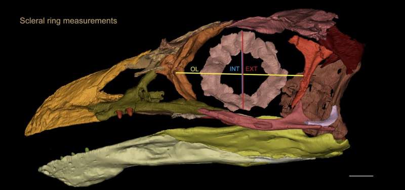
Reconstruction of Jeholornis's skull, showing the bony rings around its eye. 
Credit: Han Hu et al

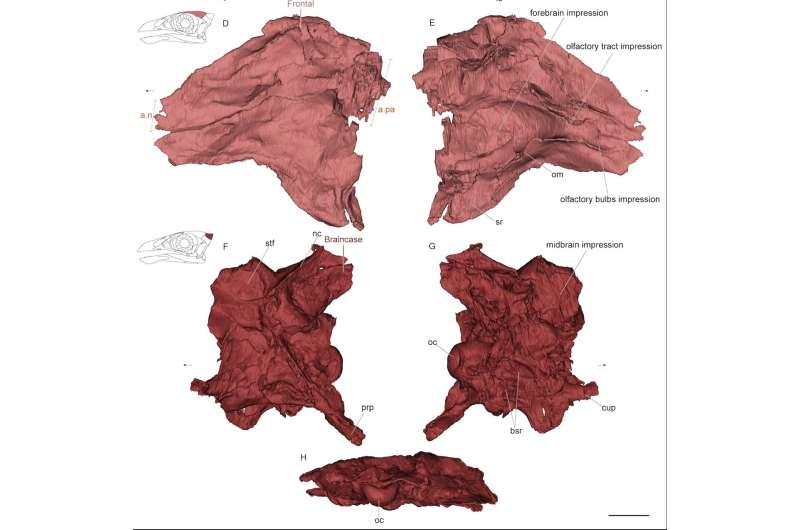
"We were able to see different features of the skull that had never been seen before in Jeholornis, and we were even able to extrapolate what its brain looked like," says co-author and Field Museum postdoctoral researcher Matteo Fabbri.

The brain itself isn't preserved—soft tissues rarely are—but bird and dinosaur brains tend to nest neatly within their skulls. Knowing the shape and dimensions of a fossil bird's skull, therefore, tells us a lot about its brain, kind of like how a glove gives a decent approximation of how a hand is shaped. What's more, brain structures are conserved across species and over time—things like olfactory bulbs and the cerebellum in the same general spots whether you're looking at the brain of a frog, a human, or a fossil bird.

Thanks to the long-standing placements of these structures, the researchers were able to determine how Jeholornis's brain compares with modern birds and dinosaurs (or, strictly speaking, ; all birds, including Jeholornis, are dinosaurs, but not all dinosaurs are birds).

"Jeholornis's brain morphology is transitional, in-between what we see in non-avian dinosaurs and what we see in modern birds," says Fabbri. "If you look at the skulls of dinosaurs, what you see is a spot for a very reptile-like brain, meaning that they have very large olfactory bulbs, and the optic lobes that are in the midbrain are reduced. They probably had a very good sense of smell and not great sight, which is very reptilian. And on the other hand, if you look at modern birds, they do the reverse. They have small olfactory bulbs, and very large optic lobes. Jeholornis falls in the middle."

Fossil bird's skull reconstruction reveals a brain made for smelling and eyes made for daylight
3D reconstruction of Jeholornis's brain. Credit: Han Hu et al

Jeholornis had bigger  than most modern birds, meaning that it probably relied more on its sense of smell than birds today (with the exception of a few keen-smellers, like vultures). Jeholornis's strong sense of smell makes sense in the context of another recent study by the team, showing that Jeholornis is the earliest-known fruit-eating animal. "As fruits ripen, they release lots of chemicals," says O'Connor. "We can't prove it yet, but having a better  might have helped Jeholornis find fruit."

In addition to a  adapted for smelling, the researchers found that Jeholornis was likely better at seeing in the daytime than at night. Birds have bones called scleral rings that help determine how much light goes into their eyes. Species that need to see at night, like owls, have wider scleral ring openings relative to their eye sockets, to let in more light; birds that are active during the day have narrower openings for light to go through, like the aperture on a camera. Jeholornis's scleral rings seem to indicate that it was most active during the day.

All of these skull features provide a better understanding of this early bird's lifestyle and the role it played in its ecosystem. "Reconstructing a skull is painstaking work, and as people are starting to put in the time to do it, It's becoming more and more clear that the evolution of birds was more complicated than what we expected," says Fabbri. "It's not just different from dinosaurs and modern birds, it's different from other early birds too. It's not a straightforward evolutionary story."

"The same as Jingmai, Jeholornis is also one of my favorite birds. Its special position as one of the most primitive birds during the dinosaur-bird transition determines that completing its story will reveal the true scenery of that critical evolutionary period, and also, tell us why and how the —the only living dinosaurs—evolved to be what we see now," says HuThe early bird gets the fruit: Fossil provides earliest evidence of fruit-eating by any animal

More information: Han Hu et al, Cranial osteology and palaeobiology of the Early Cretaceous bird Jeholornis prima (Aves: Jeholornithiformes), Zoological Journal of the Linnean Society (2022). DOI: 10.1093/zoolinnean/zlac089

Journal information: Zoological Journal of the Linnean Society 

Provided by Field Museum 


Analysis of gases recovered from asteroid Ryugu by Hayabusa2 spacecraft

Analysis of gases recovered from asteroid Ryugu by Hayabusa2 spacecraft
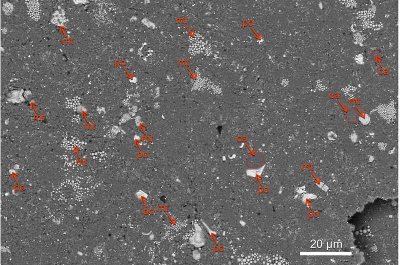
Back scattered electron image of a Ryugu pellet sample (A0105-10). The sample is mainly
 composed of phyllosilicates (dark gray regions). Spherical magnetite aggregates, thin 
magnetite plates, and iron sulfide grains are also present. Carbonates are found close to
 magnetites and sulfides. Red arrows indicate minerals labeled “mt” for magnetite, “po” for
 pyrrhotite (iron sulfide), and “ca” for carbonate (rimmed with red dotted ovals), respectively
. Credit: Science (2022). DOI: 10.1126/science.abo0431

Three international teams of researchers studying samples of gases recovered by the Hayabusa2 spacecraft from the near-Earth asteroid Ryugu have published their results. The first studied the asteroid's volatile sources and recent surface evolution. The second looked at its nucleosynthetic heritage. And the third team provided an overview of the types of gases that were returned. The first team has published their results in the journal Science; the second and third teams have both have published their results in the journal Science Advances.

The first team found that the asteroid still had  and  from the early days of the solar system, along with a nitrogen composition, which they describe as similar to Ivuna-type . They also found evidence that one of the noble gases was created by the solar wind and another via irradiation of galactic cosmic rays. And they also found what they describe as a "close" relationship been CI chondrites and gas from Ryugu.

The second team found that some of the samples from Ryugu had Fe isotopic irregularities that were the same as those found in other Ivuna-type (CI) chondrites. They also found iron isotopes that must have formed in places where there were no carbonaceous asteroids. And that, they note, suggests that Ryugu may have come from farther away in the solar system than has been theorized. They suggest it could have come from as far away as beyond the orbits of Saturn or Jupiter. They noted that the growth and migration paths of the giant planets had a destabilizing effect on planetesimals, some of which ejected material into the main belts, which could have included Ryugu.

The third team identified all of the gases that were brought back and measured the amounts of each. They also noted their . They found that some of the helium that had been captured came from the  and some of it leaked in from the Earth's atmosphere as the craft carrying it made its way home. They conclude by reiterating the fact that the gases collected on the project represent the first from a near-Earth asteroid.Scientists discover the source of one of the rarest groups of meteorites

More information: Ryuji Okazaki et al, Noble gases and nitrogen in samples of asteroid Ryugu record its volatile sources and recent surface evolution, Science (2022). DOI: 10.1126/science.abo0431

Ryuji Okazaki, First asteroid gas sample delivered by the Hayabusa2 mission: A treasure box from Ryugu, Science Advances (2022). DOI: 10.1126/sciadv.abo7239www.science.org/doi/10.1126/sciadv.abo7239

Timo Hopp, Ryugu's nucleosynthetic heritage from the outskirts of the Solar System, Science Advances (2022). DOI: 10.1126/sciadv.add8141www.science.org/doi/10.1126/sciadv.add8141


Journal information: Science Advances 


© 2022 Science X Network

VELIKOVSKY WAS RIGHT

Geomagnetic fields reveal the truth behind Biblical narratives

Geomagnetic fields reveal the truth behind Biblical narratives
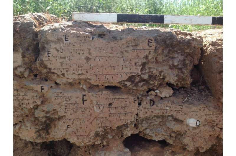
Burnt mud brick wall from Tel Batash (Biblical Timnah) with markings of the field orientation
. Credit: Yoav Vaknin.

A joint study by TAU and the Hebrew University, involving 20 researchers from different countries and disciplines, has accurately dated 21 destruction layers at 17 archaeological sites in Israel by reconstructing the direction and/or intensity of the earth's magnetic field recorded in burnt remnants. The new data verify the Biblical accounts of the Egyptian, Aramean, Assyrian, and Babylonian military campaigns against the Kingdoms of Israel and Judah.

Findings indicate, for example, that the army of Hazael, King of Aram-Damascus, was responsible for the destruction of several cities—Tel Rehov, Tel Zayit, and Horvat Tevet, in addition to Gath of the Philistines, whose destruction is noted in the Hebrew Bible. At the same time, the study refutes the prevailing theory that Hazael was the conqueror who destroyed Tel Beth-Shean.

Other geomagnetic findings reveal that the cities in the Negev were destroyed by the Edomites, who took advantage of the destruction of Jerusalem and the Kingdom of Judah by the Babylonians.

The groundbreaking interdisciplinary study was published in the Proceedings of the National Academy of Sciences and is based on the doctoral thesis of Yoav Vaknin, supervised by Prof. Erez Ben-Yosef and Prof. Oded Lipschits of TAU's Institute of Archaeology and Prof. Ron Shaar from the Institute of Earth Sciences at the Hebrew University.

Yoav Vaknin explaining about the research. Credit: Tel Aviv University

The researchers explain that geophysicists, attempting to understand the mechanism of earth's , track changes in this field throughout history. To this end they use archaeological findings containing magnetic minerals which, when heated or burned, record the magnetic field at the time of the fire.

Thus, in a 2020 study, researchers reconstructed the magnetic field as it was on the 9th of the month of Av, 586 BCE, the Hebrew date of the destruction of the First Temple and the City of Jerusalem by Nebuchadnezzar and his Babylonian army.

Now, using archaeological findings unearthed over several decades at 17 sites throughout Israel, alongside historical information from ancient inscriptions and Biblical accounts, the researchers were able to reconstruct the magnetic fields recorded in 21 destruction layers. They used the data to develop a reliable new scientific tool for archaeological dating.

Geomagnetic fields reveal the truth behind Biblical narratives
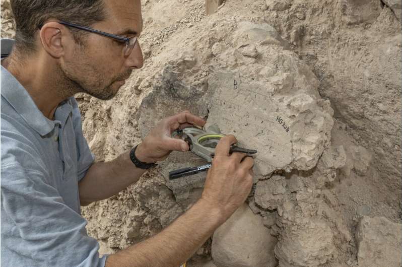
Yoav Vaknin measuring at the site. Credit: Shai Halevi, Israel Antiquities Authority.

Yoav Vaknin explains that "based on the similarity or difference in intensity and direction of the magnetic field, we can either corroborate or disprove hypotheses claiming that specific sites were burned during the same military campaign. Moreover, we have constructed a variation curve of field intensity over time which can serve as a scientific dating tool, similar to the radiocarbon dating method."

One example given by the researchers is the destruction of Gath of the Philistines (identified today as Tel Tzafit in the Judean foothills) by Hazael, King of Aram-Damascus. Various dating methods have placed this event at around 830 BCE, but were unable to verify that Hazael was also responsible for the destruction of Tel Rehov, Tel Zayit and Horvat Tevet.

Now the new study, identifying full statistical synchronization between the magnetic fields recorded at all of these four sites at the time of destruction, makes a very strong case for their destruction during the same campaign.

A destruction level at Tel Beth-Shean, on the other hand, recording a totally different magnetic field, refutes the prevailing hypothesis that it too was destroyed by Hazael. Instead, the magnetic data from Beth-Shean indicate that this city, along with two other sites in northern Israel, was probably destroyed 70-100 years earlier, a date which could correspond with the military campaign of the Egyptian Pharaoh Shoshenq.

Shoshenq's campaign is described in the Hebrew Bible and in an inscription on a wall of the Temple of Amun in Karnak, Egypt, which mentions Beth-Shean as one of his conquests.

One of the most interesting findings revealed by the new dating method has to do with the end of the Kingdom of Judah. Prof. Erez Ben Yosef says, "The last days of the Kingdom of Judah are widely debated. Some researchers, relying on archaeological evidence, argue that Judah was not completely destroyed by the Babylonians.

"While Jerusalem and frontier cities in the Judean foothills ceased to exist, other towns in the Negev, the southern Judean Mountains and the southern Judean foothills remained almost unaffected. Now, the magnetic results support this hypothesis, indicating that the Babylonians were not solely responsible for Judah's ultimate demise.

"Several decades after they had destroyed Jerusalem and the First Temple, sites in the Negev, which had survived the Babylonian campaign, were destroyed—probably by the Edomites who took advantage of the fall of Jerusalem. This betrayal and participation in the destruction of the surviving cities may explain why the Hebrew Bible expresses so much hatred for the Edomites—for example, in the prophecy of Obadiah."

Geomagnetic fields reveal the truth behind Biblical narratives
Map of the studied destruction layers and the different military campaigns. Credit: Itamar Ben-Ezra

Prof. Oded Lipschits adds that "the new dating tool is unique because it is based on geomagnetic data from sites, whose exact destruction dates are known from historical sources. By combining precise  with advanced, comprehensive archaeological research, we were able to base the magnetic method on reliably anchored chronology."

A separate paper, presenting the scientific principles of the novel archaeomagnetic dating method, is in preparation. Prof. Ron Shaar, who led the geophysical aspects of the study, as well as the development of the geomagnetic dating method, explains that "Earth's magnetic field is critical to our existence. Most people don't realize that without it there could be no life on earth—since it shields us from cosmic radiation and the solar wind. In addition, both humans and animals use it to navigate. The geomagnetic field is generated by earth's outer core, at a depth of 2,900 km, by currents of liquid iron."

Geomagnetic fields reveal the truth behind Biblical narratives
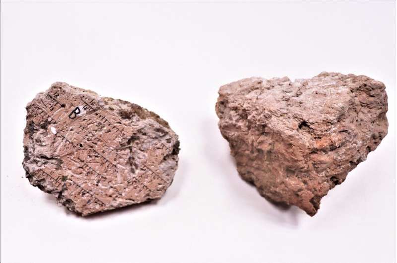
Burnt mud stones. Credit: Tel Aviv University.

"Due to the chaotic motion of this iron, the magnetic field changes over time. Until recently scientists believed that it remains quite stable for decades, but archaeomagnetic research has contradicted this assumption by revealing some extreme and unpredictable changes in antiquity. Our location here in Israel is uniquely conducive to archaeomagnetic research, due to an abundance of well-dated . Over the past decade we have reconstructed magnetic fields recorded by hundreds of archaeological items."

"By combining this dataset with the data from Yoav's investigation of historical destruction layers we were able to form a continuous variation curve showing rapid, sharp changes in the geomagnetic field. This is wonderful news, both for archaeologists who can now use geomagnetic data to determine the age of ancient materials and for geophysicists studying the earth's core."Chicken bones and snail shells help archaeologists to date ancient town's destruction

More information: Vaknin, Yoav, Reconstructing biblical military campaigns using geomagnetic field data, Proceedings of the National Academy of Sciences (2022). DOI: 10.1073/pnas.2209117119. doi.org/10.1073/pnas.2209117119

Journal information: Proceedings of the National Academy of Sciences

Provided by Tel-Aviv University