Showing posts sorted by relevance for query BACTERIOPHAGE. Sort by date Show all posts

Sunday, December 18, 2022

Atomic structure of a staphylococcal bacteriophage using cryo-electron microscopy

High-resolution knowledge of structure is a key link between viral biology and potential therapeutic use of the virus to quell bacterial infections.

Peer-Reviewed Publication

UNIVERSITY OF ALABAMA AT BIRMINGHAM

The Andhra phage 

IMAGE: THE ANDHRA PHAGE view more 

CREDIT: UAB, DOKLAND LAB

BIRMINGHAM, Ala. – Cryo-electron microscopy by University of Alabama at Birmingham researchers has exposed the structure of a bacterial virus with unprecedented detail. This is the first structure of a virus able to infect Staphylococcus epidermidis, and high-resolution knowledge of structure is a key link between viral biology and potential therapeutic use of the virus to quell bacterial infections.

Bacteriophages or “phages” is the terms used for viruses that infect bacteria. The UAB researchers, led by Terje Dokland, Ph.D., in collaboration with Asma Hatoum-Aslan, Ph.D., at the University of Illinois Urbana-Champaign, have described atomic models for all or part of 11 different structural proteins in phage Andhra. The study is published in Science Advances.

Andhra is a member of the picovirus group. Its host range is limited to S. epidermidis. This skin bacterium is mostly benign but also is a leading cause of infections of indwelling medical devices. “Picoviruses are rarely found in phage collections and remain understudied and underused for therapeutic applications,” said Hatoum-Aslan, a phage biologist at the University of Illinois.

With emergence of antibiotic resistance in S. epidermidis and the related pathogen Staphylococcus aureus, researchers have renewed interest in potentially using bacteriophages to treat bacterial infections. Picoviruses always kill the cells they infect, after binding to the bacterial cell wall, enzymatically breaking through that wall, penetrating the cell membrane and injecting viral DNA into the cell. They also have other traits that make them attractive candidates for therapeutic use, including a small genome and an inability to transfer bacterial genes between bacteria.

Knowledge of protein structure in Andhra and understanding of how those structures allow the virus to infect a bacterium will make it possible to produce custom-made phages tailored to a specific purpose, using genetic manipulation.

“The structural basis for host specificity between phages that infect S. aureus and S. epidermidis is still poorly understood,” said Dokland, a professor of microbiology at UAB and director of the UAB Cryo-Electron Microscopy Core. “With the present study, we have gained a better understanding of the structures and functions of the Andhra gene products and the determinants of host specificity, paving the way for a more rational design of custom phages for therapeutic applications. Our findings elucidate critical features for virion assembly, host recognition and penetration.”

Staphylococcal phages typically have a narrow range of bacteria they can infect, depending on the variable polymers of wall teichoic acid on the surface of different bacterial strains. “This narrow host range is a double-edged sword: On one hand, it allows the phages to target only the specific pathogen causing the disease; on the other hand, it means that the phage may need to be tailored to the patient in each specific case,” Dokland said.

The general structure of Andhra is a 20-faced, roundish icosahedral capsid head that contains the viral genome. The capsid is attached to a short tail. The tail is largely responsible for binding to S. epidermidis and enzymatically breaking the cell wall. The viral DNA is injected into the bacterium through the tail. Segments of the tail include the portal from the capsid to the tail, and the stem, appendages, knob and tail tip.

The 11 different proteins that make up each virus particle are found in multiple copies that assemble together. For instance, the capsid is made of 235 copies each of two proteins, and the other nine virion proteins have copy numbers from two to 72. In total, the virion is made up of 645 protein pieces that include two copies of a 12th protein, whose structure was predicted using the protein structure prediction program AlphaFold.

The atomic models described by Dokland, Hatoum-Aslan, and co-first authors N’Toia C. Hawkins, Ph.D., and James L. Kizziah, Ph.D., UAB Department of Microbiology, show the structures for each protein — as described in molecular language like alpha-helix, beta-helix, beta-strand, beta-barrel or beta-prism. The researchers have described how each protein binds to other copies of that same protein type, such as to make up the hexameric and pentameric faces of the capsid, as well as how each protein interacts with adjacent different protein types.

Electron microscopes use a beam of accelerated electrons to illuminate an object, providing much higher resolution than a light microscope. Cryo-electron microscopy adds the element of super-cold temperatures, making it particularly useful for near-atomic structure resolution of larger proteins, membrane proteins or lipid-containing samples like membrane-bound receptors, and complexes of several biomolecules together.

In the past eight years, new electron detectors have created a tremendous jump in resolution for cryo-electron microscopy over normal electron microscopy. Key elements of this so-called “resolution revolution” for cryo-electron microscopy are:

  • Flash-freezing aqueous samples in liquid ethane cooled to below -256 degrees F. Instead of ice crystals that disrupt samples and scatter the electron beam, the water freezes to a window-like “vitreous ice.”
  • The sample is kept at super-cold temperatures in the microscope, and a low dose of electrons is used to avoid damage to the proteins.
  • Extremely fast direct electron detectors are able to count individual atoms at hundreds of frames per second, allowing sample movement to be corrected on the fly.
  • Advanced computing merges thousands of images to generate three-dimensional structures at high resolution. Graphics processing units are used to churn through terabytes of data.
  • The microscope stage that holds the sample can also be tilted as images are taken, allowing construction of a three-dimensional tomographic image, similar to a CT scan at the hospital.

The analysis of Andhra virion structure by the UAB researchers started with 230,714 particle images. Molecular reconstruction of the capsid, tail, distal tail and tail tip started with 186,542, 159,489, 159,489 and 159,489 images, respectively. Resolution ranged from 3.50 to 4.90 angstroms.

Support for the study, “Structure and host specificity of Staphylococcus epidermidis bacteriophage Andhra,” came from National Institutes of Health phage therapy grant R21 AI156636.

The UAB Department of Microbiology is part of the Marnix E. Heersink School of Medicine.

Tuesday, April 11, 2023

Your baby’s gut is crawling with unknown viruses

Peer-Reviewed Publication

UNIVERSITY OF COPENHAGEN - FACULTY OF SCIENCE

Dennis Sandris Nielsen 

IMAGE: PROFESSOR DENNIS SANDRIS NIELSEN, UNIVERSITY OF COPENHAGEN view more 

CREDIT: EMILIE THEJLL-MADSEN / UNIVERSITY OF COPENHAGEN

Viruses are usually associated with illness. But our bodies are full of both bacteria and viruses that constantly proliferate and interact with each other in our gastrointestinal tract. While we have known for decades that gut bacteria in young children are vital to protect them from chronic diseases later on in life, our knowledge about the many viruses found there is minimal.

A few years back, this gave University of Copenhagen professor Dennis Sandris Nielsen the idea to delve more deeply into this question. As a result, a team of researchers from COPSAC (Copenhagen Prospective Studies on Asthma in Childhood) and the Department of Food Science at UCPH, among others, spent five years studying and mapping the diaper contents of 647 healthy Danish one-year-olds.

"We found an exceptional number of unknown viruses in the faeces of these babies. Not just thousands of new virus species – but to our surprise, the viruses represented more than 200 families of yet to be described viruses. This means that, from early on in life, healthy children are tumbling about with an extreme diversity of gut viruses, which probably have a major impact on whether they develop various diseases later on in life," says Professor Dennis Sandris Nielsen of the Department of Food Science, senior author of the research paper about the study, now published in Nature Microbiology.

The researchers found and mapped a total of 10,000 viral species in the children's faeces – a number ten times larger than the number of bacterial species in the same children. These viral species are distributed across 248 different viral families, of which only 16 were previously known. The researchers named the remaining 232 unknown viral families after the children whose diapers made the study possible. As a result, new viral families include names like SylvesterviridaeRigmorviridae and Tristanviridae.

Bacterial viruses are our allies

"This is the first time that such a systematic an overview of gut viral diversity has been compiled. It provides an entirely new basis for discovering the importance of viruses for our microbiome and immune system development. Our hypothesis is that, because the immune system has not yet learned to separate the wheat from the chaff at the age of one, an extraordinarily high species richness of gut viruses emerges, and is likely needed to protect against chronic diseases like asthma and diabetes later on in life," states Shiraz Shah, first author and a senior researcher at COPSAC.

Ninety percent of the viruses found by the researchers are bacterial viruses – known as bacteriophages. These viruses have bacteria as their hosts and do not attack the children's own cells, meaning that they do not cause disease. The hypothesis is that bacteriophages primarily serve as allies:

"We work from the assumption that bacteriophages are largely responsible for shaping bacterial communities and their function in our intestinal system. Some bacteriophages can provide their host bacterium with properties that make it more competitive by integrating its own genome into the genome of the bacterium. When this occurs, a bacteriophage can then increase a bacterium's ability to absorb e.g. various carbohydrates, thereby allowing the bacterium to metabolise more things," explains Dennis Sandris Nielsen, who continues:

"It also seems like bacteriophages help keep the gut microbiome balanced by keeping individual bacterial populations in check, which ensures that there are not too many of a single bacterial species in the ecosystem. It's a bit like lion and gazelle populations on the savannah."

Shiraz Shah adds:

"Previously, the research community mostly focused on the role of bacteria in relation to health and disease. But viruses are the third leg of the stool and we need to learn more about them. Viruses, bacteria and the immune system most likely interact and affect each other in some type of balance. Any imbalance in this relationship most likely increases the risk of chronic disease."

The remaining ten percent of viruses found in the children are eukaryotic – that is, they use human cells as hosts. These can be both friends and foes for us:

"It is thought-provoking that all children run around with 10-20 of these virus types that infect human cells. So, there is a constant viral infection taking place, which apparently doesn’t make them sick. We just know very little about what’s really at play. My guess is that they’re important for training our immune system to recognise infections later. But it may also be that they are a risk factor for diseases that we have yet to discover," says Dennis Sandris Nielsen.

Could play an important role in inflammatory diseases

The researchers have yet to discover where the many viruses in the one-year-olds come from. Their best answer thus far is the environment:

"Our gut is sterile until we are born. During birth, we are exposed to bacteria from the mother and environment. It is likely that some of the first viruses come along with these initial bacteria, while many others are introduced later via dirty fingers, pets, dirt that kids put in their mouths and other things in the environment," says Dennis Sandris Nielsen.

As Shiraz Shah points out, the entire field of research speaks to a huge global health problem:

"A lot of research suggests that the majority of chronic diseases that we’re familiar with – from arthritis to depression – have an inflammatory component. That is, the immune system is not working as it ought to – which might be because it wasn’t trained properly. So, if we learn more about the role that bacteria and viruses play in a well-trained immune system, it can hopefully lead us to being able to avoid many of the chronic diseases that afflict so many people today."

The research groups have begun investigating the role of gut viruses in relation to a number of different diseases that occur in childhood, such as asthma and ADHD.


FACT BOX: ABOUT BACTERIOPHAGES

  • There are generally two types of bacteriophages. Virulent bacteriophages take over the bacterium and produce 30-100 new virus particles inside it. After this, the bacterial cell explodes from the inside and the new virus particles escape into the environment. Virulent bacteriophages help to keep the intestinal ecosystem in balance.
     
  • So-called temperate bacteriophages can reproduce by integrating their genetic material into the genome of the host bacterial cell. When the cell divides, so does the bacteriophage.  Temperate bacteriophages help transfer new genes to the bacteria so it becomes more competitive. However, there are also studies suggesting that an imbalance in the temperate bacteriophage population is associated with various diseases, e.g., inflammatory bowel disease.


FACT BOX: ABOUT VIRUSES

  • A virus is a microorganism consisting of a genome – either DNA or RNA – encapsulated in a protein membrane. Viruses cannot multiply. Instead, a virus attacks a host cell, which it uses to make copies of itself.
     
  • Viruses are classified into viral families, which are then divided into a larger number of viral genera and viral species. A more well-known example of a viral family is coronavirus, to which the viruses Covid-19, MERS, SARS and several common cold viruses belong.


FACT BOX: ABOUT THE STUDY

  • The research team mapped the gut "viromes" from the guts of 647 healthy Danish one-year-old children. "Virome" is an umbrella term for all viruses found in a given environment. This includes both viruses that attack bacteria (bacteriophages), as well as those that go after human cells (eukaryotic virus).
     
  • The 647 infants are all part of the mother-child cohort Copenhagen Prospective Studies on Asthma in Childhood (COPSAC2010), that has been followed very closely clinically throughout childhood at COPSAC. The children are now 13 years old.
     
  • This interactive atlas allows you to see the diversity of viruses in the children and download information about the individual viral families.
     
  • The results have been published in the renowned scientific journal Nature Microbiology.
     
  • The researchers behind the study come from COPSAC, University of Copenhagen; Department of Food Science, University of Copenhagen; Novo Nordisk Foundation Center for Basic Metabolic Research, University of Copenhagen; Department of Health Technology, DTU; Université Laval, Canada; Université Paris-Saclay, France; Université Clermont, France and the University of Copenhagen’s Department of Biology.


    Tuesday, February 15, 2022

    New Yale Center to Advance Phage Understanding, Treatments, Training, Education

    February 14, 2022
    by Julie Parry

    The Center for Phage Biology and Therapy at Yale held its launch event on Thursday, January 27, 2022, by hosting a panel discussion centered around the context of the documentary, Salt In My Soul.

    Salt In My Soul chronicles the life of Mallory Smith, a woman with cystic fibrosis, by using her own words from her diary, audio and video clips that her mother Diane Shader-Smith discovered after her daughter’s passing. Smith’s doctors tried experimental phage therapy to treat a multi-antibiotic-resistant infection, but this occurred too late to prevent her death at age 25 from infectious complications after bilateral lung transplant.

    Photo by Robert Lisak
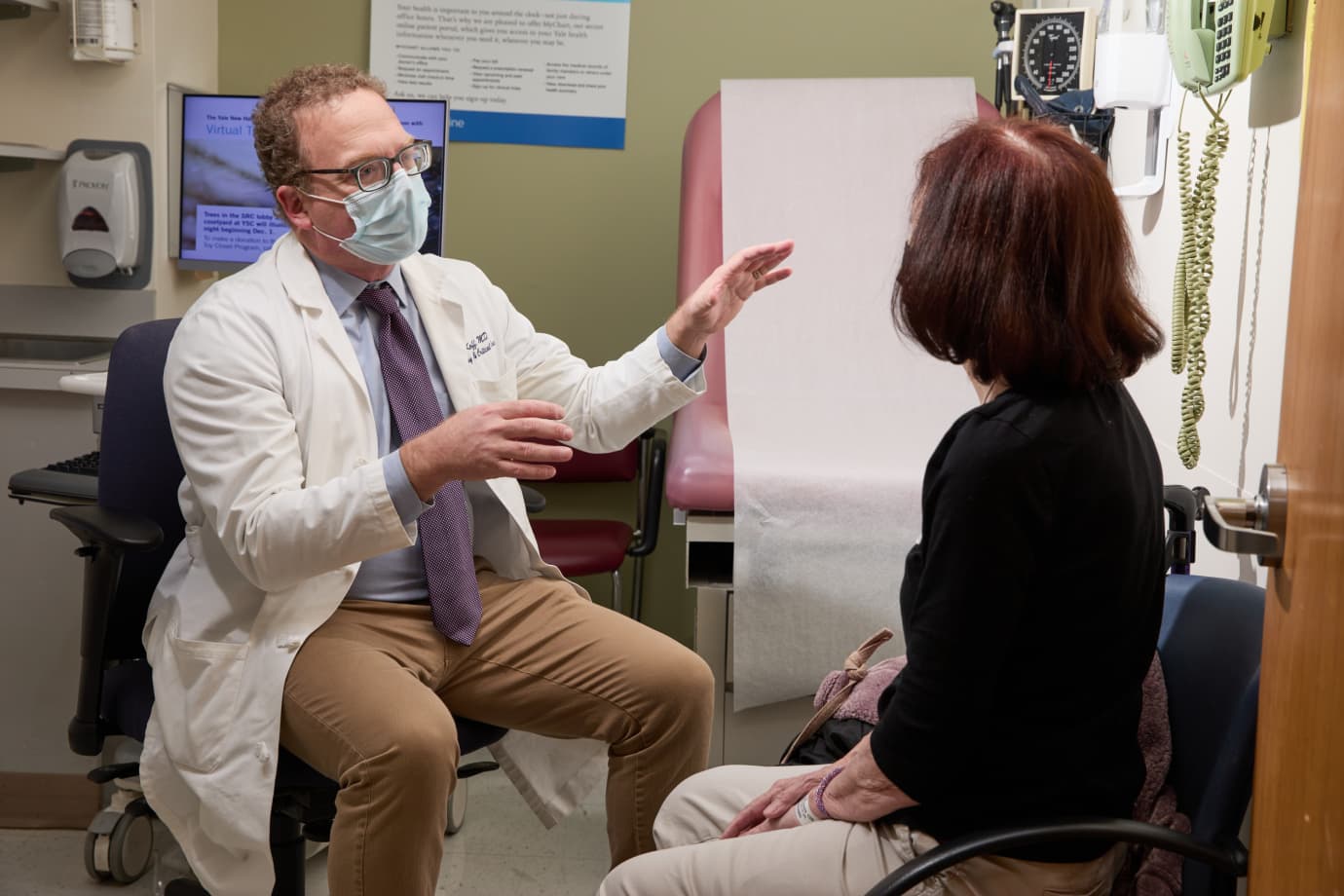
    Dr. Jon Koff meets with patient.

    Director of the Center for Phage Biology and Therapy at Yale, Paul Turner, PhD, Rachel Carson Professor of Ecology and Evolutionary Biology, discussed the antibiotic resistance crisis and how phage therapy can combat this crisis.

    The antibiotic resistance crisis is a “sobering reminder that not too far off in the future, the expected rates of death from antimicrobial resistance in the human population around the world may exceed the rates of deaths from common diseases, such as cancer,” said Turner.

    “What can we do?” Turner asked. “We can harness an old technology, phage therapy, and update it for modern times. At Yale, we have had successful therapeutic use of lytic phages.”

    The new Center for Phage Biology and Therapy at Yale will work to create solutions for antimicrobial resistance. Through their mission to advance and support phage research; to translate these advancements into new clinical therapies; and to train and educate students, scientists, healthcare professionals, and the community, the new Center combines the work of faculty, researchers, trainees, and staff to further develop phage for therapeutic use. These efforts bridge across Yale University and Yale School of Medicine, especially the Departments of Ecology and Evolutionary Biology and Internal Medicine.

    The Center has informally existed at Yale since 2016 when a case of multi-drug resistant Pseudomonas aeruginosa was treated successfully with phage OMKO1, discovered in a water sample from Dodge Pond, a small lake in Conn.

    Ella Balasa, an individual with cystic fibrosis and advocate, feels incredibly lucky to have received phage therapy at Yale in 2019. “I believe that without it [phage therapy], at this point, I would have been transplanted because of the severe lung infection that I was facing at the time,” she said.

    Balasa had been struggling with antibiotic resistant infections for many years when she came across a story about Yale researchers’ work in phage therapy. She then contacted Research Scientist Benjamin Chan, PhD, scientific director of the Center for Phage Biology and Therapy at Yale. Chan started the process to obtain approval to treat Balasa via the U.S. Food & Drug Administration (FDA) investigational new drug expanded access program, or what is often called compassionate use.

    “At that point, my lung function was about 20%,” Balasa said. “Phage did clear my infection. I want everyone to know that this therapy can be a viable option for antimicrobial resistance. I am excited that there are groups like you all [Yale] that are bringing clinical trials to people.”

    The Center for Phage Biology and Therapy at Yale is making a multi-million-dollar investment in phage biology research and phage applications. They are currently targeting pulmonary infections in cystic fibrosis and other relevant clinical conditions. In addition, the Center will expand to address causes of sepsis, prosthetic-joint infections, post-COVID pneumonia, and other antibiotic-resistant bacterial infections in the future.

    Jon Koff, MD, associate professor (Pulmonary, Critical Care, and Sleep Medicine); director, Adult Cystic Fibrosis Program; and medical director of the Center for Phage Biology and Therapy at Yale, serves as the principal investigator on the CYstic fibrosis bacterioPHage study at Yale (CYPHY), an FDA-approved human clinical phase I/II trial, developed at Yale School of Medicine and funded through Yale University, the Blavatnik Fund for Innovation at Yale, and the Cystic Fibrosis Foundation.

    “[The trial] is an exemplar of using our strategy of looking more towards treating patients chronically over the long term with phages to see if we can affect their multi-drug resistant infections and see clinical improvements. It is similar to introducing an inhaled antibiotic,” explained Koff.

    “The trial has been very rewarding and a great opportunity for me to engage with our patients, our community, and providers around the country. We’ve seen patients come to Yale from all over and this has allowed me to communicate to folks from multiple communities about our phage and our phage strategies,” said Koff.

    In addition to Turner, Chan, Koff, and Balasa, other presenters in the one-hour virtual event were Gunnar Esiason, cystic fibrosis advocate, and graduate student at Dartmouth in the MBA/MPH program; Will Battersby, film director and producer of Salt In My Soul; and Diane Shader-Smith, Mallory Smith’s mother.

    Battersby spoke about making the documentary, the themes throughout, and how people have embraced the work. “If you tell the story of somebody going through extraordinary things, extraordinary themes and lessons will emerge. I had no intention of making a film that would be picked up by the phage community in the way that it has, but it is very moving because of the possibilities in the film, […] it makes you ask so many things about phage.”

    Shader-Smith shared in Battersby’s sentiment. “Mallory was willing to try phage therapy. We had many, many long talks about it. I think the main reason that people love the film, despite the tragedy, is that they leave feeling hopeful. And phage therapy is that hope,” she said.

    Chan has devoted his career to furthering phage research and developing phage therapies. Despite the long history of phage usage, until recently, it hasn’t gained steam in the 0077saestern biomedical community. He gathers his strength to continue with his efforts from cystic fibrosis patients like Balasa and Esiason, parents like Shader-Smith, and supporters like Battersby.

    “The cystic fibrosis community is the best. We are in it together. We help each other move through stuff. If it wasn’t for them, I would have burned out a long time ago,” he said.

    Koff believes that Chan diminishes his commitment to this work. “Ben minimized his unbelievable energy to follow through on this and to have a vision for translating what he is seeing in the laboratory to the clinic. It has been an absolute pleasure working with him and seeing that level of effort,” said Koff.

    “What is awesome is that I am able to be part of this process in the clinic, in the research in collaboration with Paul [Turner] and Ben [Chan] and the research group, and we can see this happening in the clinical trial context. It has been a wonderful experience for me to cross all of these aspects and I think they make our Center pretty unique,” said Koff.

    The Center for Phage Biology and Therapy at Yale is funded by Yale University and philanthropic contributors who share the vision for phage therapy.

    To learn more about the phage research at Yale, visit Center for Phage Biology and Therapy at Yale. For more information on the CYPHY trial, visit CYstic Fibrosis bacterioPHage Study at Yale (CYPHY). To learn more about the documentary, watch the trailer, or the film, go to Salt In My Soul.

    Submitted by Julie Parry on February 13, 2022

    Thursday, September 21, 2023


    A newly identified virus emerges from the deep


    Peer-Reviewed Publication

    AMERICAN SOCIETY FOR MICROBIOLOGY


    Highlights:

    • Organisms, including viruses, live in the deepest, darkest places on the planet
    • Marine virologists analyzed sediment from the Mariana Trench, the deepest place on Earth, and identified a new bacteriophage
    • The phage infects Halomonas bacteria, which have been found in deep-sea environments and near hydrothermal vents
    • The study helps probe how phages and hosts evolve together in secluded, hostile environments

    Washington, D.C. —  The Mariana Trench, the deepest place on Earth, plunges nearly 11,000 meters at its lowest point on the floor of the Pacific Ocean. Life persists in the deep and cold darkness, and “wherever there’s life, you can bet there are regulators at work,” said marine virologist Min Wang, Ph.D, at the Ocean University of China, in Qingdao. “Viruses, in this case.”
     
    This week in Microbiology Spectrum, Wang and an international group of researchers report the discovery of a new virus isolated from sediment brought up from a depth of 8,900 meters. The virus is a bacteriophage, or a virus that infects and replicates inside bacteria, and bacteriophages are believed to be the most abundant life forms on the planet. “To our best knowledge, this is the deepest known isolated phage in the global ocean,” said Wang.
     
    The newly found phage infects bacteria in the phylum Halomonas, which are often found in sediments from the deep seas and from hydrothermal vents, geyser-like openings on the seafloor that release streams of heated water. Wang said the group’s analysis of the viral genetic material points to existence of a previously unknown viral family in the deep ocean, as well as new insights into the diversity, evolution and genomic features of deep-sea phages and phage-host interactions.
     
    In previous work, the researchers have used metagenomic analysis to study viruses that infect bacteria in the order Oceanospirallales, which includes Halomonas. For the new study, Wang’s team looked for viruses in bacterial strains collected and isolated by a team led by marine virologist Yu-Zhong Zhang, Ph.D, also at the Ocean University of China, in Qingdao. Zhang’s research explores microbial life in extreme environments, including polar regions and the Mariana Trench.
     
    The genomic analysis of the new virus, identified as vB_HmeY_H4907, suggests that it is distributed widely in the ocean and has a similar structure to its host. Wang said the study points to new questions and research areas focused on the survival strategies of viruses in harsh, secluded environments—and how they co-evolve with their hosts. The new virus is lysogenic, which means it invades and replicates inside its host, but usually without killing the bacterial cell. As the cell divides, the viral genetic material is also copied and passed on.
     
    In future studies, Wang said, the group plans to investigate the molecular machinery that drives interactions between deep-sea viruses and their hosts. They’re also searching for other new viruses in extreme places, “which would contribute to broadening our comprehension of the virosphere,” Wang said. “Extreme environments offer optimal prospects for unearthing novel viruses.”
     

    ###

    The American Society for Microbiology is one of the largest professional societies dedicated to the life sciences and is composed of 30,000 scientists and health practitioners. ASM's mission is to promote and advance the microbial sciences. 

    ASM advances the microbial sciences through conferences, publications, certifications, educational opportunities and advocacy efforts. It enhances laboratory capacity around the globe through training and resources. It provides a network for scientists in academia, industry and clinical settings. Additionally, ASM promotes a deeper understanding of the microbial sciences to diverse audiences. 

    Wednesday, September 09, 2020

    Wrocław clinic uses ‘super viruses’ to battle rebellious bacteria
    JOANNA JASIŃSKA MARCH 22, 2020
    The therapy being pioneered in Wrocław could help end the threat posed by super-bugs.Kalbar /TFN

    A pioneering institute in Wrocław is working on experimental therapy to combat antibiotic-resistant bacterial infections.

    For 15 years, the Polish Academy of Sciences Phage Therapy Unit at the Institute of Immunology and Experimental Therapy’s Medical Centre has been looking for a cure for patients who have lost all hope that their ailments could be treated.

    Doctor Ryszard Międzybrodzki with Anna Kabała, one of the Medical Unit's patients. Kalbar/TFN

    TFN travelled to Wrocław to discover how a revolutionary therapy the clinic is working on could be the answer to an ever-mutating threats to our health.

    In short, phage therapy utilises bacteriophages – bacterial viruses which attack only bacterial cells. A patient is treated with individually matched viruses, which are able to destroy different bacteria including those which are resistant to antibiotics and which cause life-threatening infections.

    The Institute of Immunology and Experimental Therapy’s Medical Centre has been looking for a cure for patients who have lost all hope that their ailments could disappear.Kalbar/TFN

    Professor Andrzej Górski, the institute’s director, told TFN: “Our centre treats antibiotic-resistant infections. The great problem in medicine today is that we are becoming defenceless against the bacteria that cause them.

    “Although the market is big, as it is estimated that about $30 to 40 billion are spent on antibiotics annually, antibiotic resistance crisis is growing,” he continued.

    With both bacteria becoming harder to kill and pharmaceutical companies finding providing newer and newer drugs unprofitable, a change in approach will be necessary.


    “Our centre treats antibiotic-resistant infections. The great problem in medicine today is that we are becoming defenceless against the bacteria that cause them,” says Professor Andrzej Górski.Kalbar/TFN

    “Poland has a long tradition in this sphere, because, in fact, the first attempts to use phage therapy were made soon after regaining independence,” explained the professor. Phages were first observed in 1896 in the River Ganges by Ernest Hanbury Hankin, who noticed their antibacterial properties.

    The first tries at phage therapy were attempted before World War II, but the medical documentation from that time isn’t credible according to today’s requirements. After the war, research on phages in Wrocław resumed in the 1960s.

    The Institute’s Medical Unit is supported by the Bacteriophage Laboratory, which stores over 600 different phages, carries out phage typing procedures, prepares the phage formulations for patients and performs some other necessary tests.

    The Institute is supported by the Bacteriophage Laboratory, which stores over 600 different phages.Kalbar/TFN

    Doctor Beata Weber-Dąbrowska, the principal specialist at the institute’s laboratory, said: “Phages naturally occur everywhere where bacteria are found, so even in the most extreme conditions such as hot springs, Arctic waters or the sands of the Sahara.”

    To expand their collection of phages and be able to combat a wider variety of bacteria, the researchers are constantly working on obtaining new strains.

    Phages, just like other viruses, are selective when it comes to which bacteria they’re effective against. The virus will attach itself to a bacteria and inject it with its own genome. The genome replaces the bacterial one and halts the infection, making the bacteria unable to reproduce.

    Phages can spring up anywhere: from Artic waters to desert sands.Kalbar/TFN

    The main advantage of using phage therapy is its precision. By choosing the right type of phages doctors can be sure it will attack only the bacteria they want to counter. Doctor Weber-Dąbrowska said: “It doesn't have any effect on the body (...). We all carry phages, as I said they are everywhere, so, for example, we have a very large amount of phages isolated from samples taken from patients.

    “The best environment for phages is the gastrointestinal tract because there are lots of bacteria,” she added. “Therefore, phages breed wonderfully there, but mainly for those gut bacteria.”

    The Institute’s Medical Unit is not a clinic. As a research centre first and foremost, they accept only a handful of cases – patients who often have been struggling with recurring infections for years and were unable to find help elsewhere.

    “This is an experimental therapy. We don’t have indisputable scientific proof that it will work,” says Doctor Ryszard Międzybrodzki.Kalbar/TFN

    Since the therapy is experimental and not yet officially approved by European law for common use, the requirements are very strict. So far, they have only admitted 700 patients.

    Doctor Ryszard Międzybrodzki is one of the physicians working with patients from all over the world who come to the Institute. The doctor stressed: “This is an experimental therapy. We don’t have indisputable scientific proof that it will work.

    “Our group of patients are in a difficult situation because they aren’t people who are suffering from the infection for the first time and decide to use phages instead of antibiotics.”

    The future of the clinic remains uncertain.Kalbar/TFN

    According to Polish and European law, as well as medical ethics, to attempt the therapy, doctors have to be convinced the therapy has a chance to work. During the qualification process, they carry out an extensive examination of the strains of viruses and bacteria the patient is carrying, to know what they are dealing with.

    “After qualifying, the most important factors are to grow the patient’s bacteria, check if the laboratory has the corresponding bacteriophages, and prepare the phage compound. And after that we can start the therapy,” explained Doctor Międzybrodzki.

    The patients suffer from a variety of infections – respiratory, urinary, genital, infected wounds – so the compound is always directly applied where it’s needed and sometimes it can be swallowed. After a while the patients' microbiology is examined once again, to find out which strains were eliminated and which reproduced.

    Doctor Beata Weber-Dąbrowska, the principal specialist at the institute’s laboratory.Kalbar/TFN

    As such, the treatment is always individually prepared for each case. To reinforce the cure, it is sometimes combined with antibiotics.

    Anna Kabała from Wrocław is one of the institute’s patients. Ten years ago, when pregnant, she contracted the E.coli bacteria. “During these 10 years, I've exhausted all possibilities of regular treatment. I was sent from doctor to doctor, at first unaware of how serious the infection was. After years of intensive antibiotic therapy, the options for administering antibiotics in hospitals and elsewhere ended.”

    For 10 years Anna had to deal with the pain in the urinary tract and even kidney infection. The long-term illness had a severe impact on her daily life. She even had strange reactions from the people around her, who would be afraid to shake her hand, even though they couldn't contract the infection from her.

    We carry phages in our bodies says Doctor Beata Weber-Dąbrowska.Kalbar/TFN

    Now, after year and a half of therapy, Anna is feeling much better, and most importantly, the painful symptoms of her illness are gone.

    “I regret I didn’t come here earlier,” Anna said. “There are great professionals here, specialists in very difficult cases. Thanks to them, I got my life back.”

    More and more patients have been healed thanks to the phage therapy and two branches of the unit opened in Kraków and Czestochowa, but its future remains uncertain. Without a proper clinical trial, it remains a therapeutic experiment and cannot be used as a fully-fledged alternative to antibiotics.

    “Everything is very promising, but it's not proof in the sense of science, according to legal and scientific standards,” states Professor Górski.Kalbar/TFN

    The institute’s resources, both in terms of finances and personnel, are meagre in comparison to the potential uses of their research, which could be far more than just therapy against bacteria.

    “It's just a matter of a lack of resources. The clinical trial would allow the registration of the experimental therapy for general use,” stated Professor Górski.

    “The bottom line is that it has great potential. We don't know yet if the phages will break through as a therapy for drug-resistant bacterial infections. It's not clear right now. Everything is very promising, but it's not proof in the sense of science, according to legal and scientific standards.”