Showing posts sorted by relevance for query PHAGES. Sort by date Show all posts

Friday, September 06, 2024

 Harnessing bacteriophages as targeted treatments for drug-resistant bacteria

As antibiotic resistance becomes an increasingly serious threat to our health, the scientific and medical communities are searching for new medicines to fight infections. Researchers at Gladstone Institutes have just moved closer to that goal with a novel technique for harnessing the power of bacteriophages.

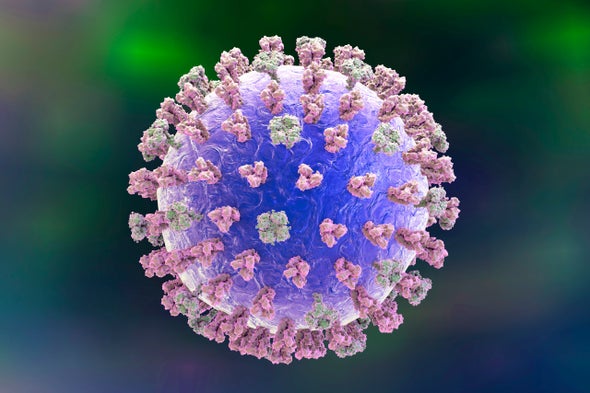
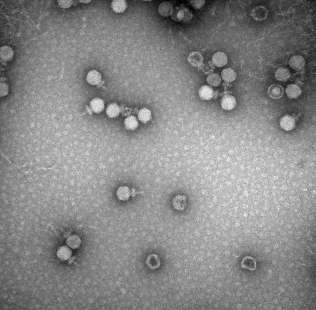
Bacteriophages, or phages for short, are viruses that naturally take over and kill bacteria. Thousands of phages exist, but using them as treatments to fight specific bacteria has so far proven to be challenging. To optimize phage therapy and make it scalable to human disease, scientists need ways to engineer phages into efficient bacteria-killing machines. This would also offer an alternative way to treat bacterial infections that are resistant to standard antibiotics.

Now, Gladstone scientists have developed a technology that lets them edit the genomes of phages in a streamlined and highly effective way, giving them the ability to engineer new phages and study how the viruses can be used to target specific bacteria.

"Ultimately, if we want to use phages to save the lives of people with infections that are resistant to multiple drugs, we need a way to make and test lots of phage variants to find the best ones," says Gladstone Associate Investigator Seth Shipman, PhD, the lead author of a study published in Nature Biotechnology. "This new technique lets us successfully and rapidly introduce different edits to the phage genome so we can create numerous variants."

The new approach relies on molecules called retrons, which originate from bacterial immune systems and act like DNA-production factories inside bacterial cells. Shipman's team has found ways to program retrons so they make copies of a desired DNA sequence. When phages infect a bacterial colony containing retrons, using the technique described in the team's new study, the phages integrate the retron-produced DNA sequences into their own genomes.

The enemy of your enemy

Unlike antibiotics, which broadly kill many types of bacteria at once, phages are highly specific for individual strains of bacteria. As rates of antibiotic-resistant bacterial infections rise-;with an estimated 2.8 million such infections in the United States each year-;researchers are increasingly looking at the potential of phage therapy as an alternative to combat these infections.

"They say that the enemy of your enemy is your friend," says Shipman, who is also an associate professor in the Department of Bioengineering and Therapeutic Sciences at UCSF, as well as a Chan Zuckerberg Biohub Investigator. "Our enemies are these pathogenic bacteria, and their enemies are phages."

Already, phages have been successfully used in the clinic to treat a small number of patients with life-threatening antibiotic-resistant infections, but developing the therapies has been complex, time-consuming, and difficult to replicate at scale. Doctors must screen collections of naturally-occurring phages to test whether any could work against the specific bacteria isolated from an individual patient.

Shipman's group wanted to find a way to modify phage genomes to create larger collections of phages that can be screened for therapeutic use, as well as to collect data on what makes some phages more effective or what makes them more or less specific to bacterial targets.

"As the natural predators of bacteria, phages play an important role in shaping microbial communities," says Chloe Fishman, a former research associate at Gladstone and co-first author of the new study, now pursuing her graduate degree at Rockefeller University. "It's important to have tools to modify their genomes in order to better study them. It's also important if we want to engineer them so that we can shape microbial communities to our benefit-;to kill antibiotic-resistant bacteria, for example."

Continuous phage editing

To precisely engineer phage genomes, the scientists turned to retrons. In recent years, Shipman and his group pioneered the development and use of retrons to edit the DNA of human cells, yeast, and other organisms.

Shipman and his colleagues began by creating retrons that produce DNA sequences specifically designed to edit invading phages-;a system the team dubbed "recombitrons." Then, they put those retrons into colonies of bacteria. Finally, they let phages infect the bacterial colonies. As the phages infected bacteria after bacteria, they continuously acquired and integrated the new DNA from the recombitrons, editing their own genome as they went along.

The research team showed that the longer they let phages infect a recombitron-containing bacterial colony, the greater the number of phage genomes were edited. Moreover, the researchers could program different bacteria within the colony with different recombitrons, and the phages would acquire multiple edits as they infected the colony.

As a phage is bouncing from bacterium to bacterium, it picks up different edits. Making multiple edits in phages is something that was previously incredibly hard to do; so much so that, most of the time, scientists simply didn't do it. Now, you basically throw some phages into these cultures, wait a while, and get your multiple-edited phages."

Seth Shipman, PhD, lead author

A platform to screen phages

If scientists already knew exactly what edits they wanted to make to a given phage to optimize its therapeutic potential, the new platform would let them easily and effectively carry out those edits. However, before researchers can predict the consequence of a genetic change, they first need to better understand what makes phages work and how variations to their genomes impact their effectiveness. The recombitron system helps makes progress here, too.

If multiple recombitrons are put into a bacterial colony, and phages are allowed to infect the colony for only a short time, different phages will acquire different combinations of edits. Such diverse collections of phages could then be compared.

"Scientists now have a way to edit multiple genes at once if they want to study how these genes interact or introduce modifications that could make the phage a more potent bacterial killer," says Kate Crawford, a graduate student in the Shipman lab and co-first author of the new study.

Shipman's team is working on increasing the number of different recombitrons that can be put into a single bacterial colony-;and then passed along to phages. They expect that eventually, millions of combinations of edits could be introduced to phages to make huge screening libraries.

"We want to scale this high enough, with enough phage variants, that we can start to predict which phage variants will work against what bacterial infections," says Shipman.

Source:
Journal reference:

Fishman, C. B., et al. (2024). Continuous multiplexed phage genome editing using recombitrons. Nature Biotechnologydoi.org/10.1038/s41587-024-02370-5.

Thursday, September 04, 2025

BACTERIOPHAGES

1.5 million euros for research into “bacterial killers”


ERC Starting Grant awarded to Jens Hör from the Helmholtz Institute for RNA-based Infection Research


Helmholtz Centre for Infection Research


Infections caused by antibiotic-resistant bacteria pose a serious threat to global health. Consequently, there is an urgent need for new antibacterial compounds. One promising avenue of research is bacteriophages, commonly known as phages, which are viruses that infect bacteria. They attach to a bacterium and inject their genetic material into it. This allows them to hijack the cell machinery and transform the bacterium into a “phage factory”. The bacterium then produces new phages until the infected microbe bursts and releases them. Each new phage can infect other bacteria, creating a chain reaction that destroys all the microbes.

These “bacteria eaters”—a meaning derived from the original Greek term “bacteriophage”—are the focus of Jens Hör's research at the Helmholtz Institute for RNA-based Infection Research (HIRI) in Würzburg. The institute is a site of the Braunschweig Helmholtz Centre for Infection Research (HZI) in cooperation with the Julius-Maximilians-Universität Würzburg (JMU). Hör has now received a Starting Grant of 1.5 million euros from the European Research Council (ERC) for his research project “RIBO-PHAGE”.

“The rising antibiotic resistance is one of the most pressing challenges of our time. This makes it all the more gratifying that the ERC awarded a Starting Grant to junior professor Jens Hör. Thanks to this prestigious funding award, he will be able to continue his pioneering research at the highest level,” says Jörg Vogel, Managing Director of the HIRI.

“Phages are much more than fascinating biological tools—they have the potential to become a game changer in everyday clinical practice, especially in the fight against multi-resistant bacteria. To exploit this potential, we need research like that conducted by Jens Hör at our HZI institute HIRI in Würzburg. ERC grants are among the most important and prestigious sources of funding in the international scientific community and create the freedom to pursue such innovative approaches, thereby laying the foundation for future therapies,” states Josef Penninger, Scientific Director of the HZI. “I warmly congratulate Jens Hör on this great success.”

Focus on RNA phages

The therapeutic use of phages dates back to the early 20th century. Nevertheless, it gradually faded into obscurity due to the development and widespread success of conventional antibiotics. Recent discoveries have brought phages back into the spotlight, highlighting their tremendous potential in basic and applied research. For example, phage research has shown that certain aspects of the human innate immune system can be traced back to bacterial defense mechanisms. In addition, researchers made significant progress in developing phages as a therapeutic agent for treating infections caused by multi-resistant bacteria.

However, this renaissance neglected an understudied group: phages with ribonucleic acid (RNA) as their genetic material. “Their biology is fundamentally different from that of DNA phages, which are based on deoxyribonucleic acid (DNA). To start, their appearance differs: While DNA phages have a head and a tail, RNA phages consist only of a head,” explains Hör. Currently, only very few RNA phages have been discovered. Through his ERC-funded project, Hör aims to further our understanding of this unique group of phages.

To reproduce efficiently, RNA phages use unique regulatory mechanisms that determine which proteins are produced and when. Hosts—bacteria infected by phages—have their own defense systems to stop the intruders. These processes are carefully coordinated. The goal of the phages is to reproduce as quickly as possible. The host cell, on the other hand, aims to ward off the phages to protect the bacterial population.

“Understanding these mechanisms of action facilitates the effective use of RNA phages as therapeutic agents. It also bears the potential to discover entirely new biotechnological tools,” says Jens Hör. “In my RIBO-PHAGE project, I want to elucidate the unique lifestyle of RNA phages and provide a detailed picture of the molecular processes involved in RNA phage infection.” Hör is particularly interested in deciphering how RNA phages efficiently regulate their replication and which host factors they hijack in the process. In addition, he intends to shed more light on how bacteria defend themselves against RNA phage infections. He will use, amongst others, RNA phages from the Fiersviridae and Cystoviridae families, which infect bacteria of the genera Escherichia and Pseudomonas, as model systems.

Jens Hör is already the fifth HIRI group leader to receive ERC funding. Jörg Vogel concludes that “the successful acquisition of this new grant is not only impressive proof of the HIRI's outstanding position in the international research landscape. This also shows that phage research has reached a critical mass, making it an integral part of future medicine.”

About Jens Hör

Jens Hör studied life and medical sciences at the University of Bonn (Germany) and received his PhD from the University of Würzburg (Germany) in 2020. During these studies, he focused on the global analysis of bacterial RNA-protein complexes. He then joined the Weizmann Institute of Science (Rehovot, Israel) as a postdoc, where he worked on the mechanisms of bacterial anti-phage defense systems. Since 2024, he has been a research group leader at HIRI and a junior professor at JMU.

You can learn more about Jens Hör, his research on RNA phages, and the steps still needed to enable widespread use of phage therapies in Germany in today's episode of “InFact – The HZI Podcast. Science that is contagious”: https://infacthzi.podigee.io/34-phages-bacteria-eaters-against-dangerous-infections.

About HIRI

The Helmholtz Institute for RNA-based Infection Research (HIRI) is the first institution of its kind worldwide to combine ribonucleic acid (RNA) research with infection biology. Based on novel findings from its strong basic research program, the institute’s long-term goal is to develop innovative therapeutic approaches to better diagnose and treat human infections.

HIRI is a site of the Braunschweig Helmholtz Centre for Infection Research (HZI) in cooperation with the Julius-Maximilians-Universität Würzburg (JMU) and is located on the Würzburg Medical Campus. More information at www.helmholtz-hiri.de

The ERC Starting Grants

ERC Starting Grants are funding instruments of the European Research Council to support young scientists in their efforts to become independent top researchers. At the time of application, a maximum of seven years may have passed since the candidates have obtained their doctoral degree. The only explicit evaluation criterion is the scientific excellence of the researchers and the proposed project. The successful projects are funded for up to five years with a total amount of up to 1.5 million euros.

The European Research Council

The European Research Council, established by the European Union in 2007, is the premier European funding organization for excellent cutting-edge research. Each year, it selects the best and most creative researchers of any nationality and funds projects based in Europe. The ERC offers four core-funding programs: Starting, Consolidator, Advanced, and Synergy Grants. With its additional Proof of Concept Grant Program, the ERC helps grant holders to bridge the gap between their frontier research and the early stages of commercialization. https://erc.europa.eu/

Helmholtz Centre for Infection Research:

Scientists at the Helmholtz Centre for Infection Research (HZI) in Braunschweig and its other sites in Germany are engaged in the study of bacterial and viral infections and the body’s defense mechanisms. They have a profound expertise in natural compound research and its exploitation as a valuable source for novel anti-infectives. As member of the Helmholtz Association and the German Center for Infection Research (DZIF) the HZI performs translational research laying the ground for the development of new treatments and vaccines against infectious diseases. www.helmholtz-hzi.de/en

Wednesday, July 30, 2025


New insights could help phages defeat antibiotic resistant bacteria


University of Southampton
Bacteria infected by phage 

image: 

Bacteria infected by phage. The dots show phage replicating.

view more 

Credit: University of Southampton


Researchers at the University of Southampton have worked out how bacteria defend themselves against viruses called phages and the new insights could be key to tackling antibiotic resistance.

Phages are seen as a promising alternative treatment to antibiotics. Unpicking how bacteria protect themselves, and how phages might overcome these defences, could be a significant step in defeating antibiotic resistant bacteria.

Phages, known as bacteria eaters, look like a syringe with spider legs. They work by attaching themselves to bacteria. Once locked on, they inject their DNA into the bacterial cell, hijacking it to produce more copies of the virus before the cell bursts open and releases the new phages to attack other bacteria.

Crucially, phages only attack bacteria and are harmless to human cells.

The new research published today [28 July] in the journal Cell is the first to describe how a bacterial defence mechanism against phages, called Kiwa, works.

“In Māori mythology, Kiwa is a divine guardian of the ocean and its creatures,” says Dr Franklin Nobrega, Associate Professor at the University of Southampton and National Institute for Health and Care Research (NIHR) Southampton Biomedical Research Centre (BRC) Unit. “In bacteria, Kiwa also acts as a guardian, defending against phages, and are one of the most common defence mechanisms bacteria have.”

Researchers used advanced imaging techniques to study the interaction between phages and Kiwa at a molecular level.

They found Kiwa is made up of two components called KwaA and KwaB. This duo works together to form a kind of chainmail around the bacteria, preventing the phage DNA from entering.  KwaA acts like a sensor detecting the presence of a phage. Once this sensor is tripped, KwaB is alerted which binds to the phage DNA and turns it off before it can take over the cell.

But some phages have evolved a clever way to break through this two-step security system. They release a ‘decoy’ protein called Gam which tricks KwaB into attacking them while the real phage DNA slips through to complete the hijack.

Unfortunately for the phages, and us, Kiwa is one of many defence mechanisms bacteria have. Another is called RecBCD which also detects and attacks phage DNA. While the decoys work well against both systems independently, when they combine phages can’t break through.

Dr Nobrega explains: “In a similar way to how hackers are constantly looking for ways to bypass security systems, phages have evolved ways to breach the defences of bacteria. But just as tech companies adapt by releasing their latest update with improved security features, bacteria have evolved their own molecular firewalls in the shape of Kiwa and RecBCD.”

Finding new ways to fight bacteria is a pressing concern due to the growing threat of antibiotic resistance, which could kill ten million people a year by 2050 and costs the NHS £180m every year.

Dr Nobrega and his team at the University of Southampton are collecting phages which have the potential to overcome bacterial defences, and have identified over 600 different types to date.

They are inviting people to collect samples of dirty water (the perfect breeding ground for bacteria and phages) and post it into the lab for analysis.

“By improving our understanding of how these defence mechanisms operate, we can work out how to exploit weaknesses and select phages which have the best chance of breaking down the bacteria,” says Dr Nobrega.

“The more samples we are able to obtain, the better our chances of finding the best phages for the job.”

The paper Kiwa is a membrane-embedded defence supercomplex activated at phage attachment sites is published in Cell and is available online.

The research was funded by The Royal Society, Wessex Medical Research, Welch Foundation, National Institutes of Health and Simons Foundation. The Phage Collection Project is supported by the NIHR Southampton BRC Unit.

Ends

Contact

Steve Williams, Media Manager, University of Southampton, press@soton.ac.uk or 023 8059 3212.

Notes for editors

  1. The paper Kiwa is a membrane-embedded defence supercomplex activated at phage attachment sites will be published in Cell. An advanced copy is available upon request.
  2. For Interviews with Dr Franklin Nobrega please contact Steve Williams, Media Manager, University of Southampton press@soton.ac.uk or 023 8059 3212.
  3. Images available here: https://safesend.soton.ac.uk/pickup?claimID=M2Xzjeo6XNzFUp5c&claimPasscode=oBit3JrkEGgPAhNX 

Additional information

The University of Southampton drives original thinking, turns knowledge into action and impact, and creates solutions to the world’s challenges. We are among the top 100 institutions globally (QS World University Rankings 2025). Our academics are leaders in their fields, forging links with high-profile international businesses and organisations, and inspiring a 22,000-strong community of exceptional students, from over 135 countries worldwide. Through our high-quality education, the University helps students on a journey of discovery to realise their potential and join our global network of over 200,000 alumni. www.southampton.ac.uk

www.southampton.ac.uk/news/contact-press-team.page

Follow us on X: https://twitter.com/UoSMedia

About the NIHR
The mission of the National Institute for Health and Care Research (NIHR) is to improve the health and wealth of the nation through research. We do this by:

  • Funding high quality, timely research that benefits the NHS, public health and social care;
  • Investing in world-class expertise, facilities and a skilled delivery workforce to translate discoveries into improved treatments and services;
  • Partnering with patients, service users, carers and communities, improving the relevance, quality and impact of our research;
  • Attracting, training and supporting the best researchers to tackle complex health and social care challenges;
  • Collaborating with other public funders, charities and industry to help shape a cohesive and globally competitive research system;
  • Funding applied global health research and training to meet the needs of the poorest people in low and middle income countries.

NIHR is funded by the Department of Health and Social Care. Its work in low and middle income countries is principally funded through UK international development funding from the UK government.


On the left, bacterial cells are uninfected and Kiwa is inactive. On the right, bright spots appear inside the cells—these show Kiwa being activated after detecting a phage, helping to stop the infection before it takes hold.


This illustration shows a phage (virus) attaching to a bacterial cell. The Kiwa defence system (shown in yellow, green, and blue) detects the threat and binds the invading DNA, preventing the phage from hijacking the cell.


Credit

University of Southampton

Monday, November 24, 2025


Fishing for phages in Lund University’s Botanical Gardens


Lund University
Vasili Hauryliuk 

image: 

Vasili Hauryliuk

view more 

Credit: Kennet Ruona


Kompetensportalen, Lucat, Lupin, Lubas and LUCRIS. Those are the names of some of Lund University’s administrative systems. They are now also the names of five new bacteriophages that have recently been discovered in the ponds of Lund University’s Botanical Gardens.

Bacteriophages – often abbreviated to phages – are viruses that attack bacteria. Phages are astonishingly effective assassins – these viruses wipe out 20 percent of all bacteria on Earth every day. The ongoing battle with bacteria has made phages humanity’s natural ally when it comes to treating bacterial infections The growing urgency of combating antibiotic resistance has made phage research – particularly the development of phage-basered therapies – more relevant than ever.

“Bacteria are under constant attack from phages. Phages are picky about their prey – different phages infect different species of bacteria, sometimes only a specific strain. The challenge lies in assembling the right “collection” of phages, each one a precision weapon calibrated to infect and obliterate only the intended strain of bacteria,” says Vasili Hauryliuk, professor of medical biochemistry at Lund University.

Finding the right bacteriophage for the right bacterial strain is a major challenge. Natural bacterial strains are also constantly changing, thanks to mutations among other things. This means that a phage that has previously been effective may become ineffective.

At Lund University, Sweden’s first international course in phage biology has been completed. Doctoral students from across Europe came to attend lectures by leading phage researchers, exchange ideas, and, of course, to hunt for new phages and find the right precision weapons with which to attack various bacteria. Phages thrive wherever bacteria are found, which often means ponds and watercourses that are rich in organic material. The ponds in Lund University’s Botanical Gardens – both indoors and out – therefore proved to be perfect locations for phage fishing. However, to catch phages requires the right “bait”, which means the right bacterial strain to attract the virus.

“Collecting phages is like fishing in that you never know what you will end up with on the hook. Since it is fairly simple to isolate bacteriophages from ponds – and Lund has several – we combined research and education and went fishing for phages,” says Marcus Johansson, associate researcher at Lund University and one of the course coordinators. He is also last author on the study.

The researchers used a strain of E. coli, a common gut bacterium that can become a lethal pathogen. When a laboratory E. coli strain is grown in flasks without shaking, it becomes motile by developing a so-called flagellum – a “tail” that the bacterium uses to propel itself and explore the environment. Some phages specifically recognise the “tail” to infect. Using a motile E. coli strain, researchers managed to catch a new “tail-loving” phage from the Botanical Gardens’ ponds. Remarkably, this phage can kill not only E. coli, but also another motile bacterial species –Salmonella.

“One fun part about phage fishing is that you can name the new viruses – and phage names can be pretty weird! We wanted our phages to have names that were linked to Lund University and the tail-loving phage was named “Kompetensportalen”. We named two other phages Lucat and Lupin, after the University’s staff directory and its purchasing and invoicing tool, respectively” explains Vasili Hauryliuk.

The total of five newly-discovered bacteriophages from the Botanical Gardens are now serving as ambassadors for Lund University in the world of international phage research. The phage, “Kompetensportalen” has quickly attracted attention and phage researchers from outside Sweden have already expressed an interest in it.

“The diversity of bacteriophages discovered in the Botanical Gardens’ ponds is particularly fascinating as the Gardens’ greenhouses are currently being renovated. It underlines the great diversity in biology and our role as a centre for education and research. It is exciting to discover that our ponds are home to more than just plants,” says Allison Perrigo, director of Lund University’s Botanical Gardens.

Monday, April 13, 2020

Scientists Program CRISPR to Fight Viruses in Human Cells

A common gene-editing enzyme could be used to disable RNA viruses such as flu or Ebola


By Tanya Lewis on October 23, 2019
Scientists Program CRISPR to Fight Viruses in Human Cells

Researchers modified the enzyme Cas13 to target and inactivate viruses such as influenza (shown here). Credit: Kateryna Kon Getty Images

CRISPR is usually thought of as a laboratory tool to edit DNA in order to fix genetic defects or enhance certain traits—but the mechanism originally evolved in bacteria as a way to fend off viruses called bacteriophages. Now scientists have found a way to adapt this ability to fight viruses in human cells.

In a recent study, Catherine Freije, Cameron Myhrvold and Pardis Sabeti at the Broad Institute of the Massachusetts Institute of Technology and Harvard University, and their colleagues programmed a CRISPR-related enzyme to target three different single-stranded RNA viruses in human embryonic kidney cells (as well as human lung cancer cells and dog kidney cells) grown in vitro and chop them up, rendering them largely unable to infect additional cells. If further experiments show this process works in living animals, it could eventually lead to new antiviral therapies for diseases such as Ebola or Zika in humans.

Viruses come in many forms, including DNA and RNA, double-stranded and single-stranded. About two thirds of the ones that infect humans are RNA viruses, and many have no approved treatment. Existing therapies often use a small molecule that interferes with viral replication—but this approach does not work for newly emerging viruses or ones that are evolving rapidly.

“CRISPR” refers to a series of DNA sequences in bacterial genomes that were left behind from previous bacteriophage infections. When the bacteria encounter these pathogens again, enzymes called CRISPR-associated (Cas) proteins recognize and bind to these sequences in the virus and destroy them. In recent years, researchers (including study co-author Feng Zhang) have reengineered one such enzyme, called Cas9, to cut and paste DNA in human cells. The enzyme binds to a short genetic tag called a guide RNA, which directs the enzyme to a particular part of the genome to make cuts. Previous studies have used Cas9 to prevent replication of double-stranded DNA viruses or of single-stranded RNA viruses that produce DNA in an intermediate step during replication. Only a small fraction of RNA viruses that infect humans produce such DNA intermediates—but another CRISPR enzyme, called Cas13, can be programmed to cleave single-stranded RNA viruses.

“The nice thing about CRISPR systems and systems like Cas13 is that their initial purpose in bacteria was to defend against viral infection of bacteria, and so we sort of wanted to bring Cas13 back to its original function—and apply this to mammalian viruses in mammalian cells,” says Freije, who is a doctoral student in virology at Harvard. “Because CRISPR systems rely on guide RNAs to specifically guide the CRISPR protein to a target, we saw this as a great opportunity to use it as a programmable antiviral.”

Freije and her colleagues programmed Cas13 to target three different viruses: lymphocytic choriomeningitis virus (LCMV), influenza A virus (IAV) and vesicular stomatitis virus (VSV). LCMV is an RNA virus that mostly infects mice—but it is in the same family as the virus that causes Lassa fever, which is found in West Africa and is much more dangerous to study in the lab. IAV is a flu virus; although some antiviral medications for flu already exist, such viruses evolve rapidly, so there is a need for better options. Finally, VSV is a model for many other single-stranded RNA viruses.

To determine how effective Cas13 was at destroying the viruses, the researchers also used it as a diagnostic tool to see how much viral RNA was being released from infected cells. They saw a twofold to 44-fold reduction in RNA, depending on which virus they were looking at and the time point. They also looked at how well the released RNA was able to go on and infect new cells. In most cases, they saw a 100-fold reduction in infectivity—and in some cases, more than 300-fold—according to Freije. The findings were published online on October 10 in Molecular Cell.

“The results are very impressive,” says Chen Liang, a professor at the Lady Davis Institute at Jewish General Hospital and the department of microbiology and immunology at McGill University in Montreal, who was not involved in the study. His own laboratory has used the Cas9 enzyme to deactivate DNA viruses. The concept is very similar, but Cas13 has a few advantages, he says. For one, Cas13 can be used to target one virus using several guide RNAs, making it difficult for the virus to “escape.” Secondly, the new study also used Cas13 to detect how much viral RNA was left over to infect cells. The amount of viral knockdown the group achieved is “very significant,” Liang says. “If you can target and inactivate all three [of these] viruses, in principle, you can inactivate any virus.


As with any approach, there are limitations. One is the question of how to deliver the Cas13 to target a virus in a living person, Liang notes, and the researchers have not yet done any animal studies. Another is the fact that viruses will eventually develop resistance. But Cas13 has an advantage here: when Cas9 cuts viral DNA, mammalian cells repair it and can cause mutations that make the virus more resistant. Yet with Cas13, these cells do not have the mechanism to repair the RNA and introduce errors that would help the virus escape being destroyed. Even if a virus does evolve resistance, or if a new virus is encountered, the method could be quickly adapted.

“One of the things that’s most exciting about this approach is the programmability,” says Myhrvold, a postdoctoral fellow at Harvard. “Once you figure out how to do this well for one virus it’s not that hard to design sequences against another virus—or another one. Furthermore, if the virus changes its own sequence—as viruses are known to do, just during an outbreak or in response to therapy—you can very easily update the CRISPR RNA sequence and keep up with the virus.”

Freije agrees. “We are definitely excited about future prospects of optimizing the system and trying it out in mouse models,” she says. Beyond therapeutics, the team hopes to understand more about how viruses operate—how they replicate and what parts of their genomes are most important. Using approaches like this, “you can really start to get a better picture of what parts of these viruses are and, most importantly, what really makes them tick.”


How do bacteria defend themselves against viruses?

The CRISPR-Cas system in some bacteria helps to form an effective barrier to invading viruses.


DIGEST Apr 3, 2019


A transmission electron microscopy image of bacteriophages taken at The University of Alabama’s Optical Analysis Facility. Image credit: Chou-Zheng and Hatoum-Aslan, 2019 (CC BY 4.0)

Just as humans are susceptible to viruses, bacteria have their own viruses to contend with. These viruses – known as phages – attach to the surface of bacterial cells, inject their genetic material, and use the cells’ enzymes to multiply while destroying their hosts.

To defend against a phage attack, bacteria have evolved a variety of immune systems. For example, when a bacterium with an immune system known as CRISPR-Cas encounters a phage, the system creates a ‘memory’ of the invader by capturing a small snippet of the phage’s genetic material. The pieces of phage DNA are copied into small molecules known as CRISPR RNAs, which then combine with one or more Cas proteins to form a group called a Cas complex. This complex patrols the inside of the cell, carrying the CRISPR RNA for comparison, similar to the way a detective uses a fingerprint to identify a criminal. Once a match is found, the Cas proteins chop up the invading genetic material and destroy the phage.

There are several different types of CRISPR-Cas systems. Type III systems are among the most widespread in nature and are unique in that they provide a nearly impenetrable barrier to phages attempting to infect bacterial cells. Medical researchers are exploring the use of phages as alternatives to conventional antibiotics and so it is important to find ways to overcome these immune responses in bacteria. However, it remains unclear precisely how Type III CRISPR-Cas systems are able to mount such an effective defense.

Chou-Zheng and Hatoum-Aslan used genetic and biochemical approaches to study the Type III CRISPR-Cas system in a bacterium called Staphylococcus epidermidis. The experiments showed that two enzymes called PNPase and RNase J2 played crucial roles in the defense response triggered by the system. PNPase helped to generate CRISPR RNAs and both enzymes were required to help to destroy genetic material from invading phages.

Previous studies have shown that PNPase and RNase J2 are part of a machine in bacterial cells that usually degrades damaged genetic material. Therefore, these findings show that the Type III CRISPR-Cas system in S. epidermidis has evolved to coordinate with another pathway to help the bacteria survive attack from phages. CRISPR-Cas immune systems have formed the basis for a variety of technologies that continue to revolutionize genetics and biomedical research. Therefore, along with aiding the search for alternatives to antibiotics, this work may potentially inspire the development of new genetic technologies in the future.

Phage-Encoded Anti-CRISPR Defenses
Annual Review of Genetics


Vol. 52:445-464 (Volume publication date November 2018)
First published as a Review in Advance on September 12, 2018
https://doi.org/10.1146/annurev-genet-120417-031321

Sabrina Y. Stanley1 and Karen L. Maxwell2
1Department of Molecular Genetics, University of Toronto, Toronto, Ontario M5S 1A8, Canada
2Department of Biochemistry, University of Toronto, Toronto, Ontario M5G 1M1, Canada; email: karen.maxwell@utoronto.ca


Abstract

The battle for survival between bacteria and bacteriophages (phages) is an arms race where bacteria develop defenses to protect themselves from phages and phages evolve counterstrategies to bypass these defenses. CRISPR-Cas adaptive immune systems represent a widespread mechanism by which bacteria protect themselves from phage infection. In response to CRISPR-Cas, phages have evolved protein inhibitors known as anti-CRISPRs. Here, we describe the discovery and mechanisms of action of anti-CRISPR proteins. We discuss the potential impact of anti-CRISPRs on bacterial evolution, speculate on their evolutionary origins, and contemplate the possible next steps in the CRISPR-Cas evolutionary arms race. We also touch on the impact of anti-CRISPRs on the development of CRISPR-Cas-based biotechnological tools.

FULL ARTICLE HERE


Biological Sciences
RESEARCH ARTICLE
Temperate and lytic bacteriophages programmed to sensitize and kill antibiotic-resistant bacteria
Ido Yosef, Miriam Manor, Ruth Kiro, and View ORCID Profile Udi Qimron
PNAS June 9, 2015 112 (23) 7267-7272; first published May 18, 2015 https://doi.org/10.1073/pnas.1500107112

Edited by Jennifer A. Doudna, University of California, Berkeley, CA, and approved April 28, 2015 (received for review January 25, 2015)

Significance

Antibiotic resistance of pathogens is a growing concern to human health, reviving interest in phage therapy. This therapy uses phages (natural bacterial enemies) to kill pathogens. However, it encounters many obstacles such as delivery barriers into the tissues and bacterial resistance to phages. Here, we use phages for delivering a programmable DNA nuclease, clustered regularly interspaced short palindromic repeats (CRISPR)–CRISPR-associated (Cas), to reverse antibiotic resistance and eliminate the transfer of resistance between strains. This approach combines CRISPR-Cas delivery with lytic phage selection of antibiotic-sensitized bacteria. The strategy may reduce the prevalence of antibiotic-resistant bacteria in treated surfaces and on skin of medical personnel, as it uses phages in a unique way that overcomes many of the hurdles encountered by phage therapy.

Abstract

The increasing threat of pathogen resistance to antibiotics requires the development of novel antimicrobial strategies. Here we present a proof of concept for a genetic strategy that aims to sensitize bacteria to antibiotics and selectively kill antibiotic-resistant bacteria. We use temperate phages to deliver a functional clustered regularly interspaced short palindromic repeats (CRISPR)–CRISPR-associated (Cas) system into the genome of antibiotic-resistant bacteria. The delivered CRISPR-Cas system destroys both antibiotic resistance-conferring plasmids and genetically modified lytic phages. This linkage between antibiotic sensitization and protection from lytic phages is a key feature of the strategy. It allows programming of lytic phages to kill only antibiotic-resistant bacteria while protecting antibiotic-sensitized bacteria. Phages designed according to this strategy may be used on hospital surfaces and hand sanitizers to facilitate replacement of antibiotic-resistant pathogens with sensitive ones.
DOWNLOAD WHOLE PAPER AS PDF FROM HERE 

   Genetically Engineered Phages: a Review of Advances over the Last Decade

Diana P. PiresSara CletoSanna SillankorvaJoana AzeredoTimothy K. Lu
DOI: 10.1128/MMBR.00069-15


SUMMARY
Soon after their discovery in the early 20th century, bacteriophages were recognized to have great potential as antimicrobial agents, a potential that has yet to be fully realized. The nascent field of phage therapy was adversely affected by inadequately controlled trials and the discovery of antibiotics. Although the study of phages as anti-infective agents slowed, phages played an important role in the development of molecular biology. In recent years, the increase in multidrug-resistant bacteria has renewed interest in the use of phages as antimicrobial agents. With the wide array of possibilities offered by genetic engineering, these bacterial viruses are being modified to precisely control and detect bacteria and to serve as new sources of antibacterials. In applications that go beyond their antimicrobial activity, phages are also being developed as vehicles for drug delivery and vaccines, as well as for the assembly of new materials. This review highlights advances in techniques used to engineer phages for all of these purposes and discusses existing challenges and opportunities for future work.

INTRODUCTION

Bacteriophages (phages) are among the most abundant biological particles on earth. They are also highly versatile and adaptable to a great number of applications. Phages are viruses that infect bacteria; their self-replication depends on access to a bacterial host. Phages were discovered independently by Frederick Twort in 1915 (1) and by Félix d'Hérelle in 1917 (2), and they were used early on as antimicrobial agents. Although the initial results of phage therapy were promising (34), poorly controlled trials and inconsistent results generated controversy within the scientific community about the efficacy and reproducibility of using phages to treat bacterial infections (57). The discovery of penicillin in 1928 and the subsequent arrival of the antibiotic era further cast a shadow on phage therapy (56). As a result, phage therapy was discontinued in Western countries, even as its use continued in Eastern Europe and the former Soviet Union (810).
Despite the important success of antibiotics in improving human health, antibiotic resistance has emerged with steadily increasing frequency, rendering many antibiotics ineffective (1114). Multidrug-resistant bacteria currently constitute one of the most widespread global public health concerns (1517). More than 2 million people are sickened every year in the United States alone as a result of antibiotic-resistant infections, resulting in at least 23,000 deaths per year (16). The rising tide of antibiotic resistance coupled with the low rate of antibiotic discovery (1718) has revived interest in phages as antibacterial agents (1921).
Unlike most antibiotics, phages are typically highly specific for a particular set of bacterial species or strains and are thus expected to have fewer off-target effects on commensal microflora than antibiotics do (22). The self-replicating nature of phages and the availability of simple, rapid, and low-cost phage production processes are additional advantages for their use as antimicrobials (22). Phages have been used not only to treat and prevent human bacterial infections (92325) but also to control plant diseases (2629), detect pathogens (3033), and assess food safety (3437).
READ/DOWNLOAD THE ARTICLE 

REVIEW ARTICLE
Microbiol., 03 May 2019 | https://doi.org/10.3389/fmicb.2019.00954


Genetic Engineering of Bacteriophages Against Infectious Diseases

Yibao Chen1,2, Himanshu Batra3, Junhua Dong1,2, Cen Chen1,2, Venigalla B. Rao3 and Pan Tao1,2,3*

Bacteriophages (phages) are the most abundant and widely distributed organisms on Earth, constituting a virtually unlimited resource to explore the development of biomedical therapies. The therapeutic use of phages to treat bacterial infections (“phage therapy”) was conceived by Felix d’Herelle nearly a century ago. However, its power has been realized only recently, largely due to the emergence of multi-antibiotic resistant bacterial pathogens. Progress in technologies, such as high-throughput sequencing, genome editing, and synthetic biology, further opened doors to explore this vast treasure trove. Here, we review some of the emerging themes on the use of phages against infectious diseases. In addition to phage therapy, phages have also been developed as vaccine platforms to deliver antigens as part of virus-like nanoparticles that can stimulate immune responses and prevent pathogen infections. Phage engineering promises to generate phage variants with unique properties for prophylactic and therapeutic applications. These approaches have created momentum to accelerate basic as well as translational phage research and potential development of therapeutics in the near future.

Introduction

Bacteriophages (phages), discovered in the early 20th century independently by Frederick Twort and Felix d’Herelle, are the most abundant organisms on earth with up to 2.5 × 10⁸ phages per ml in natural waters (Bergh et al., 1989). It is well accepted that phages specifically infect bacteria and, therefore, were considered for the development of natural approaches to treat bacterial infections since their discovery (Wittebole et al., 2014Salmond and Fineran, 2015). However, due to the discovery of antibiotics that provided greater breadth and potency, phage therapy lagged behind although research continued in some Eastern European countries (Chanishvili, 20122016Wittebole et al., 2014). Therefore, in the following several decades, phages were mainly used as model organisms to explore the basic mechanisms of life and led to the birth of modern molecular biology. One classical example is the demonstration of a central biological question in the early 20th century, the nature of a gene, by “Hershey-Chase experiment” (also called “Waring blender experiment”) (Salmond and Fineran, 2015). This elegant experiment demonstrated that DNA, not protein, is the genetic material of T2 phage.

Recently, the emergence of multi-antibiotic resistant bacterial pathogens and the low rate of new antibiotic discovery brought new urgency to develop phage-based therapies (Lu and Koeris, 2011Viertel et al., 2014Domingo-Calap and Delgado-Martinez, 2018). A striking example is the recent San Diego patient who was infected by multi-drug resistant Acinetobacter baumannii stain during travelling to Egypt. The patient went into a coma for nearly 2 months but awoke 2 days after intravenous injection of a phage cocktail that lyses this bacterium and finally completely recovered (Schooley et al., 2017). With recent advances, particularly the genome engineering (Martel and Moineau, 2014Ando et al., 2015Lemay et al., 2017Tao et al., 2017bKilcher et al., 2018), the applications of phages have greatly expanded. In addition to its use in antibacterial therapy, phages were used in synthetic biology (Lemire et al., 2018), material science (Cao et al., 2016), and biomedical fields (Cao et al., 2018Tao et al., 2018c). Considering the abundance and diversity, there is vast potential to engineer phages for different applications. In this review, we will focus on the applications of phages in infectious disease, in particular, vaccine development and phage therapy. We will discuss the phage engineering strategies and how these can equip the phages with the ability to advance the vaccine and phage therapy fields.


1College of Veterinary Medicine, Huazhong Agricultural University, Wuhan, China
2The Cooperative Innovation Center for Sustainable Pig Production, Huazhong Agricultural University, Wuhan, China
3Department of Biology, The Catholic University of America, Washington, DC, United States


READ / DOWNLOAD THE REST OF THE ARTICLE HERE 

Allosteric inhibition of CRISPR-Cas9 by bacteriophage-derived peptides
Yan-ru Cui,
Shao-jie Wang,
Jun Chen,
Jie Li,
Wenzhang Chen,
Shuyue Wang,
Bing Meng,
Wei Zhu,
Zhuhong Zhang,
Bei Yang,
Biao Jiang,
Guang Yang,
Peixiang Ma &
Jia Liu 


Genome Biology volume 21, Article number: 51 (2020) Cite this article

Research
Open Access
Published: 26 February 2020

Abstract


Background


CRISPR-Cas9 has been developed as a therapeutic agent for various infectious and genetic diseases. In many clinically relevant applications, constitutively active CRISPR-Cas9 is delivered into human cells without a temporal control system. Excessive and prolonged expression of CRISPR-Cas9 can lead to elevated off-target cleavage. The need for modulating CRISPR-Cas9 activity over time and dose has created the demand of developing CRISPR-Cas off switches. Protein and small molecule-based CRISPR-Cas inhibitors have been reported in previous studies.
ResultsWe report the discovery of Cas9-inhibiting peptides from inoviridae bacteriophages. These peptides, derived from the periplasmic domain of phage major coat protein G8P (G8PPD), can inhibit the in vitro activity of Streptococcus pyogenes Cas9 (SpCas9) proteins in an allosteric manner. Importantly, the inhibitory activity of G8PPD on SpCas9 is dependent on the order of guide RNA addition. Ectopic expression of full-length G8P (G8PFL) or G8PPD in human cells can inactivate the genome-editing activity of SpyCas9 with minimum alterations of the mutation patterns. Furthermore, unlike the anti-CRISPR protein AcrII4A that completely abolishes the cellular activity of CRISPR-Cas9, G8P co-transfection can reduce the off-target activity of co-transfected SpCas9 while retaining its on-target activity.

Conclusion

G8Ps discovered in the current study represent the first anti-CRISPR peptides that can allosterically inactivate CRISPR-Cas9. This finding may provide insights into developing next-generation CRISPR-Cas inhibitors for precision genome engineering.

READ/DOWNLOAD HERE

Heterogeneous Diversity of Spacers within CRISPR (Clustered Regularly Interspaced Short Palindromic Repeats)
Article (PDF Available) in Physical Review Letters 105(12):128102 · September 2010 with 231
Download full-text PDF

DOI: 10.1103/PhysRevLett.105.128102 · Source: PubMedCite this publication

Jiankui he
Southern University of Science and Technology


Michael Deem
Rice University

Abstract
Clustered regularly interspaced short palindromic repeats (CRISPR) in bacterial and archaeal DNA have recently been shown to be a new type of antiviral immune system in these organisms. We here study the diversity of spacers in CRISPR under selective pressure. We propose a population dynamics model that explains the biological observation that the leader-proximal end of CRISPR is more diversified and the leader-distal end of CRISPR is more conserved. This result is shown to be in agreement with recent experiments. Our results show that the CRISPR spacer structure is influenced by and provides a record of the viral challenges that bacteria face.
https://www.researchgate.net/publication/46424214_Heterogeneous_Diversity_of_Spacers_within_CRISPR_Clustered_Regularly_Interspaced_Short_Palindromic_Repeats


Volume 366
Issue 9

May 2019

Article Contents
ABSTRACT
INTRODUCTION
BIOLOGICAL RELEVANCE OF ANTI-CRISPR PROTEINS
MECHANISMS AND STRUCTURES OF ANTI-CRISPR PROTEINS
APPLICATIONS OF ANTI-CRISPR PROTEINS
OUTLOOK
FUNDING
REFERENCES


MINI REVIEW

Keeping CRISPR in check: diverse mechanisms of phage-encoded anti-CRISPRS 

Despoina Trasanidou, Ana Sousa Gerós, Prarthana Mohanraju, Anna Cornelia Nieuwenweg, Franklin L Nobrega, Raymond H J Staals


FEMS Microbiology Letters, Volume 366, Issue 9, May 2019, fnz098, https://doi.org/10.1093/femsle/fnz098

Published: 11 May 2019

ABSTRACT

CRISPR-Cas represents the only adaptive immune system of prokaryotes known to date. These immune systems are widespread among bacteria and archaea, and provide protection against invasion of mobile genetic elements, such as bacteriophages and plasmids. As a result of the arms-race between phages and their prokaryotic hosts, phages have evolved inhibitors known as anti-CRISPR (Acr) proteins to evade CRISPR immunity. In the recent years, several Acr proteins have been described in both temperate and virulent phages targeting diverse CRISPR-Cas systems. Here, we describe the strategies of Acr discovery and the multiple molecular mechanisms by which these proteins operate to inhibit CRISPR immunity. We discuss the biological relevance of Acr proteins and speculate on the implications of their activity for the development of improved CRISPR-based research and biotechnological tools.


The physicist's guide to one of biotechnology's hottest new topics: CRISPR-Cas


Melia E Bonomo1,3 and Michael W Deem1,2,3,4

Published 30 April 2018 • © 2018 IOP Publishing Ltd
Physical BiologyVolume 15Number 4


DownloadArticle PDF

Article information

Abstract

Clustered regularly interspaced short palindromic repeats (CRISPR) and CRISPR-associated proteins (Cas) constitute a multi-functional, constantly evolving immune system in bacteria and archaea cells. A heritable, molecular memory is generated of phage, plasmids, or other mobile genetic elements that attempt to attack the cell. This memory is used to recognize and interfere with subsequent invasions from the same genetic elements. This versatile prokaryotic tool has also been used to advance applications in biotechnology. Here we review a large body of CRISPR-Cas research to explore themes of evolution and selection, population dynamics, horizontal gene transfer, specific and cross-reactive interactions, cost and regulation, non-immunological CRISPR functions that boost host cell robustness, as well as applicable mechanisms for efficient and specific genetic engineering. We offer future directions that can be addressed by the physics community. Physical understanding of the CRISPR-Cas system will advance uses in biotechnology, such as developing cell lines and animal models, cell labeling and information storage, combatting antibiotic resistance, and human therapeutics.

Export citation and abstract BibTeX RIS


Original content from this work may be used under the terms of the Creative Commons Attribution 3.0 licence. Any further distribution of this work must maintain attribution to the author(s) and the title of the work, journal citation and DOI.


1. Introduction


In 1987, Ishino and colleagues had set out to identify the encoded protein and primary structure of a particular gene in Escherichia coli by analyzing its chromosomal DNA segment and flanking regions [1]. They found an interesting sequence structure at the gene's 3'-end flanking region, in which five homologous sequences of 29 nucleotides were arranged as direct repeats with 32-nucleotide sequences spaced between them. Little did they know that their discovery would prove to have critical immunological significance. It was not until 2000 that these mysterious repeated genomic elements were revisited when Mojica and colleagues searched the available microbial genome database and found many organisms that contained partially palindromic sequences of 24–40 basepairs with 20–58 basepair sequences spaced between them [2]. These were found in almost all archaea, about half of bacteria, no viruses, and no eukaryotes. Related and unrelated species had nearly identical structure in these repeat sequence units. The sequences in between, called 'spacers', were unique to an individual locus and were not found in other genomes [3]. After many suggested abbreviations, including SRSRs, short regularly spaced repeats, and SPIDR, spacers interspersed direct repeats, the scientific community settled on calling these elements clustered regularly interspaced short palindromic repeats, or CRISPR.


SEE 
https://plawiuk.blogspot.com/search?q=BACTERIOPHAGES

https://plawiuk.blogspot.com/search?q=PHAGES