Showing posts sorted by relevance for query BACTERIOPHAGE. Sort by date Show all posts

Saturday, October 14, 2023


Funding will help further development of bacteriophages to combat disease on a commercial scale


Pioneering work to develop effective and safe bacteriophages to combat disease has received an £800,000 boost.

Grant and Award Announcement

UNIVERSITY OF LEICESTER

Professor Martha Clokie (left) and Dr Anisha Thanki 

IMAGE: 

PROFESSOR MARTHA CLOKIE (LEFT) AND DR ANISHA THANKI

view more 

CREDIT: UNIVERSITY OF LEICESTER


Pioneering work to develop effective and safe bacteriophages to combat disease has received an £800,000 boost.

The grant from the Biotechnology and Biological Sciences Research Council (BBSRC), is aimed at advancing the production of phages to combat disease in the veterinary field and bring them to market.

It has been awarded to Professor Martha Clokie, the Director of the Leicester Centre of Phage Research, and Dr Anisha Thanki who earlier this year successfully developed a bacteriophage ‘liquid’ product to prevent Salmonella in broiler chickens.

The latter will now be used as a case study to advance ways in which this novel medicine can successfully and safely be produced in larger scales to meet UK guidelines.

Bacteriophage are viruses that infect bacteria and kill them. They are naturally occurring in the environment around us and can be found where high numbers of bacteria lurk. They have been identified by the UK Government and World Health Organisation as having great potential to prevent and treat infections.

Researcher, Dr Anisha Thanki helped develop the product to prevent Salmonella and will continue with this next stage.

She said: “We know that the development of bacteriophages will help counter growing resistance to existing antimicrobials. If a product such as this was eventually commercialised, it could save the farming industry billions of pounds each year while preventing Salmonella from entering our food chain – something which infects around 91,000 people in the EU every year.

“However, at present we have an effective product but no known way to bring it into wider commercial use. The work we’re doing is so novel that protocols and regulations don’t yet exist to allow that to happen. We’re very excited that this funding will allow us to translate this work to establish how to use phages effectively at a much larger scale and within UK regulation guidelines.

“Once we do this, we aim to have a successful blueprint to enable other effective phage products to be brought to the commercial market.”

Work on the two-year project begins early next year and will take place in collaboration with Dr Robert Atterbury from the University of Nottingham’s School of Veterinary Medicine and Science.

Dr Thanki added: “Working with the school will allow us to develop further models to study phage production on a larger scale and test production protocols to ensure its efficacy and safety.”

Dr Robert Atterbury, Associate Professor in Microbiology at the University of Nottingham said: “Antimicrobial resistance is one of the key global public health challenges of the 21st century. Bacteriophages show great promise in the treatment of infections caused by multidrug resistant bacteria in animals and people. This exciting project, supported by the BBSRC, will allow us to address some of the key hurdles currently preventing their wider use in the agrifood sector and beyond.”    

Bacteriophage used within the Salmonella trial, published in scientific journal, Emerging Microbes and Infections, was developed in the University’s pioneering new Leicester Centre for Bacteriophage Research which is studying bacteriophage-based products to prevent and treat bacterial infections in humans, animals and agriculture. 

 DOI  10.1080/22221751.2023.2217947 

SEE

https://plawiuk.blogspot.com/search?q=PHAGES


Tuesday, March 02, 2021

A materials science approach to combating coronavirus

New cerium molybdate material could be a game-changer in managing SARS-CoV-2

TOKYO INSTITUTE OF TECHNOLOGY

Research News

IMAGE

IMAGE: (A) ANTIVIRAL ACTIVITY OF PREPARED POWDERS AGAINST CORONAVIRUS AND PHOTOGRAPHS SHOWING THE CHANGE IN PLAQUE NUMBER OF CORONAVIRUS AFTER FOUR HOURS: (B) CONTROL AND (C) WITH CMO. view more 

CREDIT: MATERIAL LETTERS

Researchers at Tokyo Institute of Technology working in collaboration with colleagues at the Kanagawa Institute of Industrial Science and Technology and Nara Medical University in Japan have succeeded in preparing a material called cerium molybdate (γ-Ce2Mo3O13 or CMO), which exhibits high antiviral activity against coronavirus.

The ongoing coronavirus pandemic has highlighted the urgency not only of vaccine development and rollout but also of developing innovative materials and technologies with antiviral properties that could play a vital role in helping to contain the spread of the virus.

Conventional inorganic antimicrobial materials are often prepared with metals such as copper or photocatalysts such as titanium dioxide. However, metal-based materials can be prone to corrosion, and the effects of photocatalysts are usually limited under dark conditions.

Now, a research team led by Akira Nakajima of Tokyo Institute of Technology's Department of Materials Science and Engineering proposes a new type of an antiviral material that can overcome these drawbacks. The team successfully combined a relatively low-cost rare earth element cerium (Ce) with molybdenum (Mo), which is well known for its antibacterial effects, to prepare two types of cerium molybdate (Ce2Mo3O12 and γ-Ce2Mo3O13) in powder form.

Both powders exhibited antiviral activity against bacteriophage Φ6[1]. Notably, γ-Ce2Mo3O13 also exhibited high antiviral activity against SARS-CoV-2, the virus that causes COVID-19.

The researchers infer that an effective combination of cerium with the molybdate ion as well as the specific surface area[2] are key factors contributing to the observed antiviral activity.

The study builds on earlier work led by Nakajima which demonstrated the antiviral activity of a material named LMO (La2Mo2O9), composed of lanthanum (La) oxide and molybdenum oxide. LMO's activity, however, was found to be better against non-envelope-type (bacteriophage Qβ) than against envelope-type (bacteriophage Φ6) viruses. Subsequent tests showed that incorporating cerium into the material to make La1.8Ce0.2Mo2O9 (LCMO) improved antiviral activity against bacteriophage Φ6. It was this remarkable finding that spurred further investigations into cerium molybdates (CMO) as promising materials with high antiviral activity against envelope-type viruses such as influenza and SARS-CoV-2.

To obtain the desired CMO powder samples with an almost single-crystal phase, the team conducted many trial experiments before successfully preparing Ce2Mo3O12 using the polymerizable complex method and γ-Ce2Mo3O13 through hydrothermal processing[3].

If standardized and mass-produced, CMO could be used in a wide range of materials such as resins, paper, thin films and paints. This would open up the possibility of using CMO coatings for high-contact surfaces such as door handles, straps inside vehicles, elevator buttons and escalator belts as well as walls, tiles and windows. Nakajima envisions that materials incorporating CMO could also be used in everyday items such as smartphones and clothing. He notes that applications for eye and face ware such as glasses and masks may take a little longer time to develop, but be on the horizon.

Scanning electron microscope image of CMO powder (IMAGE)


Technical terms

[1] bacteriophage Φ6: A member of the virus family Cystoviridae that has the rare distinction of having a lipid envelope. It is thus considered a useful surrogate for enveloped viruses and is often used as a model in studies investigating antiviral activity.

[2] specific surface area: Here referring to the total available surface area for adsorption of the virus.

[3] hydrothermal processing: A method harnessing the chemistry of hot water under pressure that enables effective dissolution, which can yield high-quality inorganic products.

Related links

Living in a world with COVID-19 - Future technology for prevention, diagnosis, and treatment
https://www.titech.ac.jp/english/research/stories/with_corona_healthcare.html

Nakajima-Matsushita-Isobe Lab
http://www.rmat.ceram.titech.ac.jp/staff_e.html

Preparation of hydrophobic La2Mo2O9 ceramics with antibacterial and antiviral properties
https://doi.org/10.1016/j.jhazmat.2019.05.003

About Tokyo Institute of Technology

Tokyo Tech stands at the forefront of research and higher education as the leading university for science and technology in Japan. Tokyo Tech researchers excel in fields ranging from materials science to biology, computer science, and physics. Founded in 1881, Tokyo Tech hosts over 10,000 undergraduate and graduate students per year, who develop into scientific leaders and some of the most sought-after engineers in industry. Embodying the Japanese philosophy of "monotsukuri," meaning "technical ingenuity and innovation," the Tokyo Tech community strives to contribute to society through high-impact research.

https://www.titech.ac.jp/english/

About Kanagawa Institute of Industrial Science and Technology

We work as a reliable public experimental and research institute, by means of supporting creation of innovation and promoting local industry, science and technology.

About Nara Medical University (NMU)

Located in Kashihara, Nara, the ancient capital of Japan at around 7th century, NMU has opened as a prefectural University since 1948. As one of unique activities, we have been promoting the concept of medicine-based town (MBT), which aims to contribute to future society by medical approach, in order to utilize our knowledge and skills not only for medical practice but also for all things related to industrial creation and regional revitalization.

Tuesday, September 07, 2021

These fridge-free COVID-19 vaccines are grown in plants and bacteria


Peer-Reviewed Publication

UNIVERSITY OF CALIFORNIA - SAN DIEGO

Nanoengineers at the University of California San Diego have developed COVID-19 vaccine candidates that can take the heat. Their key ingredients? Viruses from plants or bacteria.

The new fridge-free COVID-19 vaccines are still in the early stage of development. In mice, the vaccine candidates triggered high production of neutralizing antibodies against SARS-CoV-2, the virus that causes COVID-19. If they prove to be safe and effective in people, the vaccines could be a big game changer for global distribution efforts, including those in rural areas or resource-poor communities.

“What’s exciting about our vaccine technology is that is thermally stable, so it could easily reach places where setting up ultra-low temperature freezers, or having trucks drive around with these freezers, is not going to be possible,” said Nicole Steinmetz, a professor of nanoengineering and the director of the Center for Nano-ImmunoEngineering at the UC San Diego Jacobs School of Engineering.

The vaccines are detailed in a paper published Sept. 7 in the Journal of the American Chemical Society.

The researchers created two COVID-19 vaccine candidates. One is made from a plant virus, called cowpea mosaic virus. The other is made from a bacterial virus, or bacteriophage, called Q beta.

Both vaccines were made using similar recipes. The researchers used cowpea plants and E. coli bacteria to grow millions of copies of the plant virus and bacteriophage, respectively, in the form of ball-shaped nanoparticles. The researchers harvested these nanoparticles and then attached a small piece of the SARS-CoV-2 spike protein to the surface. The finished products look like an infectious virus so the immune system can recognize them, but they are not infectious in animals and humans. The small piece of the spike protein attached to the surface is what stimulates the body to generate an immune response against the coronavirus.

The researchers note several advantages of using plant viruses and bacteriophages to make their vaccines. For one, they can be easy and inexpensive to produce at large scales. “Growing plants is relatively easy and involves infrastructure that’s not too sophisticated,” said Steinmetz. “And fermentation using bacteria is already an established process in the biopharmaceutical industry.”

Another big advantage is that the plant virus and bacteriophage nanoparticles are extremely stable at high temperatures. As a result, the vaccines can be stored and shipped without needing to be kept cold. They also can be put through fabrication processes that use heat. The team is using such processes to package their vaccines into polymer implants and microneedle patches. These processes involve mixing the vaccine candidates with polymers and melting them together in an oven at temperatures close to 100 degrees Celsius. Being able to directly mix the plant virus and bacteriophage nanoparticles with the polymers from the start makes it easy and straightforward to create vaccine implants and patches. 

The goal is to give people more options for getting a COVID-19 vaccine and making it more accessible. The implants, which are injected underneath the skin and slowly release vaccine over the course of a month, would only need to be administered once. And the microneedle patches, which can be worn on the arm without pain or discomfort, would allow people to self-administer the vaccine.

“Imagine if vaccine patches could be sent to the mailboxes of our most vulnerable people, rather than having them leave their homes and risk exposure,” said Jon Pokorski, a professor of nanoengineering at the UC San Diego Jacobs School of Engineering, whose team developed the technology to make the implants and microneedle patches.

“If clinics could offer a one-dose implant to those who would have a really hard time making it out for their second shot, that would offer protection for more of the population and we could have a better chance at stemming transmission,” added Pokorski, who is also a founding faculty member of the university’s Institute for Materials Discovery and Design.

In tests, the team’s COVID-19 vaccine candidates were administered to mice either via implants, microneedle patches, or as a series of two shots. All three methods produced high levels of neutralizing antibodies in the blood against SARS-CoV-2.

Potential pan-coronavirus vaccine

These same antibodies also neutralized against the SARS virus, the researchers found.

It all comes down to the piece of the coronavirus spike protein that is attached to the surface of the nanoparticles. One of these pieces that Steinmetz’s team chose, called an epitope, is almost identical between SARS-CoV-2 and the original SARS virus.

“The fact that neutralization is so profound with an epitope that’s so well conserved among another deadly coronavirus is remarkable,” said co-author Matthew Shin, a nanoengineering Ph.D. student in Steinmetz’s lab. “This gives us hope for a potential pan-coronavirus vaccine that could offer protection against future pandemics.”

Another advantage of this particular epitope is that it is not affected by any of the SARS-CoV-2 mutations that have so far been reported. That’s because this epitope comes from a region of the spike protein that does not directly bind to cells. This is different from the epitopes in the currently administered COVID-19 vaccines, which come from the spike protein’s binding region. This is a region where a lot of the mutations have occurred. And some of these mutations have made the virus more contagious.

Epitopes from a nonbinding region are less likely to undergo these mutations, explained Oscar Ortega-Rivera, a postdoctoral researcher in Steinmetz’s lab and the study’s first author. “Based on our sequence analyses, the epitope that we chose is highly conserved amongst the SARS-CoV-2 variants.”

This means that the new COVID-19 vaccines could potentially be effective against the variants of concern, said Ortega-Rivera, and tests are currently underway to see what effect they have against the Delta variant, for example.

Plug and play vaccine

Another thing that gets Steinmetz really excited about this vaccine technology is the versatility it offers to make new vaccines. “Even if this technology does not make an impact for COVID-19, it can be quickly adapted for the next threat, the next virus X,” said Steinmetz.

Making these vaccines, she says, is “plug and play:” grow plant virus or bacteriophage nanoparticles from plants or bacteria, respectively, then attach a piece of the target virus, pathogen, or biomarker to the surface.

“We use the same nanoparticles, the same polymers, the same equipment, and the same chemistry to put everything together. The only variable really is the antigen that we stick to the surface,” said Steinmetz.

The resulting vaccines do not need to be kept cold. They can be packaged into implants or microneedle patches. Or, they can be directly administered in the traditional way via shots.

Steinmetz and Pokorski’s labs have used this recipe in previous studies to make vaccine candidates for diseases like HPV and cholesterol. And now they’ve shown that it works for making COVID-19 vaccine candidates as well.

Next steps

The vaccines still have a long way to go before they make it into clinical trials. Moving forward, the team will test if the vaccines protect against infection from COVID-19, as well as its variants and other deadly coronaviruses, in vivo.

###

Paper: “Trivalent subunit vaccine candidates for COVID-19 and their delivery devices.” Co-authors include Angela Chen, Veronique Beiss, Miguel A. Moreno-Gonzalez, Miguel A. Lopez-Ramirez, Maria Reynoso and Joseph Wang, UC San Diego; Hong Wang and Brett L. Hurst, Utah State University.

This work was funded in part by a National Science Foundation both through a RAPID grant (CMMI-2027668) and through the UC San Diego Materials Research Science and Engineering Center (MRSEC, grant DMR-2011924).

Disclosure: Nicole Steinmetz and Jon Pokorski are co-founders of and have a financial interest in Mosaic ImmunoEngineering Inc. All other authors declare no competing interests.

Tuesday, April 23, 2024


Odor-causing bacteria in armpits targeted using bacteriophage-derived lysin


Bacteriophage therapy could be developed based on study’s results


OSAKA METROPOLITAN UNIVERSITY

A source of body odor 

IMAGE: 

NATIVE BACTERIA METABOLIZE SWEAT IN THE ARMPITS, CAUSING ODOR TO ARISE.

view more 

CREDIT: OSAKA METROPOLITAN UNIVERSITY


Body odor from the armpits comes from bacteria metabolizing sweat produced by the apocrine glands. These bacteria are native to our skin, but the odors produced differ among people. Generally, people use deodorants on their armpits, but perhaps there is a way to get rid of the bacteria.

To find out, a research team led by Osaka Metropolitan University Professor Satoshi Uematsu and Associate Professor Kosuke Fujimoto at the Graduate School of Medicine collected body fluid samples from the armpits of 20 men that were deemed healthy. In advance, a subjective olfactory panel classified them into two types of odors, with 11 having a more noticeable smell. The researchers analyzed the matter produced from bacterial metabolism and the DNA of the skin microflora and found an increased presence of odor-causing precursors in those 11 samples along with a proliferation of Staphylococcus hominis bacteria.

The team then synthesized a lysin from a bacteriophage, or virus that attacks bacteria, that infects S. hominis. During in vitro experiments, this lysin was found to target only S. hominis, not other bacteria normally present on the skin.

“We performed a large-scale metagenomic analysis of the skin microflora using the SHIROKANE supercomputer at the University of Tokyo and found that S. hominis is important in the development of odor,” said Assistant Professor Miho Uematsu in the Department of Immunology and Genomics. “The identification of the lysin that attacks S. hominis is also the result of the comprehensive genome analysis.”

Dr. Miki Watanabe, who is part of the Department of Immunology and Genomics and the Department of Dermatology added: “Axillary [armpit] odors are one of the few dermatological disorders in which bacteria are the primary cause. Although many patients suffer from axillary odors, there are few treatment options. We believe that this study will lead to a new therapy.”

The study was published in the Journal of Investigative Dermatology.

###

About OMU

Established in Osaka as one of the largest public universities in Japan, Osaka Metropolitan University is committed to shaping the future of society through “Convergence of Knowledge” and the promotion of world-class research. For more research news, visit https://www.omu.ac.jp/en/ and follow us on social media: XFacebookInstagramLinkedIn.

Saturday, April 27, 2024

Study suggests host response needs to be studied along with other bacteriophage research

Study suggests host response needs to be studied along with other bacteriophage research
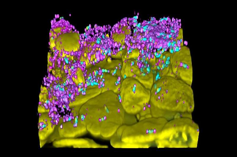
Bacteriophage (magenta) attack Pseudomonas aeruginosa (teal) biofilms grown in 
association with respiratory epithelial cells (nuclei, yellow). Credit: Paula Zamora, 

A team of micro- and immunobiologists from the Dartmouth Geisel School of Medicine, Yale University, and the University of Pittsburgh has found evidence suggesting that future research teams planning to use bacteriophages to treat patients with multidrug-resistant bacterial infections need to also consider how cells in the host's body respond to such treatment.

In their paper published in the open-access journal PLOS Biology, the group describes experiments they conducted that involved studying the way epithelial cells in the lungs respond to bacteriophages.

Over the past decade, medical scientists have found that many of the antibiotics used to treat bacterial infections are becoming resistant, making them increasingly useless. Because of this, other scientists have been looking for new ways to treat such infections. One possible approach has involved the use of bacteriophages, which are viruses that parasitize bacteria by infecting and reproducing inside of them, leaving them unable to reproduce.

To date, most of the research involving use of bacteriophages to treat infections has taken place in Eastern Europe, where some are currently undergoing . But such trials, the researchers involved in this new study note, do not take into consideration how cells in the body respond to such treatment. Instead, they are focused on determining which phages can be used to fight which types of bacteria, and how well they perform once employed.

The reason so little attention is paid to host cell interaction, they note, is that prior research has shown that phages can only replicate inside of the  they invade; thus, there is little opportunity for them to elicit a response in human cells.

In this new study, the research team suggests such thinking is misguided because it fails to take into consideration the  in the host. To demonstrate their point, the team conducted a series of experiments involving exposing human epithelial cells from the lungs (which are the ones that become infected as part of lung diseases) to bacteriophages meant to eradicate the bacteria causing an .

They found that in many cases, the immune system responded by producing proinflammatory cytokines in the . They noted further that different phages elicited different responses, and there exists the possibility that the unique properties of some phages could be used to improve the results obtained from such therapies. They conclude by suggesting that future  research involve inclusion of host cell response.

More information: Paula F. Zamora et al, Lytic bacteriophages induce the secretion of antiviral and proinflammatory cytokines from human respiratory epithelial cells, PLOS Biology (2024). DOI: 10.1371/journal.pbio.3002566


Journal information: PLoS Biology 


© 2024 Science X NetworkMammalian cells may consume bacteria-killing viruses to promote cellular health

Study details a common bacterial defense against viral infection


Complex of 2 proteins enhances blockage of phage replication


Peer-Reviewed Publication

OHIO STATE UNIVERSITY


COLUMBUS, Ohio – One of the many secrets to bacteria’s success is their ability to defend themselves from viruses, called phages, that infect bacteria and use their cellular machinery to make copies of themselves.

Technological advances have enabled recent identification of the proteins involved in these systems, but scientists are still digging deeper into what those proteins do.

In a new study, a team from The Ohio State University has reported on the molecular assembly of one of the most common anti-phage systems – from the family of proteins called Gabija – that is estimated to be used by at least 8.5%, and up to 18%, of all bacteria species on Earth.

Researchers found that one protein appears to have the power to fend off a phage, but when it binds to a partner protein, the resulting complex is highly adept at snipping the genome of an invading phage to render it unable to replicate.

“We think the two proteins need to form the complex to play a role in phage prevention, but we also believe one protein alone does have some anti-phage function,” said Zhangfei Shen, co-lead author of the study and a postdoctoral scholar in biological chemistry and pharmacology at Ohio State’s College of Medicine. “The full role of the second protein needs to be further studied.”

The findings add to scientific understanding of microorganisms’ evolutionary strategies and could one day be translated into biomedical applications, researchers say.

Shen and co-lead author Xiaoyuan Yang, a PhD student, work in the lab of senior author Tianmin Fu, assistant professor of biological chemistry and pharmacology at Ohio State.

The study was published April 16 in Nature Structural & Molecular Biology.

The two proteins that make up this defense system are called Gabija A and Gabija B, or GajA and GajB for short.

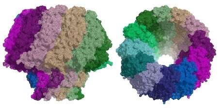
Researchers used cryo-electron microscopy to determine the biochemical structures of GajA and GajB individually and of what is called a supramolecular complex, GajAB, created when the two bind to form a cluster consisting of four molecules from each protein.

In experiments using Bacillus cereus bacteria as a model, researchers observed the activity of the complex in the presence of phages to gain insight into how the defense system works.

Though GajA alone showed signs of activity that could disable a phage’s DNA, the complex it formed with GajB was much more effective at ensuring phages would not be able take over the bacterial cell.

“That’s the mysterious part,” Yang said. “GajA alone is sufficient to cleave the phage nucleus, but it also does form the complex with GajB when we incubate them together. Our hypothesis is that GajA recognizes the phage’s genomic sequence, but GajB enhances that recognition and helps to cut the phage DNA.”

The large size and elongated configuration of the complex made it difficult to get the full picture of GajB’s functional contributions when bound to GajA, Shen said, leaving the team to make some assumptions about protein roles that have yet to be confirmed.

“We only know GajB helps enhance GajA activity, but we don’t yet know how it works because we only see about 50% of it on the complex,” Shen said.

One of their hypotheses is that GajB may influence the concentration level of an energy source, the nucleotide ATP (adenosine triphosphate), in the cellular environment – specifically, by driving ATP down upon detection of the phage’s presence. That would have the dual effect of expanding GajA’s phage DNA-disabling activity and stealing energy that a phage would need to start replicating, Yang said.

There is more to learn about bacterial anti-phage defense systems, but this team has already shown that blocking virus replication isn’t the only weapon in the bacterial arsenal. In a previous study, Fu, Shen, Yang and colleagues described a different defense strategy: bacteria programming their own death rather than letting phages take over a community.

This work was supported by the National Institute of General Medical Sciences.

Additional co-authors are Jiale Xie, Jacelyn Greenwald, Ila Marathe, Qingpeng Lin and Vicki Wysocki of Ohio State, and Wenjun Xie of the University of Florida.

#

Contact: Tianmin Fu, Fu.978@osu.edu

Written by Emily Caldwell, Caldwell.151@osu.edu; 614-292-8152

Friday, February 14, 2020

POLITICAL SCIENCE= EPSTEIN BARR

Cryo-electron microscopy opens a door to fight Epstein-Barr

Cryo-EM opens a door to fight Epstein-Barr
The Epstein Barr virus portal structure as found at eBIC. Credit: Diamond Light Source
The Epstein-Barr virus is one of the most widespread human viruses. Part of the herpesvirus family, it causes glandular fever (infectious mononucleosis), cancer and autoimmune diseases. At present, there is no treatment for infections caused by this virus. In work recently published in Nature Communications, scientists from the Institute for Research in Biomedicine (IRB Barcelona) and the Molecular Biology Institute of Barcelona (IBMB-CSIC) in Spain used cryo-electron microscopy (cryo-EM) to reveal the structure of a key protein, known as a portal, in the Epstein-Barr virus. Similarities between herpesviruses and tailed bacteriophages (viruses that infect bacteria) suggest that these two types of organism may be related. In a second paper published in the same journal, the team solved the structure of the portal protein in bacteriophage T7, using a combination of cryo-EM and X-ray crystallography. These results allowed them to infer how the Epstein-Barr virus portal works and may help in the development of a treatment for this virus.
In 2018, we brought you the news that high-resolution cryo-EM at eBIC had uncovered new information about a critical feature of the Herpes Simplex Virus. Cryo-EM has now worked its magic on the related Epstein-Barr virus, paving the way towards ways to defeat this untreatable virus.
The herpesvirus family is enormous and includes eight human pathogens. The Epstein-Barr virus infects B-cells (a type of white blood cell) and also the epithelial cells that make up skin and also line the inside of the throat, blood vessels and organs. It causes glandular fever (infectious mononucleosis) and can cause several kinds of cancer and autoimmune diseases.
All herpesviruses infect in a similar way. Once the virus has entered a cell and reached the nucleus, it releases its DNA. This DNA can lie dormant for many years until specific conditions trigger replication. When the virus replicates, the DNA is introduced into a new viral shell (capsid), forming a new virus capable of attacking other cells. The virus uses a protein called a portal for packaging its DNA into the viral capsid and to release it to the host cell during infection. As the portal plays a critical role in replication and infection, it makes an attractive target for the development of new anti-viral drugs.
The portal: an open and shut case?
The similarities between the capsid structure and viral DNA packaging mechanism of herpesviruses and tailed bacteriophages suggest that they may be related. Although researchers have been able to determine the structure of portal proteins from bacteriophages successfully, the study of herpesvirus portals has been more challenging. Scientists from the Institute for Research in Biomedicine (IRB Barcelona) and the Molecular Biology Institute of Barcelona (IBMB-CSIC) have now used cryo-EM at eBIC to reveal the structure of the portal protein in the Epstein-Barr virus at a resolution of 3.5 Å.
In a second study, the same team used a combination of cryo-EM and X-ray crystallography to characterise the structure of the portal protein in bacteriophage T7. Their work illustrates the power of using these techniques in conjunction to solve challenging molecular structures.
The bacteriophage also uses its portal to package its DNA inside a pro-capsid. The tail components then assemble on the portal to make an infectious virus. The ejection conduit remains tightly sealed until infection, when the channel opens to deliver the DNA to the host cell. All of the portals analysed to date for the Caudovirales family of bacteriophages share common structural features.
In search of antivirals
Miquel Coll is head of the Structural Biology of Protein & Nucleic Acid Complexes and Molecular Machines Lab at IRB Barcelona and a professor at IBMB-CSIC. He says;
"Understanding the structure of the portal protein could aid the design of inhibitors for the treatment of herpesvirus infections such as Epstein-Barr. As this protein is only found in herpesviruses, these inhibitors would be virus-specific and may be less toxic for humans."
Cristina Machón and Montserrat Fàbrega, postdoctoral fellows at IRB Barcelona and IBMB-CSIC are first authors on both papers. They say that "solving the structure of the portal  of bacteriophage T7 has allowed us to infer how the portal from Epstein-Barr virus works."
The drugs currently used to treat herpesvirus infections target the viral DNA polymerase. They are not very efficient, with serious side effects and the appearance of viral resistance after prolonged treatment. There is no specific treatment for the Epstein-Barr . Knowledge of the atomic structures of  proteins will be extremely valuable, allowing the -driven design of compounds targeting their function—highly specific anti-virals that should cause fewer side effects.

Tuesday, May 23, 2023

Public aware of and accept use of bacteria-killing viruses as alternative to antibiotics, study shows


Peer-Reviewed Publication

UNIVERSITY OF EXETER

The public are in favour of the development of bacteria-killing viruses as an alternative to antibiotics – and more efforts to educate will make them significantly more likely to use the treatment, a new study shows.

The antimicrobial resistance (AMR) crisis means previously treatable infections can kill. This has revitalised the development of antibiotic alternatives, such as phage therapy, which was first explored over a century ago but abandoned in many countries in favour of antibiotics.

The study shows public acceptance of phage therapy is already moderately high, and priming people to think about novel medicines and antibiotic resistance significantly increases their likelihood of using it.

There is a higher acceptance of phage therapy when described without using perceived harsh words, such as “kill” and “virus” but instead “natural bacterial predator”.

Those who took part in the survey had a high awareness of antibiotic resistance – 92 per cent had heard of antibiotic resistance, but only 13 per cent reported that they had heard about phage therapy prior to the survey.

Success and side effect rate, treatment duration, and where the medicine has been approved for use, influenced their treatment preferences.

The study was conducted by Sophie McCammon, Kirils Makarovs, Susan Banducci and Vicki Gold from the University of Exeter.

Dr Banducci said: “While phage therapy remains poorly understood by the UK public our research suggests there is extensive acceptance and support for its development. Exposure to only very limited information about antibiotic resistance and alternative treatments to antibiotics greatly increases the public acceptance of phage therapy.”

Dr Gold said: “Those involved in the research wanted to know more about phage therapy and were inspired to research this topic after completing our survey. Exposure to only a very limited amount of information about phage therapy significantly increases acceptance.”

Researchers held a workshop with experts and a review of phage research. They also fielded a survey assessing the UK public’s acceptance, opinions and preferences regarding phage therapy. A total of 787 people completed the survey, distributed in December 2021.

One group was given two scenarios; in the first they presented with a minor infection, and in the second they presented with an infection that did not respond well to antibiotics for three months. In each scenario, the group ranked the selected attributes based on their importance in deciding whether to accept a treatment or not.

Participants were randomly assigned one of four descriptions of phage therapy and then surveyed to assess their acceptance of the treatment. The acceptance of phage therapy was high across the board. However, describing phage therapy using perceived harsh words, such as “kill and “virus”, resulted in lower acceptance rates than alternative descriptions. Additionally, participants who had recent exposure to information about antibiotic resistance and alternative treatments were more accepting of phage therapy.

From the 787 participants who completed the survey, 213 left written responses expressing their opinions on the potential of phage therapy. Of this group, 38 per cent showed a specific interest in phage therapy development, while a further 17 per cent supported the development of antibiotic alternatives generally.

Sophie McCammon said: “An advantage of phage therapy is often the minimal side effects. Emphasising this through education and marketing may increase public acceptance of phage therapy.

“Even though phage therapy may be some years away from routine clinical use in the UK, increasing pressures from the AMR crisis require evaluation of the UK public’s acceptance of alternative treatments.

“The public desire for increased education is apparent. Expanding schemes which are interactively involving children in phage research not only generates excitement for the therapy now, but also promotes awareness in the generation likely to be treated with antibiotic alternatives.”