It’s possible that I shall make an ass of myself. But in that case one can always get out of it with a little dialectic. I have, of course, so worded my proposition as to be right either way (K.Marx, Letter to F.Engels on the Indian Mutiny)
Building on the momentum of the 6th World Congress on Targeting Phage Therapy, that gathered more than 150 attendees from over 30 countries and featured over 71 presentations, the highly anticipated Targeting Phage Therapy 2024 is set to unfold.
Mark Your Agendas for the 7th World Congress on Targeting Phage Therapy
Date: June 20-21, 2024
Location: Corinthia Palace, Malta
What to Expect:
Cutting-edge insights into phage therapy advancements and its potential to revolutionize medicine.
Engaging keynotes and expert panels tackling current challenges head-on.
Focused discussions on regulatory frameworks, phage selection, and the critical role of clinical trials.
Gain insights into the direction of Targeting Phage Therapy 2024 by exploring the concluding remarks of 2023.
How to contribute?
We welcome submissions for innovative sessions and talks. If you have groundbreaking insights to share, be part of shaping tomorrow's medical landscape.
A Look Back at Targeting Phage Therapy 2023: Award Winners
1. Best Scientific Contribution
Martha Clockie, Editor in Chief of PHAGE Journal, University of Leicester, United Kingdom
Topic: Challenges and Opportunities for Bacteriophage Therapy
2. Best Scientific Innovation
Amanda (Curtis) Burkardt, CEO of PHIOGEN, USA
Topic: Creating Patient Ready Products in a Remedy Ready World.
3. Best Short Oral:
Brieuc Van Nieuwenhuyse, UC Louvain, Belgium
Topic: Bacteriophage-Antibiotic Combination to Allow Liver Transplantation
4. Best Poster:
Pantiora Panagiota, Agricultural University of Athens, Greece
Revisit Targeting Phage Therapy 2023: Replay is Available
Explore the Targeting Phage Therapy 2023 replay to preview what's in store for 2024. Whether you missed the event or want to rewatch it, the replay is available. Access 40+ talks and innovations from key industries like Phiogen, Armata Pharmaceuticals, BiomX, Cellexus, and more.
The Abstracts Book is also accessible for additional insights.
Wishing you a joyous holiday season, we anticipate the pleasure of meeting you at Targeting Phage Therapy 2024 in Malta. For more information about the event, please visit our website.
DR VIJINI MALLAWAARACHCHI, RESEARCH ASSOCIATE IN BIOINFORMATICS, FLINDERS ACCELERATOR FOR MICROBIOME EXPLORATION (FAME) LAB, COLLEGE OF SCIENCE AND ENGINEERING, FLINDERS UNIVERSITY
A new bioinformatics software program at Flinders University is paving the way for a rapid expansion of research into bacteriophages, the viruses or phages that play key roles in controlling bacteria.
Experts at the Flinders University College of Science and Engineering have released a computational tool for researchers around the world to find‘bacteriophages’ or phages through more accurate genome sequencing.
Research into isolating and harnessing bacteriophages paves the way for progress in the emerging field of ‘phage therapy’, a more natural way to target specific bacteria which post a constant risk to immune-compromised, young and elderly patients, as well as ‘super’ bacteria which has become resistant to regular antibiotics.
Antimicrobial resistance (AMR) is a major global risk when broad-spectrum antibiotics no longer work on ‘superbugs’ created when common bacteria goes through multiple genetic changes. The WHO has warned that AMR is one of the top public health threats facing humanity in the 21st century and was associated with the death of close to 5 million people in 2019.
“Traditional methods of studying phages from environmental sequencing data have been limited, often failing to fully capture the complete genetic information of phages. This incomplete picture has been a barrier to fully understanding their roles and impacts.”
FAME Lab director Professor Robert Edwards, a coauthor of the latest article, says the Phables software can computationally reconstruct the genetic content of phages from environmental sequencing data.
“This marks a major advancement in phage bioinformatics, allowing us to computationally reconstruct complete phage genomes,” says Professor Edwards, from the College of Science and Engineering.
“It will facilitate the discovery of novel phages and enable their laboratory isolation, which will lead to advancements in medical treatments, environmental management, and a deeper understanding of microbial life.
“This revolutionary tool not only enhances our understanding of the microbial world but also paves the way for innovative solutions to some of the most pressing health and environmental challenges of our time.”
Phables uses a new, more effective approach to piece together the genetic information of phages with tests on various datasets showing the new tool can identify more complete contiguous genomes of phages than existing state-of-the-art software tools.
Phables has almost 9000 downloads across different software repositories. The tool was launched at the Australian Society for Microbiology Annual National Meeting 2023 and the Australian Bioinformatics and Computational Biology Society Conference 2023.
Next year, the Flinders University research team aims to use the Phables tool to discover novel phages, and potentially use these isolated phages in therapies, including new treatment options for individuals with conditions such as cystic fibrosis and inflammatory bowel disease.
Read the article – Phables: From fragmented assemblies to high-quality bacteriophage genomes (2023) by Vijini Mallawaarachchi, Michael J Roach, Przemyslaw Decewicz, Bhavya Papudeshi, Sarah K Giles, Susanna R Grigson, George Bouras, Ryan D Hesse, Laura K Inglis, Abbey L K Hutton, Elizabeth A Dinsdale, and Robert A Edwards has been published in Bioinformatics (Oxford University Press).First published 21 September 2023.DOI: 10.1093/bioinformatics/btad586
Acknowledgements: This work was supported by the National Institutes of Health (NIH), the Australian Research Council [DP220102915], and the Polish National Agency for Academic Exchange.
OKINAWA INSTITUTE OF SCIENCE AND TECHNOLOGY (OIST) GRADUATE UNIVERSITY
VIDEO:
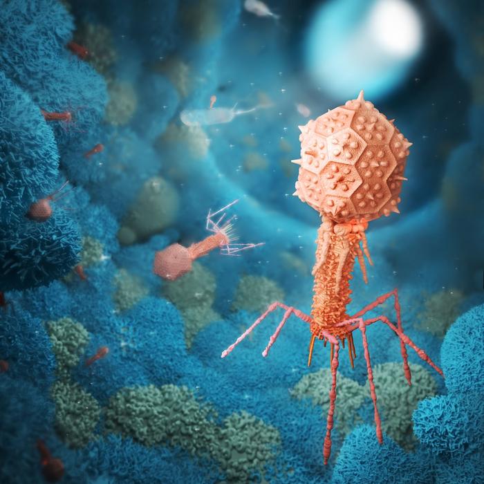
STRUCTURE OF AN ENTIRE DT57C BACTERIOPHAGE. THE ENTIRE VIRUS WAS RECONSTRUCTED IN THREE DIMENSIONS AT NEAR-ATOMIC RESOLUTION AND VISUALIZED IN THIS ANIMATION.
The word “virus” is often associated with negative connotations. However, it is important to note that not all viruses are harmful. In fact, there are many viruses that live inside our bodies and play important roles in our health. One example is bacteriophages, viruses that infect bacteria and can be used to keep bacterial infections under control.
These viruses are known to have more complex shapes and have not been studied in full detail at the atomic level before. They can be engineered to better suit applications of human interest, such as providing an alternative to the use of antibiotics.
Scientists at the Okinawa Institute of Science and Technology (OIST) together with their international collaborators at MSU Moscow and Shenzhen, and Academia Sinica in Taiwan, have studied the molecular architecture of the tequintavirus, also known as T5-like bacteriophages, to understand how these viruses are organized at a molecular level. T5 viruses are nonenveloped viruses with a head that has an icosahedral shape and contains the viral DNA, and a non-contractile flexible tail which acts as the channel for DNA injection into the bacterial host cell.
The scientists obtained atomic models for all structural components of the virus. This is the first time that a tailed virus with a flexible tail has been revealed in its entirety at this level of detail. The results of their study have been published in the journal Nature Communications and set the basis for future studies on the mechanism of infection of these viruses.
“To engineer and modify these viruses efficiently for specific purposes, we must know their organization at an atomic level and the mechanisms through which they infect their target bacteria. For these reasons, we decided to use cryo-electron microscopy to visualize the DT57C bacteriophage at high-resolution in its entirety,” Prof. Matthias Wolf, head of the Molecular Cryo-Electron Microscopy Unit, explained.
Researchers working on phage therapies which use bacteriophages to treat bacterial infections in agricultural crops, fish aquaculture and other fields, can benefit from the results of this study. “The structural knowledge we have obtained can enable the engineering of bacteriophages with improved ability to kill these bacterial pathogens,” Prof. Wolf added.
Does this mean that bacteriophages are ‘good’ viruses? Dr. Rafael Ayala, lead author of the research paper, explained that these viruses are ‘good’ when their actions benefit us and ‘bad’ when they cause us harm, as is the case with bacteria.
An example of how bacteriophages can benefit us is their use in gene therapy. “One of the ways to distribute genes to cells is to put them into a human virus that has been modified in two ways, first to not cause disease, and second to also carry the genes that you want to introduce to cure a specific disease. In this way the virus is used as a vehicle to introduce a cure,” Dr. Ayala said.
One of the main challenges of the research was to reconstruct the bacteriophage as a whole from electron micrographs in significant detail, and not just some of its components. The DT57C bacteriophage comprises a head, a neck, a tail and a baseplate at the end of the tail. Many of these components are flexible and can move freely, which makes it difficult to visualize their molecular architecture in detail, similar to how difficult it is to take a good photo of an object that is moving fast.
To deal with this, the researchers developed new methods that they plan to apply to other viruses with complex shapes. “We had to think of new ways to tackle the problems we encountered, and we believe that the methods developed in this study will be of interest to many researchers studying viruses,” Dr. Ayala explained. “Phage therapy is an active area of research, and it is very likely that we are going to see these treatments in our lifetime.”
Using viruses to modify bacteria is a huge area of interest because bacteria are at the core of many natural and engineered processes, including nutrient recycling, symbiosis, bioremediation (bacteria are used to clean up environmental pollutants), and food production. This research will be useful in designing viruses to combat bacterial diseases that affect humans, plants and other organisms.
An electron micrograph of the DT57C bacteriophage (IMAGE)
OKINAWA INSTITUTE OF SCIENCE AND TECHNOLOGY (OIST) GRADUATE UNIVERSITY
An electron micrograph — a photograph taken by means of a transmission electron microscope — of the DT57C bacteriophage. These types of images have been used to obtain a three-dimensional structure of the entire virus. Scale bar: 80 nanometers
Scientists have discovered what may be the first 'vampire' virus
SHOULD NAME IT BELALUGOSIUM
Carolyn Y. Johnson Updated Tue, November 14, 2023 The discovery started with an undergraduate class designed to teach students basic laboratory techniques, asking them to isolate phages from soil samples and study them using genetics.
(Getty Images) (Getty Images)
In March 2020, Tagide deCarvalho saw something truly strange - something she thinks no other scientist has ever seen before: a virus with another, smaller virus latched onto its "neck." The backstory of this viral attachment is like a master class in how wild and weird biology can be.
The two microbes are both bacteriophages, viruses that infect bacteria, that were harvested from a clump of dirt in Poolesville, Md. Bacteriophages, also called simply phages, are among the most abundant organisms on Earth. There can be millions in a gram of dirt.
But with a special kind of microscope that uses a beam of electrons to capture images, deCarvalho witnessed a truly bizarre moment - kind of like a wildlife photographer who captures an animal behavior that no one had anticipated.
"I could see literally hundreds of them had this little guy attached at the neck, and it was clearly not random," said deCarvalho, who manages the Keith R. Porter Imaging Facility at University of Maryland at Baltimore County. "We know that viruses can do some amazing, interesting things. But this is just another new thing that no one could have predicted we would see."
In a recent study in the Journal of the International Society for Microbial Ecology, deCarvalho and colleagues explain how the viral odd couple likely came to be. The small virus, called MiniFlayer, lost the ability to make copies of itself inside cells, which is how viruses reproduce. So evolution devised a clever, parasitic workaround. MiniFlayer takes advantage of another virus, dubbed MindFlayer, by grabbing onto its neck, and when they enter cells together, MiniFlayer utilizes its companion's genetic machinery to proliferate.
Is it an embrace? A stranglehold? DeCarvalho compares the relationship to viral hitchhiking. Her collaborator, Ivan Erill, a computational biologist at UMBC, likens it to a vampire sinking its teeth into its prey. It's not a perfect analogy, but he notes that sometimes, when they find MindFlayer alone, they can find "bite marks" where MiniFlayer's tendrils were attached.
"Viruses will do anything. They are the most creative force of nature," Erill said. "If anything is possible, they will come up with a way to do it. But no one had anticipated that they would do something like this."
The strange universe of viruses
The discovery started with an undergraduate class designed to teach students basic laboratory techniques, asking them to isolate phages from soil samples and study them using genetics. DeCarvalho has been working with the program for seven years and says that for many of the students, seeing the phage is an exciting moment, like when expecting parents see the ultrasound of a fetus for the first time.
In this case, undergraduates Jenell Lewis and Hira Ahmed isolated and named their phage MindFlayer in 2019. But genome sequencing returned puzzling results, suggesting some kind of contamination. When deCarvalho looked at it with a microscope, she noticed not one phage, but two.
The "virosphere," as scientists call the strange universe of viruses, is known to include elements called "satellites" that have lost their ability to replicate inside cells. Usually, satellites overcome this deficiency by integrating into the genome of the cells that they infect. They lurk there until another virus, a "helper" that has the missing ingredients, happens to enter the cell. The satellites then seize the opportunity to make copies of themselves.
MiniFlayer is a satellite, but unlike the typical version, it doesn't have the ability to hide inside cells. That leaves it with a conundrum: How to make sure it ends up in the cell with its helper at the same time.
"What this virus has done is say, okay, I'm going to attach to my helper, attach to its neck - and travel with my helper until we find a new cell," Erill said.
This is par for the course in microbiology, where tactics like molecular piracy and hijacking have been honed over millions of years of evolution. Bacteria are wildly outnumbered by their viral predators, putting them in an ongoing evolutionary arms race. Bacteria develop defenses, and viral phages develop counter-defense strategies. Phages parasitize other phages.
Researchers are interested in using phages, the natural predators of bacteria, as medicine. Phage therapy can be used to target harmful infections, an approach that could become more important as antibiotic-resistant bacteria have become a growing threat.
Terje Dokland, a microbiology professor at the University of Alabama at Birmingham who was not involved in the study, said the observation of the two attached phages was "intriguing" but called for more images and research to draw firmer conclusions about the interaction, and to tease out whether the two viruses are really co-infecting cells.
The authors hope to collaborate with groups that use a different form of electron microscopy to understand what's happening more clearly. Unlike a vampire, deCarvalho points out, the MiniFlayer isn't sucking something out of MindFlayer.
"We don't know whether or not the satellite is injecting its DNA into the helper or if it's just hitchhiking along for a ride and then falling off, like a tick," deCarvalho said. "Hopefully someone else will pick up this work and figure out that really interesting question."
IN LABORATORY FLASKS CONTAINING JUST TWO TEASPOONS OF MEDIA, SCIENTISTS DOCUMENT HOW RAPID ADAPTATION BETWEEN BACTERIA AND VIRUSES PRODUCE COMPLEX ECOLOGICAL NETWORKS.
As conceived by Charles Darwin in the 1800s, evolution is a slow, gradual process during which species adaptations are inherited incrementally over generations. However, today biologists can see how evolutionary changes unfold on much more accelerated timescales.
Rather than the evocative plants and animals of the Galapagos Islands that Darwin studied in forming his theory of evolution, Postdoctoral Scholar Joshua Borin and Associate Professor Justin Meyer of UC San Diego’s School of Biological Sciences are documenting rapid evolutionary processes in simple laboratory flasks.
Borin and Meyer set bacteria and viruses together in a closed laboratory flask — just two teaspoons large — to study coevolution in action. As viruses infect their bacterial neighbors, the bacteria evolve new defensive measures to repel the attacks. The viruses then counter these adaptations with their own evolutionary changes that work around the new defensive measures.
In only three weeks, this accelerated arms race between bacteria (Escherichia coli) and viruses (bacteriophage, or “phage”) results in several generations of evolutionary adaptations. The new findings, published in the journal Science, reveal the emergence of distinct evolutionary patterns.
“In this study we show the power of evolution,” said Meyer, an associate professor in the Department of Ecology, Behavior and Evolution. “We see how coevolution between bacteria and phage drive the emergence of a highly complicated ecological network. Evolution doesn’t have to be slow and gradual as Darwin thought.”
Meyer says the new study offers fresh perspectives on how intricate ecological networks develop across disparate ecosystems, whether they are food webs across the savanna, pollinator networks in the rainforest or microbes interacting in the ocean.
As bacteria and viruses adapted to each other’s presence over time, two prominent repeating patterns emerged. These included nestedness, a development in which narrow interactions between bacteria and virus specialists are “nested” within a broader range of generalist interactions; and modularity, in which interactions between species form modules within specialized groups, but not between groups.
“We were amazed to discover that our evolution experiment in tiny flasks had recapitulated the complex patterns that had been previously observed between bacteria and viruses collected at regional and transoceanic scales,” said Borin.
“When our research team first quantified this multiscale pattern in environmental bacteria and phage interaction data, we thought the emergence of such complexity required long periods of evolution,” added study coauthor Professor Joshua Weitz from the Department of Biology at the University of Maryland.
Meyer says capturing these evolutionary developments “in action” reinforces the power of evolution, which is often underestimated. Rapid pathogenic evolution continues to shape our world in new ways. Through COVID-19 and new mutations of SARS-CoV-2, viruses have demonstrated the potent capability for evolutionary adaptations that result in new strains when they encounter antibodies, vaccines and other roadblocks that keep them from effectively infecting and spreading. Such new concepts in microbial evolution are reframing the way patients are treated.
“We show that evolution can produce complex ecological networks quickly from very little external help,” said Meyer, who indicated that examples of such external evolutionary forces include isolation via geographical distance, environmental drivers and interactions with other species. “So we can use phage and bacteria as a model system to understand general evolutionary principles and help show how life on Earth has evolved into such diverse and complex ecosystems from simple beginnings.”
In related work, Meyer and Weitz are using artificial intelligence to study how phage could be used in the growing antibiotic resistance crisis. The research includes analysis of evolutionary data to determine which mutations in phage and bacteria can lead to infection and resistance. The research also highlights a new effort supported by the Howard Hughes Medical Institute to study how “jumbo” phages could be used as new therapeutic agents.
Coauthors of the Science paper include Joshua Borin, Justin Lee, Adriana Lucia-Sanz, Krista Gerbino, Joshua Weitz and Justin Meyer.
Phage interactions with bacteria are well known, and interactions between bacteria and their mammalian host can lead to a range of symbioses. However, the impact of bacteriophages on mammalian cellular and immunological processes is not well understood. Now, a new study by researchers at Monash University suggests that mammalian cells internalize phages as a resource to promote cellular growth and survival.
“There is a growing appreciation that the direct interaction between bacteriophages and the mammalian host can facilitate diverse and unexplored symbioses,” wrote the researchers. “Yet the impact these bacteriophages may have on mammalian cellular and immunological processes is poorly understood. Here, we applied highly purified phage T4, free from bacterial byproducts and endotoxins to mammalian cells and analyzed the cellular responses using luciferase reporter and antibody microarray assays.”
In order to investigate how mammalian cells’ immune responses interact with and are modulated by interactions with phages, researchers led by Jeremy J. Barr, PhD, associate professor at Monash University, applied phage T4 to mammalian cells in vitro and analyzed the cellular responses using luciferase reporter and antibody microarray assays.
The researchers found that T4 phages did not activate DNA-mediated inflammatory pathways, but triggered a sequence of signaling pathway events that promote cellular growth and survival.
“Highly purified T4 phages were rapidly internalized by mammalian cells and accumulated within macropinosomes but did not activate the inflammatory DNA response TLR9 or cGAS-STING pathways,” noted the researchers. “Following 8 hours of incubation with T4 phage, whole cell lysates were analyzed via antibody microarray that detected expression and phosphorylation levels of human signaling proteins. T4 phage application led to the activation of AKT-dependent pathways, resulting in an increase in cell metabolism, survival, and actin reorganization, the last being critical for macropinocytosis and potentially regulating a positive feedback loop to drive further phage internalization.”
Future studies are needed to determine why cells use phage particles as resources, and whether they have specifically evolved via adaptation to benefit from this internalization.
According to the authors, “This preliminary study provides novel insights into the impact phages have on mammalian systems, with broader potential implications across the fields of immunology, phage therapy, microbiome, and human health.”
Barr added, “This work provides new insights into the additional benefits that bacteriophages may have on their mammalian hosts. This is of particular importance given the increased use of phage therapy to treat antibiotic-resistant infections.”
Pioneering work to develop effective and safe bacteriophages to combat disease has received an £800,000 boost.
The grant from the Biotechnology and Biological Sciences Research Council (BBSRC), is aimed at advancing the production of phages to combat disease in the veterinary field and bring them to market.
It has been awarded to Professor Martha Clokie, the Director of the Leicester Centre of Phage Research, and Dr Anisha Thanki who earlier this year successfully developed a bacteriophage ‘liquid’ product to prevent Salmonella in broiler chickens.
The latter will now be used as a case study to advance ways in which this novel medicine can successfully and safely be produced in larger scales to meet UK guidelines.
Bacteriophage are viruses that infect bacteria and kill them. They are naturally occurring in the environment around us and can be found where high numbers of bacteria lurk. They have been identified by the UK Government and World Health Organisation as having great potential to prevent and treat infections.
Researcher, Dr Anisha Thanki helped develop the product to prevent Salmonella and will continue with this next stage.
She said: “We know that the development of bacteriophages will help counter growing resistance to existing antimicrobials. If a product such as this was eventually commercialised, it could save the farming industry billions of pounds each year while preventing Salmonella from entering our food chain – something which infects around 91,000 people in the EU every year.
“However, at present we have an effective product but no known way to bring it into wider commercial use. The work we’re doing is so novel that protocols and regulations don’t yet exist to allow that to happen. We’re very excited that this funding will allow us to translate this work to establish how to use phages effectively at a much larger scale and within UK regulation guidelines.
“Once we do this, we aim to have a successful blueprint to enable other effective phage products to be brought to the commercial market.”
Work on the two-year project begins early next year and will take place in collaboration with Dr Robert Atterbury from the University of Nottingham’s School of Veterinary Medicine and Science.
Dr Thanki added: “Working with the school will allow us to develop further models to study phage production on a larger scale and test production protocols to ensure its efficacy and safety.”
Dr Robert Atterbury, Associate Professor in Microbiology at the University of Nottingham said: “Antimicrobial resistance is one of the key global public health challenges of the 21st century. Bacteriophages show great promise in the treatment of infections caused by multidrug resistant bacteria in animals and people. This exciting project, supported by the BBSRC, will allow us to address some of the key hurdles currently preventing their wider use in the agrifood sector and beyond.”
Bacteriophage used within the Salmonella trial, published in scientific journal, Emerging Microbes and Infections, was developed in the University’s pioneering new Leicester Centre for Bacteriophage Research which is studying bacteriophage-based products to prevent and treat bacterial infections in humans, animals and agriculture.