It’s possible that I shall make an ass of myself. But in that case one can always get out of it with a little dialectic. I have, of course, so worded my proposition as to be right either way (K.Marx, Letter to F.Engels on the Indian Mutiny)
MAGA influencer Steve Bannon is calling on President Donald Trump to ban all immigration into the United States for at least 10 years
During a Monday interview with MyPillow CEO Mike Lindell on Real America's Voice, Bannon used alleged fraud in the daycare industry to push for Somali immigrants to be deported.
"They're stealing money and laughing in your face," Bannon said. "When somebody shows up, they got a million-in-one excuse, and they play dumb: 'Oh, I don't know, you're white, I don't speak the language.' They've got to all be deported."
"If you can't figure out there's $9 billion missing, you're in the wrong business," Lindell, a candidate for Minnesota governor, agreed. "And then transparency, Steve, I'll tell you what, I'll be the most transparent governor this country's ever seen. The people are going to know where the problems lie, and then we fix them."
Bannon then told anti-immigration activist Rosemary Jenks that Trump should implement "a 10-year moratorium on immigration." "No immigration!" he insisted. "Not just getting rid of the H-1B visas, but moratoriums across the board on no immigration for a decade until we get this mess sorted out."
"I think most Americans would say, yeah, okay, what's wrong with that?" Jenks replied. "Nothing at all."
"We're done. It's over. We're not doing this anymore. We are not paying taxes so that you can throw our money away. We need a moratorium," she added. "It would go probably longer, for 10 years actually."
"You got to get rid of H-1Bs today and ship them all home and give those jobs for American citizens," Bannon agreed. "And then a 10-year moratorium on every other program."
"That's how long it's going to take to take this thing back to the basics and reconstruct it."
'Twisted homunculus' Stephen Miller nailed for his 'dumb as a box of hair' hate screeds
A series of anti-immigrant posts by Donald Trump adviser Stephen Miller over the holiday weekend got a swift dismissal by longtime conservative Charlie Sykes, who questioned the intelligence of the president’s resident “evil genius.”
On his Substack platform, Sykes noted the controversial Miller making broad-based attacks on immigrants, including writing what Sykes sarcastically characterized as a “Deep Thought.”
Miller wrote he watched a Christmas special with his family that featured Frank Sinatra and Dean Martin and commented, “Imagine watching that and thinking America needed infinity migrants from the third world.”
Critics have fired back at Miller and pointed out, “Enjoy your racism grift while it lasts. Dean Martin’s dad came here from Italy at a time when Italy was considered third-world. Both of Frank Sinatra’s parents came here from Italy.”
Sykes pointed out that Miller wasn’t done sneering.
“Someone should write an alternate historical novel where Americans are the first to master the automobile, the first in flight, the first to harness the atom, the first to land on the moon — but just keep going and never open our borders to the entire third world for sixty years,” Miller claimed — ignoring the substantial number of foreign scientists who made major contributions to the Manhattan Project.
That led Sykes to launch a broadside at Trump’s key adviser.
“My contrarian take: I hope Stephen Miller goes on tweeting, because it exposes not merely his rancid bigotry (which was known), but also his invincible ignorance, because it turns out that this twisted homunculus — the evil genius behind Trump’s mass deportations — is actually as dumb as a box of hair,” he wrote.
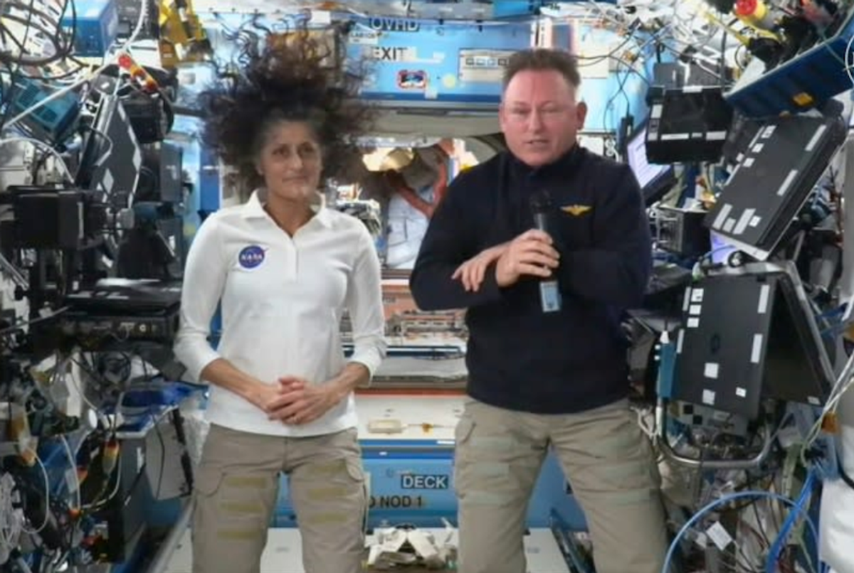
NASA again delays return of astronauts stranded on space station
Agence France-Presse December 18, 2024 U.S. astronauts Suni Williams (L) and Butch Wilmore took off aboard Boeing's Starliner in June 2024 for what was meant to be an eight-day mission -- they now will not return until spring 2025 (Handout)
Two U.S. astronauts stranded for months on the International Space Station will remain there at least until late March, NASA said Tuesday as it announced another delay in the mission to bring them home.
Veteran astronauts Butch Wilmore and Suni Williams arrived at the ISS in June aboard Boeing's Starliner spacecraft, and were due to spend eight days on the orbiting laboratory.
But problems arose with the Starliner's propulsion system during the flight there, so NASA opted for a big change in plans.
After weeks of intensive tests on the Starliner, the space agency decided to return it to Earth without its crew, and to bring the two stranded astronauts back home with the members of a SpaceX mission called Crew-9.
Crew-9's two astronauts arrived at the ISS aboard a Dragon spacecraft in late September, with two empty seats for Wilmore and Williams. The plan was for all four to return home in February 2025.
But NASA said Tuesday that Crew-10, which would relieve Crew-9 and the stranded pair, would now launch no earlier than March 2025, and both teams would remain on board for a "handover period."
"The change gives NASA and SpaceX teams time to complete processing on a new Dragon spacecraft for the mission," NASA said in a blog post.
The bottom line is that Wilmore and Williams will spend more than nine months in space, rather than eight days as initially planned.
SpaceX, the private company founded by billionaire Elon Musk, has been flying regular missions every six months to allow the rotation of ISS crews. How to catch a supernova explosion before it happens – and what we can learn from it
Stars are born, live and die in spectacular ways, with their deaths marked by one of the biggest known explosions in the Universe. Like a campfire needs wood to keep burning, a star relies on nuclear fusion — primarily using hydrogen as fuel — to generate energy and counteract the crushing force of its own gravity.
But when the fuel runs out, the outward pressure vanishes, and the star collapses under its own weight, falling at nearly the speed of light, crashing into the core and rebounding outward. Within seconds, the star is violently blown apart, hurling stellar debris into space at speeds thousands of times faster than the most powerful rocket ever built. This is a supernova explosion.
Astronomers aim to understand what types of stars produce different kinds of explosions. Do more massive stars result in brighter explosions? What happens if a star is surrounded by dust and gas when it explodes?
While we have simulations modeling a star’s death, they are difficult to validate. Observing a star’s behavior in real-time before the explosion could help answer these questions — but finding such a star is no easy task.
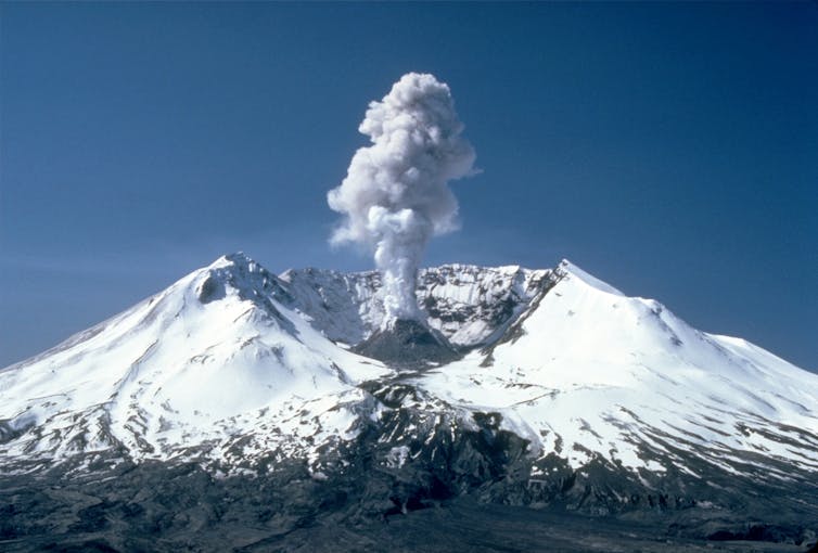
Scientists already do this with eruptions on Earth. Volcanologists monitor volcanoes, measuring changes in activity to predict an upcoming eruption. For example, in March 1980, Mount St. Helens in the US began to show some precursor events, such as seismic activity, and dozens of steam eruptions ejecting ash and gas into the atmosphere.
Plumes of steam, gas, and ash often occurred at Mount St. Helens in the early 1980s. Wikipedia
Two months later, an earthquake triggered the largest landslide ever recorded, releasing built up pressure in the magma chamber, resulting in a catastrophic eruption that devastated an area of almost 232 square miles (600 square kilometers).
Pre-supernova eruptions
Massive stars – larger than around 10 times the mass of the Sun – can do the same thing, albeit at much larger scales. In 2009, astronomers observed a bright event 65 million light years away that on first impressions resembled a supernova explosion.
Dubbed SN 2009ip, the explosion did not brighten as expected and was reclassified shortly after discovery as a “supernova impostor” – a giant eruption which ultimately does not destroy the star.
Over the next three years, the star underwent many rapid “flickering” events, a bit like quickly turning on and off a light bulb. Finally, in 2012, an unexpected supernova occurred. The evolution of the supernova explosion is still being studied to this day, and what exactly happened from 2009 to 2012 remains a mystery.
In a recent paper, published in Astronomy and Astrophysics, our team found a peculiar star in the Virgo Cluster, coincidentally also 65 million light years away. Unlike SN 2009ip, the star lacked hydrogen and was composed primarily of helium. The star was observed very slowly increasing its brightness for over five years – akin to slowly turning on a bulb using a dimmer switch – before a supernova was observed.
The supernova, labelled as SN 2023fyq, provided astronomers with a rare opportunity to capture the first light from the supernova explosion, known as shock breakout, from observatories worldwide and in space, largely due to the daily monitoring of the precursor activity.
Clash with current theory
This precursor activity offers an exciting chance to uncover the mysteries of supernova explosions, shedding light on both the conditions leading up to and following these cosmic events.
The underlying cause of this pre-supernova activity remains unclear. It is thought that an isolated massive star does not experience such rapid fluctuations in brightness. In the final moments of a star’s life, its core undergoes rapid evolution, desperately attempting to counteract the crushing force of gravity with its dwindling fuel reserves.
However, the star is so large at this stage that any activity in the core doesn’t have enough time to reach the surface. Observing these dramatic changes, occurring so close to the star’s demise, present a significant challenge to current theories.
One compelling hypothesis points to the interaction of multiple stars. Stars are born in dense clouds of gas and dust where multiple stars can form in close proximity. Neighboring stars may interact gravitationally with one another – exchanging material as they orbit each other.
This mass transfer could account for the changes in brightness observed in SN 2009ip before its explosion and the hydrogen deficiency seen in SN 2023fyq. The companion involved might be another massive star – or perhaps a more exotic object, such as a black hole.
We know not all eruptions will not end in a supernova explosion. For example, in the 1840s, Eta Carinae – a star 100 times larger than the Sun – experienced the “Great Eruption” launching 30 times the Sun’s mass into space. Although this was an extremely energetic explosion, the massive star was not destroyed.
Do all stars announce their departure? We aren’t sure. Seemingly normal supernovas have been observed with precursor eruptions, thanks in part to deep observations catching the faint precursor activity.
In 2025, the Vera C. Rubin Observatory, equipped with the world’s largest camera, will begin to study these events. At 3,200-megapixels, it is over 40 times more sensitive than cameras we have available on Earth, providing the opportunity to search for fainter precursor activity.
At Stockholm University, our team is currently using telescopes from the European Southern Observatory and the Zwicky Transient Facility, including the Nordic Optical Telescope in La Palma, Spain and the Very Large Telescope at Cerro Paranal in the Atacama Desert of northern Chile, to identify the signs that indicate a star is nearing the end of its life.
By recognising these signals, we can alert the scientific community and be ready to watch as a star experiences its final, dramatic moments.
This article is republished from The Conversation under a Creative Commons license. Read the original article. Human settlement of Mars isn’t as far off as you might think
Could humans expand out beyond their homeworld and establish settlements on the planet Mars? The idea of settling the red planet has been around for decades. However, it has been seen by sceptics as a delusion at best and mere bluster at worst.
Yet in an era where space tourism has become possible, the red planet has emerged as a dreamland for rich eccentrics and techno utopians. As is often the case with science communication, there’s a gulf between how close we are to this ultimate goal and where the general public understands it to be.
However, I believe there is a rationale for settling Mars and that this objective is not as far off as some would believe. There are actually a few good reasons to be optimistic about humanity’s future on the red planet.
First, Mars is reachable. During an optimal alignment between Earth and Mars as the two planets orbit the Sun, its possible to travel there in a spacecraft in six to eight months. Some very interesting new engine designs suggest that it could be done in two months. But based on technology that’s ready to go, it would take astronauts six months to travel to Mars and six months back to Earth.
Astronauts have already stayed for this long on the International Space Station (ISS) and on the Soviet orbiting lab Mir. We can get there safely and we have already shown that we can reliably land robots on the surface. There’s no technical reason why we couldn’t do the same with humans.
Second, Mars is abundant in the raw materials required for humans to “live off the land”, in other words, achieve a level of self sufficiency. The red planet has plentiful carbon, nitrogen, hydrogen and oxygen which can be separated and isolated, using processes developed on Earth. Mars is interesting and useful in a multitude of ways that the moon isn’t. And we have technology on Earth to enable us to stay and settle Mars by making use of its materials.
A third reason for Mars optimism is the radical new technology that we can put to use on a crewed mission to the planet. For example, Moxie (Mars Oxygen In-Situ Resource Utilization Experiment) is an project developed by scientists at the California Institute of Technology (Caltech) that sucks in Martian atmosphere and separates it into oxygen. Byproducts of the process – carbon monoxide, nitrogen and argon – can be vented.
When scaled up, similar machines would be able to separate oxygen from hydrogen to produce breathable air, rocket fuel and water. This makes it easier to travel to the planet and live on the surface because it’s not necessary to bring these commodities from Earth – they can be made once on Mars. Generating fuel on the surface would also make any future habitat less reliant on electric or solar-powered vehicles.
But how would we build the habitats for our Mars settlers? Space architect Melodie Yasher has developed ingenious plans for using robots to 3D print the habitats, landing pads and everything needed for human life on Mars. Using robots means that these could all be manufactured on Mars before humans landed. 3D printed homes have already been demonstrated on Earth.
Volunteers have also spent time living in simulated Mars habitats here on Earth. These are known as Mars analogues. The emergency medicine doctor Beth Healey spent a year overwintering in Antarctica (which offers many parallels with living on another planet) for the European Space Agency (Esa) and communicates her experience regularly.
She is not alone, as each year sees new projects in caves, deserts and other extreme environments, where long term studies can explore the physical and psychological demands on humans living in such isolated environments.
Finally, the Mars Direct plan devised by Dr Robert Zubrin has existed for more than 30 years, and has been modified to account for modern technology as the private sector has grown. The original plan was based on using a Saturn V rocket (used for the Apollo missions in the 1960s and 1970s) to launch people. However, this can now be accomplished using the SpaceX Falcon 9 rocket and a SpaceX Dragon capsule to carry crew members.
Elon Musk wants to use his Starship vehicle to establish large settlements on Mars. SpaceX, CC BY-NC
Several uncrewed launches from Earth could ferry necessary equipment to Mars. These could include a vehicle for crew members to return on. This means that everything could be ready for the first crew once they arrived.
For astronauts making the journey to Mars, radiation is the biggest problem. But using certain materials in the walls of the spacecraft or building a protective shelter inside the vehicle could shield astronauts from high energy particles. Similar ideas could apply to 3D printed habitats on the Martian surface. Alternatively, settlers could live underground or in Martian caves.
On Mars, there’s a 24-minute communication delay with Earth. This means that Martians will need to be self-sustaining and less reliant on their homeworld from the beginning. While this could pose challenges, they are not insurmountable.
Elon Musk’s Starship vehicle, which launches on the most powerful rocket ever built, could be a game changer. Starship is currently undergoing testing at SpaceX’s facility in southern Texas. It is hard to overstate what a reliable Starship, that has been cleared to carry humans, could do for exploration of the moon and Mars.
Lower costs, higher payloads and larger crews all make for a far more efficient programme of lunar and Martian exploration. Yet even without it, everything we need to travel to Mars is currently available or in exciting late stages of development. There will not be a shortage of well-suited astronauts eager to go.
First ever binary star found near our galaxy’s supermassive black hole
ESO
image:
This image indicates the location of the newly discovered binary star D9, which is orbiting Sagittarius A*, the supermassive black hole at the centre of our galaxy. It is the first star pair ever found near a supermassive black hole. The cut-out shows the binary system as detected by the SINFONI spectrograph on ESO’s Very Large Telescope. While the two stars cannot be discerned separately in this image, the binary nature of D9 was revealed by the spectra captured by SINFONI over several years. These spectra showed that the light emitted by hydrogen gas around D9 oscillates periodically towards red and blue wavelengths as the two stars orbit each other.
An international team of researchers has detected a binary star orbiting close to Sagittarius A*, the supermassive black hole at the centre of our galaxy. It is the first time a stellar pair has been found in the vicinity of a supermassive black hole. The discovery, based on data collected by the European Southern Observatory’s Very Large Telescope (ESO’s VLT), helps us understand how stars survive in environments with extreme gravity, and could pave the way for the detection of planets close to Sagittarius A*.
“Black holes are not as destructive as we thought,” says Florian Peißker, a researcher at the University of Cologne, Germany, and lead author of the study published today in Nature Communications. Binary stars, pairs of stars orbiting each other, are very common in the Universe, but they had never before been found near a supermassive black hole, where the intense gravity can make stellar systems unstable.
This new discovery shows that some binaries can briefly thrive, even under destructive conditions. D9, as the newly discovered binary star is called, was detected just in time: it is estimated to be only 2.7 million years old, and the strong gravitational force of the nearby black hole will probably cause it to merge into a single star within just one million years, a very narrow timespan for such a young system.
“This provides only a brief window on cosmic timescales to observe such a binary system — and we succeeded!” explains co-author Emma Bordier, a researcher also at the University of Cologne and a former student at ESO.
For many years, scientists also thought that the extreme environment near a supermassive black hole prevented new stars from forming there. Several young stars found in close proximity to Sagittarius A* have disproved this assumption. The discovery of the young binary star now shows that even stellar pairs have the potential to form in these harsh conditions. “The D9 system shows clear signs of the presence of gas and dust around the stars, which suggests that it could be a very young stellar system that must have formed in the vicinity of the supermassive black hole,” explains co-author Michal Zajaček, a researcher at Masaryk University, Czechia, and the University of Cologne.
The newly discovered binary was found in a dense cluster of stars and other objects orbiting Sagittarius A*, called the S cluster. Most enigmatic in this cluster are the G objects, which behave like stars but look like clouds of gas and dust.
It was during their observations of these mysterious objects that the team found a surprising pattern in D9. The data obtained with the VLT’s ERIS instrument, combined with archival data from the SINFONI instrument, revealed recurring variations in the velocity of the star, indicating D9 was actually two stars orbiting each other. “I thought that my analysis was wrong,” Peißker says, “but the spectroscopic pattern covered about 15 years, and it was clear this detection is indeed the first binary observed in the S cluster.”
The results shed new light on what the mysterious G objects could be. The team proposes that they might actually be a combination of binary stars that have not yet merged and the leftover material from already merged stars.
The precise nature of many of the objects orbiting Sagittarius A*, as well as how they could have formed so close to the supermassive black hole, remain a mystery. But soon, the GRAVITY+ upgrade to the VLT Interferometer and the METIS instrument on ESO’s Extremely Large Telescope (ELT), under construction in Chile, could change this. Both facilities will allow the team to carry out even more detailed observations of the Galactic centre, revealing the nature of known objects and undoubtedly uncovering more binary stars and young systems. “Our discovery lets us speculate about the presence of planets, since these are often formed around young stars. It seems plausible that the detection of planets in the Galactic centre is just a matter of time,” concludes Peißker.
More information
This research was presented in the paper “A binary system in the S cluster close to the supermassive black hole Sagittarius A*” published today in Nature Communications (doi: 10.1038/s41467-024-54748-3).
The team is composed of F. Peißker (Institute of Physics I, University of Cologne, Germany [University of Cologne]), M. Zajaček (Department of Theoretical Physics and Astrophysics, Masaryk University, Brno, Czechia; University of Cologne), L. Labadie (University of Cologne), E. Bordier (University of Cologne), A. Eckart (University of Cologne; Max Planck Institute for Radio Astronomy, Bonn, Germany), M. Melamed (University of Cologne), and V. Karas (Astronomical Institute, Czech Academy of Sciences, Prague, Czechia).
The European Southern Observatory (ESO) enables scientists worldwide to discover the secrets of the Universe for the benefit of all. We design, build and operate world-class observatories on the ground — which astronomers use to tackle exciting questions and spread the fascination of astronomy — and promote international collaboration for astronomy. Established as an intergovernmental organisation in 1962, today ESO is supported by 16 Member States (Austria, Belgium, Czechia, Denmark, France, Finland, Germany, Ireland, Italy, the Netherlands, Poland, Portugal, Spain, Sweden, Switzerland and the United Kingdom), along with the host state of Chile and with Australia as a Strategic Partner. ESO’s headquarters and its visitor centre and planetarium, the ESO Supernova, are located close to Munich in Germany, while the Chilean Atacama Desert, a marvellous place with unique conditions to observe the sky, hosts our telescopes. ESO operates three observing sites: La Silla, Paranal and Chajnantor. At Paranal, ESO operates the Very Large Telescope and its Very Large Telescope Interferometer, as well as survey telescopes such as VISTA. Also at Paranal ESO will host and operate the Cherenkov Telescope Array South, the world’s largest and most sensitive gamma-ray observatory. Together with international partners, ESO operates ALMA on Chajnantor, a facility that observes the skies in the millimetre and submillimetre range. At Cerro Armazones, near Paranal, we are building “the world’s biggest eye on the sky” — ESO’s Extremely Large Telescope. From our offices in Santiago, Chile we support our operations in the country and engage with Chilean partners and society.
Young exoplanet’s atmosphere unexpectedly differs from its birthplace
New study shows planet formation might be more complicated than previously thought
Northwestern University
image:
The natal disk of PDS 70 with new planet PDS 70b (bright spot on the right). By studying this system, researchers uncovered a mismatched composition of gases in the planet’s atmosphere compared to gases within the disk.
Just as some children physically resemble their parents, many scientists have long thought that developing planets should resemble the swirling disk of gas and dust that births them.
But, in a new study, a Northwestern University-led team of astrophysicists discovered the resemblance might be looser than previously thought. By studying a still-forming exoplanet and its surrounding natal disk, the researchers uncovered a mismatched composition of gases in the planet’s atmosphere compared to gases within the disk.
The surprising finding potentially confirms long-held skepticism that scientists’ current model of planet formation is too simplified.
The study will be published on Wednesday (Dec. 18) in the Astrophysical Journal Letters. It marks the first time physicists have compared information from an exoplanet, its natal disk and host star.
“For observational astrophysicists, one widely accepted picture of planet formation was likely too simplified,” said Northwestern’s Chih-Chun “Dino” Hsu, who led the study. “According to that simplified picture, the ratio of carbon and oxygen gases in a planet’s atmosphere should match the ratio of carbon and oxygen gases in its natal disk — assuming the planet accretes materials through gases in its disk. Instead, we found a planet with a carbon and oxygen ratio that is much lower compared to its disk. Now, we can confirm suspicions that the picture of planet formation was too simplified.”
All planets are born from a natal disk, a rotating disk of gas and dust that surrounds a new star. Over millions of years, gravity pulls gas and dust together to form clumps, which eventually grow into planets. Until recently, it was impossible to obtain a direct view of a natal disk in order to track a planet’s birth. Most observable exoplanets are too old, so their natal disks have already disappeared.
The exception, however, is PDS 70, a natal disk that envelopes two fledgling gas-giant exoplanets — similar to Jupiter — called PDS 70b and PDS 70c. Located just 366 million lightyears from Earth within the constellation Centaurus, the planets are, at most, a youthful 5 million years old.
“This is a system where we see both planets still forming as well as the materials from which they formed,” Wang said. “Previous studies have analyzed this disk of gas to understand its composition. For the first time, we were able to measure the composition of the still-forming planet itself and see how similar the materials are in the planet compared to the materials in the disk.”
Examining planetary fingerprints
To measure the materials, Hsu, Wang and their team examined the light emitted from PDS 70b. This light, or spectra, is like a fingerprint, revealing an object’s composition, motion, temperature and other characteristics. Each molecule or element produces its own spectrum. So, by studying these spectra, researchers can pinpoint the specific molecules or elements within an object.
In previous work, Wang co-developed new photonics technologies that enable astronomers to capture the spectrum of targeted faint objects near much brighter stars. The researchers used this technique to zero in on the faint features of the young planetary system.
“These new tools make it possible to take a really detailed spectra of faint objects next to really bright objects,” Wang said. “Because the challenge here is there’s a really faint planet next to a really bright star. It’s hard to isolate the light of the planet in order to analyze its atmosphere.”
With the spectra, the researchers obtained information about carbon monoxide and water from PDS 70b. From that, they calculated the inferred ratio of carbon and oxygen within the planet’s atmosphere. Then, they compared that ratio to previously reported measurements of gases in the disk.
“We initially expected the carbon-to-oxygen ratio in the planet might be similar to the disk,” Hsu said. “But, instead, we found the carbon, relative to oxygen, in the planet was much lower than the ratio in the disk. That was a bit surprising, and it shows that our widely accepted picture of planet formation was too simplified.”
Solid components might make the difference
To explain this mismatch, Hsu and Wang think two different scenarios might be at play. One explanation is the planet might have formed before its disk became enriched in carbon. Another explanation is the planet might have grown mostly by absorbing large amounts of solid materials in addition to gases. While the spectra show only gases, some of the carbon and oxygen initially could be accreted from solid — trapped in ice and dust.
“If the planet preferentially absorbed ice and dust, then that ice and dust would have evaporated before going into the planet,” Wang said. “So, it might be telling us that we can’t just compare gas versus gas. The solid components might be making a big difference in the carbon to oxygen ratio.”
For this study, the team only studied PDS 70b. Next, they plan to observe the spectra from the other planet in the PDS 70 system.
“By studying these two planets together, we can understand the system’s formation history even better,” Hsu said. “But, also, this is just one system. Ideally, we need to identify more of them to better understand how planets form.”
The study, “PDS 70b shows stellar-like carbon-to-oxygen ratio,” was supported by the Heising-Simons Foundation, the Simons Foundation and the National Science Foundation.
Journal
The Astrophysical Journal Letters
Method of Research
Observational study
Article Title
PDS 70b shows stellar-like carbon-to-oxygen ratio
Article Publication Date
18-Dec-2024
Brain cells remain healthy after a month on the International Space Station, but mature faster than brain cells on Earth
Scripps Research scientists reveal microgravity’s effects on brain cells
Scripps Research Institute
image:
Brain organoids were healthy and continued to grow after spending a month on the International Space Station.
LA JOLLA, CA—Microgravity is known to alter the muscles, bones, the immune system and cognition, but little is known about its specific impact on the brain. To discover how brain cells respond to microgravity, Scripps Research scientists, in collaboration with the New York Stem Cell Foundation, sent tiny clumps of stem-cell derived brain cells called “organoids” to the International Space Station (ISS).
Surprisingly, the organoids were still healthy when they returned from orbit a month later, but the cells had matured faster compared to identical organoids grown on Earth—they were closer to becoming adult neurons and were beginning to show signs of specialization. The results, which could shed light on potential neurological effects of space travel, were published on October 23, 2024, in Stem Cells Translational Medicine.
“The fact that these cells survived in space was a big surprise,” says co-senior author Jeanne Loring, PhD, professor emeritus in the Department of Molecular Medicine and founding director of the Center for Regenerative Medicine at Scripps Research. “This lays the groundwork for future experiments in space, in which we can include other parts of the brain that are affected by neurodegenerative disease.”
On Earth, the team used stem cells to create organoids consisting of either cortical or dopaminergic neurons, which are the neuronal populations impacted in multiple sclerosis and Parkinson’s disease—diseases that Loring has studied for decades. Some organoids also included microglia, a type of immune cell that is resident within the brain, to examine the impact of microgravity on inflammation.
Organoids are usually grown in a nutrient-rich liquid medium that must be changed regularly to ensure that the cells have adequate nutrition, and to remove waste products. To avoid the need for lab work on the ISS, the team pioneered a method for growing smaller-than-usual organoids in cryovials—small, airtight vials that were originally designed for deep freezing.
The organoids were prepared in labs at the Kennedy Space Station and traveled to the ISS in a miniature incubator. After a month in orbit, they returned to Earth, where the team showed that they were healthy and intact.
To examine how the space environment impacts cellular functions, the team compared the cells’ RNA expression patterns—a measure of gene activity—to identical “ground control” organoids that had remained on Earth. Surprisingly, they found that the organoids grown in microgravity had higher levels of genes associated with maturity and lower levels of genes associated with proliferation compared to the ground controls, meaning that the cells exposed to microgravity developed faster and replicated less than those on Earth.
“We discovered that in both types of organoids, the gene expression profile was characteristic of an older stage of development than the ones that were on ground,” says Loring. “In microgravity, they developed faster, but it’s really important to know these were not adult neurons, so this doesn’t tell us anything about aging.”
The team also noted that, contrary to their hypothesis, there was less inflammation and lower expression of stress-related genes in organoids grown in microgravity, but more research is needed to determine why.
Loring speculates that microgravity conditions may more closely mirror the conditions experienced by cells within the brain compared to organoids grown under conventional lab conditions and in the presence of gravity.
“The characteristics of microgravity are probably also at work in people's brains, because there's no convection in microgravity—in other words, things don't move,” says Loring. “I think that in space, these organoids are more like the brain because they're not getting flushed with a whole bunch of culture medium or oxygen. They're very independent; they form something like a brainlet, a microcosm of the brain.”
The paper describes the team’s first space mission, but since then, they have sent four more missions to the ISS. With each one, they’ve replicated the conditions from the first mission and added additional experiments.
“The next thing we plan to do is to study the part of the brain that's most affected by Alzheimer's disease,” says Loring. “We also want to know whether there are differences in the way neurons connect with each other in space. With these kinds of studies, you can't rely on earlier work to predict what the result would be because there is no earlier work. We're on the ground floor, so to speak; in the sky, but on the ground floor.”
This work was supported by funding from the National Stem Cell Foundation.
In addition to Loring, authors of the study, “Effects of microgravity on human iPSC-derived neural organoids on the International Space Station” are Jason Stein of Scripps Research; Davide Marotta, Laraib Ijaz, Lilianne Barbar, Madhura Nijsure, Nicolette Pirjanian, Ilya Kruglikov, Scott A. Noggle, and Valentina Fossati of The New York Stem Cell Foundation Research Institute; Twyman Clements and Jana Stoudemire of Space Tango; and Paula Grisanti of the National Stem Cell Foundation.
About Scripps Research
Scripps Research is an independent, nonprofit biomedical institute ranked one of the most influential in the world for its impact on innovation by Nature Index. We are advancing human health through profound discoveries that address pressing medical concerns around the globe. Our drug discovery and development division, Calibr-Skaggs, works hand-in-hand with scientists across disciplines to bring new medicines to patients as quickly and efficiently as possible, while teams at Scripps Research Translational Institute harness genomics, digital medicine and cutting-edge informatics to understand individual health and render more effective healthcare. Scripps Research also trains the next generation of leading scientists at our Skaggs Graduate School, consistently named among the top 10 US programs for chemistry and biological sciences. Learn more at www.scripps.edu.
Journal
Stem Cells Translational Medicine
Article Title
Effects of microgravity on human iPSC-derived neural organoids on the International Space Station
Astrophysics: Mystery of the ‘missing’ binary stars solved
University of Cologne
An international team of researchers led by PD Dr Florian Peißker has found the first binary star in the immediate vicinity of the supermassive black hole Sgr A* (Sagittarius A star) at the centre of our galaxy. Although it is known that most stars in the universe do not form alone, so far there are only five confirmed binary stars at a greater distance from the black hole. None of the systems are so close. The researchers assume that the binary star system they found, named D9, will merge into a single star in the near future. The discovery was published in Nature Communications under the title ‘A binary system in the S cluster close to the supermassive black hole Sagittarius A*’. The work contributes to a better understanding of the centre of our galaxy and the conditions around the supermassive black hole.
For approximately thirty years it has been possible to observe individual stars in the vicinity of the black hole using infrared telescopy. So far, many of the observations have raised more questions. The central region around the supermassive black hole Sgr A* contains millions of stars and is divided into various sub-regions. A particularly interesting region of this so-called ‘inner parsec’ is the S star cluster, which contains Sgr A*. Due to its high density of stars, there should in theory be many binary stars. However, the five known binary stars are actually located in other, more distant regions, while none have yet been detected in this star cluster.
Researchers attributed this to gravitational forces: The stars in the S star cluster move in stable orbits around the black hole, similar to the Earth orbiting the Sun. However, the conditions there are much more extreme, as Sgr A* is four million times heavier than our sun. The stars therefore can reach speeds of several thousand kilometres per second, making it an unfavourable environment for the formation of binary star systems.
The researchers discovered D9 by taking a different approach to observing certain dust sources in the S star cluster. Normally, several individual observations over the course of a year are superimposed and added together to amplify the signal of the objects. “Nobody has looked closely at individual observations of the dust sources each night,” said Florian Peißker from the University of Cologne’s Institute of Astrophysics. “That was the crux of our study: investigating and analysing every single night. The data from the recordings is noisier, but still good enough. This is how we identified the binary star.”
The discovery of D9 now opens up the possibility for researchers to investigate the processes of star formation in more detail, as the system is very likely to merge in the coming decades to millennia and thus form a new, slightly heavier star. This would solve another mystery. Because the stars in the S star cluster close to the supermassive black hole are younger than any star cluster theory has predicted. The presence of the binary star system could therefore provide new clues as to how the stars form around the central black hole. The researchers assume that some of the young stars formed from binary star systems that had previously migrated from the area of the ‘inner parsec’ to the supermassive black hole.
Co-author Dr Michael Zajaček from Masaryk University in Brno, Czech Republic, said: “Until now, it was a mystery how such young stars could form so close to Sgr A*, which in principle should prevent any gravitational collapse that is necessary for star formation. The discovery of this binary star system will significantly expand our knowledge regarding star formation.” Dr Emma Bordier, co-author and postdoc in the Collaborative Research Centre 1601 ‘Habitats of Massive Stars across Cosmic Time’ at the University of Cologne, added: “Different generations of Very Large Telescope instruments were used for these observations. The new findings clearly demonstrate how the combination of archival data and recent observations can complement each other to enable innovative studies and lead to exciting discoveries.”
Journal
Nature Communications
Method of Research
Observational study
Subject of Research
Not applicable
Article Title
A binary system in the S cluster close to the supermassive black hole Sagittarius A*
Article Publication Date
17-Dec-2024
SwRI awarded $26 million to develop NOAA magnetometers
SW-MAG data will help NOAA predict, mitigate the effects of space weather
Southwest Research Institute
image:
Southwest Research Institute is developing magnetometers for NOAA’s Space Weather Next (SW Next) program for two missions to be launched in 2029 and 2032. The magnetometers will be based on the SWFO-MAG pictured here to measure the interplanetary magnetic field carried by the solar wind.
SAN ANTONIO — December 17, 2024 —NASA and the National Oceanic and Atmospheric Administration (NOAA) recently awarded Southwest Research Institute a $26 million contract to develop magnetometers for NOAA’s Space Weather Next (SW Next) program for two missions to be launched in 2029 and 2032. The magnetometers will measure the interplanetary magnetic field carried by the solar wind.
“The instruments provide critical data to NOAA’s Space Weather Prediction Center which issues forecasts, warnings and alerts that help mitigate space weather impacts,” said Dr. Roy Torbert, a program director in SwRI’s Earth, Oceans, and Space office at the University of New Hampshire (UNH) in Durham, N.H., and principal investigator of the magnetometer. “Space weather refers to the variable conditions on the Sun and in space that can influence the performance of technology we use on Earth, such as electrical power grids, and disrupt satellite-based communication and navigation systems.”
The magnetometers will be deployed on satellites that will orbit the Sun at approximately 1.5 million kilometers from the Earth at a point known as Lagrange 1, or L1. Gravitational forces from the Sun and the Earth hold objects at L1 in a stable position and offer an uninterrupted view of the Sun. The instrument will make local measurements of the magnetic field conveyed by the solar wind as it blows towards the Earth.
“The instrument, known as SW-MAG, provides key data about the solar wind as it approaches Earth,” Torbert said. “The data will be available to the science community but are targeted to the Space Weather Prediction Center.”
SwRI will work with UNH to design, develop, fabricate, integrate, calibrate and evaluate the magnetometer instrument. The team will also support launch and on-orbit check-out of the instrument, supply and maintain the instrument’s ground support equipment, and support NOAA’s mission operations center as needed. SW-MAG includes two three-axis magnetometers and associated electronics to measure the vector interplanetary magnetic field.
“The solar wind magnetic field controls the processes that transfer energy and particles into the Earth’s magnetosphere and often initiates geomagnetic storms,” Torbert said. “These disturbances can create spectacular auroras but can also shut down electrical power grids and disrupt satellite-based communication and navigation systems.”
NASA is planning to launch the SW-NEXT as a follow-up to the SWFO-L1 mission, which also will have an SwRI-developed magnetometer in its payload, in 2025 as a rideshare with the Interstellar Mapping and Acceleration Probe (IMAP) vehicle. SwRI also plays a role in that mission, managing the payload and payload systems engineering for IMAP, which will sample, analyze and map particles streaming to Earth from the edge of interstellar space.
NASA and NOAA oversee the development, launch, testing, and operation of all the satellites in the Lagrange 1 Series project. NOAA is the program owner providing the requirements and funding along with managing the program, operations, data products, and dissemination to users. NASA and its collaborators will develop and build the instruments and spacecraft and provide launch services on behalf of NOAA. SwRI will work with NASA’s Goddard Space Flight Center and Kennedy Space Center to develop the magnetometers.
Event Horizon Telescope: Moving towards a close-up of a black hole and its jets
Chalmers University of Technology
After taking the first images of black holes, the ground-breaking Event Horizon Telescope (EHT) is poised to reveal how black holes launch powerful jets into space. Now, a research team led by Anne-Kathrin Baczko from Chalmers University of Technology in Sweden has shown that the EHT will be able to make exciting images of a supermassive black hole and its jets in the galaxy NGC 1052. The measurements, made with interconnected radio telescopes, also confirm strong magnetic fields close to the black hole’s edge.
The main research question for the project’s scientists was how do supermassive black holes launch galaxy-size streams of high-energy particles – known as jets – into space at almost light-speed? Now, scientists have taken an important step towards being able to answer this question, with intricate measurements of the centre of the galaxy NGC 1052, at a distance of 60 million light years from Earth.
The scientists made coordinated measurements using several radio telescopes, providing new insights into the workings of a galaxy and its supermassive black hole. The results are reported in a paper published in the scientific journal Astronomy & Astrophysics on 17 December 2024.
A promising yet challenging target
The work has been led by Anne-Kathrin Baczko, astronomer at Onsala Space Observatory, Chalmers University of Technology.
“The centre of this galaxy, NGC 1052, is a promising target for imaging with the Event Horizon Telescope, but it’s faint, complex and more challenging than all other sources we’ve attempted so far”, says Anne-Kathrin Baczko.
The galaxy has a supermassive black hole that is the source of two powerful jets which stretch thousands of light years outwards through space.
“We want to investigate not just the black hole itself, but also the origins of the jets which stream out from the east and west sides of the black hole as seen from Earth”, says Eduardo Ros, team member and astronomer at the Max Planck Institute for Radio Astronomy in Bonn, Germany.
The team made measurements using just five of the telescopes in the EHT’s global network – including ALMA (the Atacama Large Millimeter/submillimeter Array) in Chile, in a configuration that would allow the best possible estimate of its potential for future observations, and supplemented with measurements from other telescopes.
“For such a faint and unknown target, we were not sure if we would get any data at all. But the strategy worked, thanks in particular to the sensitivity of ALMA and complementary data from many other telescopes,” says Anne-Kathrin Baczko.
Measurements show successful imaging possible in the future
The scientists are now convinced that successful imaging will be possible in the future, thanks to two new key pieces of information: The black hole’s surroundings shine brightly at just the right frequency of radio waves to be sure that they can be measured by the EHT.
The size of the region where the jets are formed is similar in size to the ring of M 87* – easily big enough to be imaged with the EHT at full strength.
From their measurements, the scientists have also estimated the strength of the magnetic field close to the black hole’s event horizon. The field strength, 2.6 tesla, is about 40 000 times stronger than the Earth’s magnetic field. That’s consistent with previous estimates for this galaxy.
“This is such a powerful magnetic field that we think it can probably stop material from falling into the black hole. That in turn can help to launch the galaxy’s two jets”, says Matthias Kadler.
Even though the source is as challenging as this, the future looks bright as radio astronomers prepare for new generations of telescope networks, like the NRAO’s ngVLA (next generation Very Large Array) and the ngEHT (The next generation Event Horizon Telescope).
“Our measurements give us a clearer idea of how the innermost centre of the galaxy shines at different wavelengths. Its spectrum is bright at wavelengths around one millimetre, where we can make the very sharpest images today. It’s even brighter at slightly longer wavelengths, which makes it a prime target for the next generation of radio telescopes”, says team member Matthias Kadler, astronomer at the University of Würzburg in Germany.
More about the research
The research paper, The putative center in NGC 1052, by Anne-Kathrin Baczko (Chalmers University of Technolgy, Sweden) and 286 co-authors, is published in the journal Astronomy & Astrophysics. The research team also includes Chalmers scientists John Conway and Michael Lindqvist (both Onsala Space Observatory) and Chiara Ceccobello (now at AI Sweden).
The measurements were made by five telescopes in the EHT network: ALMA (the Atacama Large Millimeter/submillimeter Array) in Chile, the IRAM 30-metre telescope in Spain; the James Clerk Maxwell Telescope (JCMT) and the Submillimeter Array (SMA) in Hawaii; and the South Pole Telescope (SPT) in Antarctica. These were supplemented with measurements from 14 other radio telescopes in the GMVA network (Global Millimetre VLBI Array), in Spain, Finland and Germany, including the 20-metre telescope at Onsala Space Observatory, Sweden, and the telescopes of the VLBA (Very Long Baseline Array) in the US.
The EHT Collaboration involves more than 400 researchers from Africa, Asia, Europe, North and South America. The international collaboration aims to capture the most detailed black hole images ever obtained by creating a virtual Earth-sized telescope.


.jpg)