It’s possible that I shall make an ass of myself. But in that case one can always get out of it with a little dialectic. I have, of course, so worded my proposition as to be right either way (K.Marx, Letter to F.Engels on the Indian Mutiny)
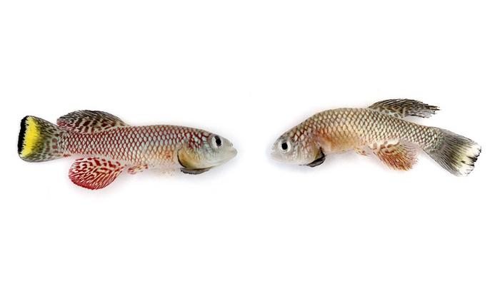
The killifish is an emerging model for investigating the genetic architecture of aging and age-related pathologies, which often exhibit sex-specific patterns between females (left) and males (right).
A new study identifies vgll3 as a key gene that promotes rapid growth and early reproduction while increasing the risk of aging and cancer later in life. The findings provide rare experimental evidence for the theory that evolution favors early-life advantages even at the expense of long-term health. Researchers say the discovery could open new paths for understanding, and potentially separating, the biological links between development, aging, and disease.
Researchers have identified a gene that directly links early-life growth and reproductive success with accelerated aging and increased cancer risk later in life, offering new insight into a longstanding theory in evolutionary biology.
Now, an international team led by Dr. Eitan Moses, Dr. Marva Bergman, and Prof. Itamar Harel at Hebrew University, in collaboration with Prof. Nabieh Ayoub (Technion) and Prof. Alexei A. Maklakov (University of East Anglia),provides experimental evidence for the theory of antagonistic pleiotropy, the idea that certain genes can provide advantages early in life while contributing to disease and decline in old age.
While widely accepted in theory, scientists have struggled to identify specific genes responsible for such trade-offs in vertebrates. Using the African turquoise killifish, a short-lived species recently pioneered by Harel and colleagues for genetic aging research, the team focused on the gene vgll3, which has been previously linked to the timing of human puberty and maturation in other species, particularly Atlantic salmon.
By modifying this gene using CRISPR technology, the researchers observed clear effects. Fish with altered vgll3 grew faster and reached sexual maturity earlier, traits that could offer a reproductive advantage in natural environments.
However, these benefits came with significant long-term costs. The same fish showed reduced lifespans and a higher incidence of age-related tumors, including melanoma-like cancers.
“We have effectively caught evolution in the act of making a trade-off. For years, we’ve asked why our bodies can’t just maintain themselves indefinitely. This gene gives us a direct answer: nature doesn’t prioritize longevity; it prioritizes continuity. We are built to sprint, not to marathon,” said Dr. Harel.
Further analysis showed that the gene influences key biological processes, including cell division,
stem cell activity, and DNA repair. Increased cellular activity may help explain both the rapid
development observed in younger fish and the accumulation of damage that leads to disease in older individuals.
The researchers also developed a new immunodeficient killifish model, enabling them to transplant and study tumor cells in ways not previously possible in this system.
“What’s fascinating—and slightly terrifying—is that the cancer we see in these fish isn’t a random accident. It’s the direct shadow of their youthful vitality. The same machinery that drives a cell to build a young body is hijacking the system to build a tumor in the old one. If we can understand this mechanism, we might finally learn how to decouple healthy growth from the disease of aging,” Dr. Harel added.
Because vgll3 is conserved in humans, the findings may have broader implications for understanding human development, aging, and age-related diseases. While previous association studies have linked the gene to puberty timing and hormone levels, functional data were missing until now.
The discovery could contribute to future efforts in cancer prevention and research aimed at extending healthy lifespan. Researchers say the next step will be to explore whether it is possible to separate the gene’s beneficial early-life effects from its harmful consequences later in life.
A 3-month-old African turquoise killifish, left, and a 5-month-old killifish, right, show aging much like that in humans.
Every cell in every organism on Earth copies DNA the same way. Except one bacterial protein — quietly doing something scientists had never seen before.
Think of it like a recipe — passed down from parent to child over countless generations, all the way back 4 billion years to the earliest life on Earth. With tweaks and changes accumulating along the way, but always copied from something that already existed.
That's the one rule that has held the entire time: to make DNA, you need existing genetic material to copy from.
Scientists just found a protein that breaks this rule.
A mechanism nobody has seen before
"It was quite a surprise!" Alex Gao, a biochemist at Stanford University in California and senior author of the study, told DW.
His team had been investigating how bacteria protect themselves from viruses when they identified something unexpected: a protein called Drt3b that builds DNA without anything to copy from. It uses its own shape as a mold to snap the right building blocks into place.
"We didn't believe it until we saw the cryo-EM structure [...] That was the moment it really clicked for us," he said — referring to cryo-electron microscopy, a technique that images molecules at near-atomic resolution.
Gao and his team used the bacterium E. coli, found in the intestines of humans and other warm‑blooded animals. It is a cornerstone of research because of its fast growth and simple, well‑mapped genetics
Image: NIH/IMAGE POINT FR/picture alliance
So how does it actually work?
DRT3 — the full system studied by Gao's team — works in two steps.
DNA is double-stranded: think of it like a zipper, with two sides that fit together.
One side is built in a familiar way, with a protein called Drt3a using a small piece of genetic material as a template to build one strand.
The other side is where things get strange. A second protein, Drt3b, needs to build the other side of that zipper — but does so without a template. Instead, specific parts of the protein itself act as the guide, locking onto the right DNA building blocks or "nucleotides" one by one until the strand is complete. And that's what we didn't think was possible — at least not like this.
Other proteins have done something similar before — but only in short fragments, like writing a sentence. Drt3b writes a whole paragraph. It's the first known protein to produce a long, sequence-specific strand of DNA using nothing but its own structure as a guide.
Why does it matter?
"The research is groundbreaking," said Philip Kranzusch, a biochemist at Harvard Medical School who was not involved in the study.
That’s because scientists have been studying DNA since the 1950s and bacteria have been quietly doing something they never imagined was possible. Which raises the question: what else are we missing?
There's also a practical angle. If scientists could engineer Drt3b to produce other DNA sequences, it might one day work as a tool for building custom DNA molecules — without needing a template to copy from.
But we're not there yet. "We do not yet know if it can be reprogrammed or engineered in a useful way," Rafael Pinilla-Redondo, an assistant professor at the Section of Microbiology at the University of Copenhagen, told DW.
Francis Crick helped uncover the structure of DNA in 1953, alongside James Watson, building on crucial experimental work by Rosalind Franklin and Maurice Wilkins
Image: UPI Photo/IMAGO
So does this break the rules of biology?
The discovery has sparked debate around what is called the "central dogma of biology" — the idea that genetic information flows from DNA to RNA to protein, but never from protein back into DNA. If a protein can write a DNA sequence, does that break the rule?
"No, I would not say the central dogma has been broken," said Pinilla-Redondo. What the study shows is a protein helping to build a short, repetitive DNA sequence in a very specific context — not proteins generally rewriting genetic code. "The exciting part is not that the rules of biology have collapsed. It is that evolution has found a very unexpected way to build a DNA molecule," he said.
But what does the DNA actually do?
Scientists don't fully know yet.
The leading hypothesis is that the DNA acts as a kind of molecular sponge — soaking up essential components of the attacking virus and neutralizing it. But Alex Gao is careful about how firmly he holds that idea. "That's currently our leading hypothesis, but we're certainly open to alternative models," he said.
Pinilla-Redondo agrees the mechanism is still far from understood. "Is the DNA a decoy, a signal, a scaffold, or a toxic molecule? That is the key mystery," he said.
Jennifer Doudna (left) and Emmanuelle Charpentier were awarded the 2020 Nobel Prize in chemistry for the development of the CRISPR method for genome editing, which allows scientists to rewrite DNA in almost any organism
Image: Alexander Heinl/dpa/picture alliance
Is this the next CRISPR?
CRISPR — the molecular scissors that allow scientists to cut and edit DNA with unprecedented precision — was itself first discovered as a quirky bacterial defense system. It has since transformed medicine, including the first approved gene therapyfor sickle cell disease in 2023.
Sounds familiar, right? But will it be a similar story with DRT3?
Probably not — at least not yet. "CRISPR is a once-in-a-generation breakthrough that revolutionized biotechnology," said Gao. "While it is early to predict applications of DRT3, we are most excited about DRT3 for expanding our understanding of the mechanisms of DNA synthesis."
A glimpse into microbial dark matter
"The field of bacterial immunity is exploding," said Pinilla-Redondo. Experimental research on these bacterial defense systems has only just begun — and the diversity of mechanisms being uncovered is striking, with multiple research groups around the world independently making similar findings.
For Gao's team, this discovery is less an ending than a beginning. Bacteria have spent billions of years fighting viruses, quietly evolving molecular tricks that we're only beginning to discover. How many more are out there?
"It points to a vast reservoir of uncharacterized biology within microbial 'dark matter,' where fundamental mechanisms likely remain undiscovered," said Gao.
Edited by: Frank Lee Esteban Pardo Science journalist and communicator
Tuesday, May 19, 2026
Profound changes in Frontiers of Medicine and promotion of MedScience
Recent decades have witnessed an unprecedented explosion of scientific innovation, driven by the deep convergence of clinical medicine, life sciences, information technology, materials science, and quantum computing. This technological wave is fundamentally reshaping industrial paradigms and societal structures while acting as a powerful catalyst for transformative advances in healthcare. Landmark achievements such as the completion of the Human Genome Project, the continuous refinement of gene-editing tools like CRISPR-Cas9, and the sophisticated interpretation of multi-omics data—including genomics, proteomics, metabolomics, transcriptomics, single-cell omics, and spatial multi-omics—have provided unparalleled insights into the molecular and cellular foundations of human life. Simultaneously, the proliferation of wearable devices, artificial intelligence, machine learning, big data analytics, cloud computing, and the Internet of Medical Things has resolved longstanding bottlenecks in processing massive medical datasets, enhancing clinical decision-making efficiency, and enabling telemedicine and remote patient monitoring systems that transcend geographical limitations.
These advances are catalyzing a comprehensive upgrade and digitalization of public health systems worldwide. Innovative drug development is accelerating dramatically, while medical diagnosis is evolving from a purely human, experience-based discipline toward one increasingly augmented by AI and other cutting-edge technologies. Detection capabilities have reached new heights of timeliness, accuracy, and personalization. Personalized precision medicine is now benefiting millions of patients, particularly those battling cancer, while regenerative medicine and tissue engineering are pioneering novel approaches to repairing damaged human tissues and organs.
Amidst this rapidly evolving landscape of medical frontiers, the global scientific community urgently requires a comprehensive medical journal that serves as both a platform for academic communication and a bridge for translating scientific breakthroughs into clinical practice and fostering international medical cooperation. With profound pride and a deep sense of responsibility, the inaugural issue of MedScience is introduced, marking a new identity for the Chinese Academy of Engineering medical journal and ushering in a fresh chapter dedicated to serving the global medical community.
The publication was originally established in January 2007 under the title Frontiers of Medicine in China. As medical science advanced rapidly across the globe, the editorial team recognized the growing necessity for a more influential platform dedicated to academic exchange of cutting-edge medical research. In 2011, the journal was renamed Frontiers of Medicine, broadening its scope to encompass the latest developments in clinical medicine, basic medical sciences, translational medicine, epidemiology, public health, health policy, and traditional Chinese medicine. Through the unwavering efforts and generous support of the editorial board, contributing authors, peer reviewers, and the broader medical community, the journal steadily elevated its academic standing and international visibility, achieving several significant milestones including indexing in Scopus in 2009, PubMed/Medline in 2010, and the Science Citation Index Expanded in 2016.
The new name MedScience reflects an evolved and refined editorial philosophy. "Med" represents medicine, underscoring the fundamental mission of serving human health, while "Science" symbolizes an unwavering commitment to originality, rigor, and innovation in scientific inquiry. Under this new identity, MedScience aspires to transcend traditional disciplinary boundaries, serving as a dynamic platform that integrates cutting-edge insights across the entire spectrum of medical sciences. Special emphasis will be placed on the convergence of clinical medicine, life sciences, and advanced technologies, with particular focus on emerging fields such as cell and gene therapy, AI-driven drug discovery and diagnostics, organoids and regenerative medicine, precision medicine, and environmental health—domains positioned to drive transformative breakthroughs in healthcare.
There is a firm conviction that MedScience, as an important platform for international academic exchange, will continue to expand its influence, broaden its scholarly reach, and better serve its community of authors, readers, and reviewers. Through these efforts, the journal aims to make meaningful contributions to the advancement of human health and to open a new and compelling chapter for medical science on the global stage..
Millet has been an important crop in East Asia for much of the Holocene, a period beginning about 11,700 years ago. To better understand how environmental conditions may have shaped the development of millet agriculture, researchers from the Institute of Earth Environment of the Chinese Academy of Sciences, and their collaborators in and aboard China investigated loess deposits from the Chinese Loess Plateau (CLP).
The study, which was published in PNAS on May 4, suggests that fluctuations in growing-season soil temperature played an important role in modulating the development and geographic spread of millet agriculture in East Asia. The researchers found that mid-Holocene soil cooling from about 7,500 to 6,000 years ago likely reduced thermally suitable zones for millet cultivation, contributing to a southward displacement of farming and a delayed large-scale agricultural expansion until subsequent soil temperature recovery after around 6,000 years ago.
To conduct the research, the scientists analyzed loess deposits from the Longgugou (LGG) section of the CLP. They combined 14 radiocarbon and 18 optically stimulated luminescence dates to develop a high‑resolution chronology spanning approximately 12,300 to 2,800 years ago. Based on this chronology, they performed biomarker analyses on 114 samples to reconstruct growing-season soil temperature and vegetation conditions. By integrating these proxy reconstructions with archaeological datasets and transient climate simulations, the researchers explored how Holocene soil temperature variations may have influenced the spatiotemporal evolution of millet agriculture in Neolithic East Asia.
The results indicate that between approximately 12,300 and 7,500 years ago, soil temperatures were relatively high, accompanied by comparatively low moisture and sparse vegetation. From 7,500 to 6,000 years ago, soil temperatures progressively declined under wetter conditions with denser vegetation. After 6,000 years ago, soil temperatures rapidly recovered and then remained relatively stable for millennia, while moisture and vegetation cover decreased.
By comparing these climate and vegetation reconstructions with archeological evidence for millet agriculture, the researchers suggest that, between 8,000 and 7,500 years ago, relatively warmer soils may have supported early millet cultivation Yanshan-Liaoning region, where communities may have faced greater subsistence pressure. In the CLP and lower-latitude regions, millet use appears to have remained limited and likely co-existed with hunting, gathering, and other subsistence practices.
In contrast, from 7,500 to 6,000 years ago, cooler soils, wetter conditions, and denser vegetation may have narrowed reliable cultivation space, particularly near the lower thermal thresholds for frost-sensitive millets. These conditions likely contributed to a southward shift of millet farming toward the CLP and surrounding regions. From 6,000 to 4,000 years ago, recovering and more stable soil temperatures, along with reduced vegetation cover, and advances in cultivation practices, might have facilitated millet expansion and broader late Neolithic development.
This study provides a high-resolution Holocene growing-season soil temperature record from a core region of early millet agriculture in East Asia and highlights soil temperature as an underappreciated climatic constraint on millet agricultural development. The findings suggest that large-amplitude soil temperature fluctuations helped modulate the geographic distribution and developmental trajectory of early millet farming, offering refined insights into climate-society interactions during the Neolithic period in East Asia.
This soil bacterium is recognised by the plant, which, following the release of a signal that spreads throughout all its organs, strengthens its defences against attack by pathogens. Bacillus produces a molecule called surfactin, capable of interacting with root cells (bottom right). More specifically, surfactin binds preferentially to certain lipids in the plant cell membrane (glucosylceramides). This interaction alters the physical properties of the membrane, leading to the opening of mechanosensitive ion channels (top right) and triggering the immune response.
Credit: Created in BioRender. Deleu, M. (2026) https://BioRender.com/qddoip5
A study led by researchers at the University of Liège reveals the mechanism by which surfactin, a molecule produced by beneficial soil bacteria, activates plants’ immune defences. This mechanism, distinct from the classical paradigm of immune recognition, relies on direct interaction with the plant cell membrane. This discovery opens up prospects for the development of next-generation biopesticides.
Plants are not defenceless against pathogens. Certain soil bacteria, far from being mere inhabitants of the roots, send chemical signals to plants that prepare them to resist pathogens. An international research consortium, led by researchers from Gembloux Agro-Bio Tech, has just elucidated the molecular mechanism behind this immunisation. This study shows that surfactin, a cyclic lipopeptide produced by bacteria of the genus Bacillus, acts not via a protein receptor, but by interacting directly with the lipids in the plant cell membrane. "Plants have sophisticated defence mechanisms against disease," explains Marc Ongena, researcher at the TERRA Research Centre. Among these, immunity induced by beneficial soil microorganisms is attracting growing interest, both in fundamental and applied research. We already knew that certain rhizosphere bacteria, particularly those of the genus Bacillus, produce cyclic lipopeptides capable of stimulating plant defences. But how these molecules were recognised by plant cells remained poorly understood until now."
The researchers focused on surfactin - one of these lipopeptides - and its interaction with Arabidopsis thaliana, a model plant commonly used in plant biology. Using a transdisciplinary approach combining cell biology, biochemistry and biophysics, they demonstrated that surfactin binds to sphingolipids - and more specifically to glucosylceramide - present in the root cell membrane. “This interaction causes a slight remodelling of the membrane, increasing its tension, which activates mechanosensitive ion channels,” explains Magali Deleu. This triggers a signalling cascade that spreads from the roots to the leaves and prepares the plant to better resist pathogens, including the fungus Botrytis cinerea, which causes grey mould.
This mechanism differs from the classical paradigm of plant innate immunity, in which the recognition of foreign molecules usually involves membrane protein receptors. Here, it is the physical modification of the membrane itself – rather than a lock-and-key interaction with a receptor protein – that acts as the triggering signal. This finding sheds new light on how plants can perceive their microbial environment and distinguish between beneficial bacteria and true pathogens.
In practical terms, this research forms part of efforts to develop a new generation of biopesticides. By understanding precisely how these bacteria or their molecules activate plant immunity, it becomes possible to envisage more targeted and effective crop protection strategies, partially replacing chemical inputs. These results thus provide a solid scientific basis for guiding the rational development of bio-based products for use in sustainable agriculture.
This study illustrates the value of interdisciplinary basic research in shedding light on concrete agronomic challenges. By deciphering the chemical dialogue between soil bacteria and host plants at the molecular level, the teams at the University of Liège and their partners are opening up new avenues for better exploiting the natural alliances that exist between microorganisms and plants, to the benefit of an agriculture less dependent on synthetic products.
When scientists think about how plants will respond to climate change, they often look north. As temperatures rise, many species are expected to shift their ranges toward cooler regions with a loss of populations in warmer habitats. But new research from the University of Virginia, published in the journal Evolution Letters, suggests the story may be more complicated and more hopeful.
The University of Virginia’s Commonwealth Professor of Biology Laura Galloway and postdoctoral research associate Antoine Perrier are studying what they call “rear-edge” populations, those found at the warmest edges of their geographic ranges. These populations, often descended from groups that survived the last ice age, have endured thousands of years of climate change.
“Because these populations have been there since the last glaciation, they’ve gone through warming in the past,” Galloway said. “We can use them as models for what we might expect in response to future warming.”
Their recent work on a native wildflower brings together multiple lines of evidence, including genomics, greenhouse experiments and field studies, to test how these populations evolved and what that might mean for the future.
Rethinking Vulnerability at the Warm Edge
Conventional ecological models predict that populations at the warm edge of a species’ range will be the first to disappear as temperatures rise. But Perrier and Galloway found something different.
“We often think that populations at the warmer edge are the ones that will go extinct,” Perrier said. “But it turns out there’s a lot that we don’t know about these populations.”
One possibility is that they harbor high genetic diversity, a legacy of their age and persistence since the last ice age, and therefore may be a resource for adapting to future change. Another is that as small, isolated populations, they might show signs of genetic drift, a process that reduces diversity and can make populations more fragile. A third possibility is that these populations have undergone local adaptation, evolving traits that allow them to thrive in conditions warmer than typical for the species.
The answer, in this case, was clear.
“We found patterns of local adaptation throughout the range,” Perrier said. “But what was very interesting is that in the deep south only the populations coming from very similar environments were able to actually grow and reproduce.”
In other words, southern populations have evolved specific traits that allow them to survive and reproduce in warmer climates. Northern populations transplanted into those same conditions failed to flower at all.
A Surprising Forecast for Climate Change
The findings challenge a central assumption about how species will respond to warming. Instead of southern populations disappearing first, the researchers’ data suggest that they are likely to persist, while populations in the middle of the range may struggle.
Many plant species use cold to cue reproduction, “As winters get warmer, populations are expected to experience a loss in reproduction,” Perrier said. “But this was not the case for the rear edge.”
Southern populations may be less affected by continued warming because they have already evolved to reproduce without relying on cold winter cues. By contrast, populations in regions like the mid-Atlantic could face new challenges.
“It’s almost the opposite of what we expect,” Galloway said, noting that both far-northern and far-southern populations may prove more resilient than those in between.
The work also points to practical applications. Traits that allow southern populations to thrive in warmer climates could potentially be introduced into more vulnerable populations through conservation strategies such as assisted gene flow.
Natural Laboratories for the Future
Beyond its immediate findings, the research highlights the value of studying long-term evolutionary history. Rear-edge populations, the researchers argue, act as “natural laboratories” for understanding how species respond to environmental change.
For Perrier, the work underscores both the urgency and the opportunity of climate research.
“We don’t often think of these populations as being the ones that might be the best adapted to future conditions,” he said. “But they could actually persist and change how we think about species responses to climate change.”
When an asteroid as big as Mount Everest struck Earth 66 million years ago, it wiped out all non-avian dinosaurs and roughly a third of life on the planet. But many plants survived the devastation.
In a new study publishing May 8 in the Cell Press journal Cell, researchers reveal that the accidental duplications of genomes—a natural phenomenon—might have helped many flowering plants survive some of the most extreme environmental upheavals in Earth’s history. This strategy could help plants adapt to the rapid climate changes unfolding today.
“Whole-genome duplication is often seen as an evolutionary dead end in stable environments,” says author Yves Van de Peer of Ghent University in Belgium. “But in harsh situations, it can provide unexpected advantages.”
Most organisms carry two sets of chromosomes, one from each parent. But in flowering plants, many species carry additional sets as a result of random whole-genome duplication. For example, most cultivated bananas have three sets of chromosomes while wheat plants can have as many as six, a condition known as polyploidy.
Whole-genome duplication occurs relatively frequently in plants, and it can be costly. Larger genomes require more nutrients to maintain, increase the risk of acquiring harmful mutations, and affect fertility. For these reasons, only a small fraction of duplicated genomes are retained and passed down through generations in the wild.
On the other hand, genome duplications can increase genetic variations, and genes can evolve new functions. These changes may help organisms better tolerate stress such as heat or drought.
To understand why some duplicated genomes persist, Van de Peer and his team analyzed the genomes of 470 species of flowering plants, constructing one of the largest datasets of its kind. They looked for blocks of genes that appear in almost identical pairs—a marker of past whole-genome duplication events. Then, they compared the data with information from 44 plant fossils to estimate when these duplications occurred.
Their analysis revealed a striking pattern. The researchers found that the genes that persist over time tend to originate from whole-genome duplications during major periods of environmental upheaval. These include the asteroid-triggered mass extinction 66 million years ago, several periods of global cooling when ecosystems collapsed, and the Paleocene-Eocene Thermal Maximum (PETM) about 56 million years ago—a period of rapid global warming.
The findings help explain a long-standing puzzle of why polyploidy is common, but only a few persevere in plant genomes over millions of years. Under these extreme conditions, polyploid plants might have gained an edge. Traits that are normally disadvantageous, such as maintaining a larger and more complex genome, can become beneficial, say the researchers.
The study also offers some clues about how plants may respond to climate change today. During the PETM, global temperatures rose by about 5 to 9°C (9 to 14°F) over roughly 100,000 years, a change comparable to the warming happening today.
“While the current climate is warming at a much faster rate, what we see from the past suggests that polyploidy may help plants cope with these stressful conditions,” Van de Peer says.
###
This work was supported by Research Foundation–Flanders, the European Research Council, and Ghent University.
Cell (@CellCellPress), the flagship journal of Cell Press, is a bimonthly journal that publishes findings of unusual significance in any area of experimental biology, including but not limited to cell biology, molecular biology, neuroscience, immunology, virology and microbiology, cancer, human genetics, systems biology, signaling, and disease mechanisms and therapeutics. Visit: http://www.cell.com/cell. To receive Cell Press media alerts, contact press@cell.com.
Photosynthesis is one of the most complex processes in nature. However, plants use only a fraction of the available light spectrum and are highly sensitive to environmental stressors such as changing light intensities, heat and drought. As climate change intensifies these stresses, safeguarding crop productivity is becoming an increasingly urgent challenge. To better understand the process of photosynthesis and at the same time identify starting points for improving it, researchers led by LMU biologist Dario Leister study model organisms such as the cyanobacterium Synechocystis.
In a study now published in Nature Communications, the scientists stressed Synechocystis by subjecting it to fluctuations in light intensity. “These kinds of conditions, in which high and low light intensities alternate at intervals ranging from one to several minutes, disrupt the process of photosynthesis and damage the photosystems,” explains Leister, who is Chair of Plant Molecular Biology at the Faculty of Biology in Martinsried. To find out how the blue-green algae can adapt to these unfavorable light conditions, the team recreated an accelerated evolutionary process in the laboratory.
Over time, this approach produced Synechocystis strains capable of tolerating light fluctuations that would normally be lethal. Genetic analysis of these adapted strains revealed mutations that influence the activity and relative quantity of biomolecules that are vital for photosynthesis. These include the protein-pigment complexes photosystem I and II and the light-harvesting antenna complexes. These evolutionary adaptations enhanced the resilience of Synechocystis to extreme variations in light intensity.
Plants growing in agricultural fields face similar challenges. Outdoor light conditions change constantly due to cloud cover, shading, and weather fluctuations, forcing crops to continuously adjust their photosynthetic machinery. “Photosynthesis works most efficiently at fairly low light intensity, whereas excessive light reduces efficiency. When the light levels change too quickly, the regulatory mechanisms cannot respond fast enough, which reduces efficiency and so also reduces the yield,” explains Dario Leister.
The findings from Synechocystis may provide new strategies for improving the ability of crops to cope with fluctuating light conditions. “The next step is to transfer the approach we used in our current study to eukaryotic algae because they are evolutionary closer to crop plants.” The biologist hopes that this will allow him to move step by step from relatively simple single-celled organisms toward applications in crops.
The study is part of the “PhotoRedesign: Redesigning the Photosynthetic Light Reactions” project, which has been funded by an ERC Synergy Grant from the European Union. In this research project, scientists from LMU are exploring new approaches for improving plant photosynthesis, using single-cell organisms like Synechocystis as model organisms because of their short generation times and the ease with which they can be genetically manipulated.
Ultimately, the goal is to produce crops that can utilize a broader range of light wavelengths. “To achieve this, it is important that optimized plants are also more robust and better able to cope with the additional absorbed light energy,” says Leister. The current study is providing new potential starting points for intervening in the complex photosynthetic apparatus of crops: “Our improved Synechocystis substrains contain point mutations that can also be transferred to related organisms using gene editing. With current legislative developments in the EU, such modifications may no longer be classified as transgenic in the future. Moreover, this strategy more closely resembles natural evolutionary processes than approaches based on overexpressing individual genes, which is a method often used by other researchers.”
Plants evolved distinct functions for two forms of a fundamental signaling molecule. These create redundancy and more robustness. Arabidopsis thaliana (mouse ear cress) plants at different developmental stages, photographed at the Plant Facility of the Institute of Science and Technology Austria (ISTA)
The molecule cAMP, which plays essential roles in mammalian cells, is less well understood in plants. In a new Science Advancespaper, researchers from the Institute of Science and Technology Austria (ISTA) and international collaborators demonstrate that plants use two forms of cAMP in parallel to regulate normal cellular processes and respond to stress, while maintaining crosstalk between them. That crosstalk provides redundancy, so that if one fails, the other can compensate, allowing plants to respond more robustly to a wider range of environmental factors. Ultimately, the findings could help improve crop resilience and productivity in a rapidly changing climate.
Plants can’t escape danger. To cope with stresses such as heat, freezing, flooding, drought, or infection, they rely on biological mechanisms evolved over millions of years.
Different life forms face unique environmental challenges, driving them to evolve distinct biological processes. Although animals, plants, and microbes share many molecular mechanisms, insights from animal models often don’t apply directly to other kingdoms.
Cyclic adenosine monophosphate, also known as cAMP, is a fundamental signaling molecule known to play essential roles in both animal and plant cells. However, although its production and role in mammalian cells are well understood, its functions in plants remain largely unknown.
Now, ISTA alum Mingyue Li and professor Jiří Friml at the Institute of Science and Technology Austria (ISTA) have teamed up with scientists in Germany, Saudi Arabia, the Czech Republic, and the United States to shed light on cAMP in the plant model Arabidopsis thaliana, commonly known as mouse ear cress or thale cress.
Twin molecules with distinct but partially overlapping properties
In animal systems, the main form of cAMP, called 3’,5’-cAMP, is involved in the transfer of signals between nerve cells, hormone signaling, and the regulation of metabolic functions. This predominant form of cAMP is derived from the cell’s energy currency, ATP. However, cAMP has a ‘twin’ form: a molecule with the same chemical formula but different atomic bonds. Concretely, the phosphate group is attached to the adenosine molecule at a different location. This other form, called 2’,3’-cAMP, is associated with RNA degradation and stress response. Its levels are tightly controlled in mammalian cells because excessive amounts can be toxic.
Li, Friml, and their colleagues now show that, while both forms of cAMP exist in plants, the levels of 2’,3’-cAMP—the ‘other’ form of the molecule—are over 60 times higher than those of 3’,5’-cAMP, the main form found in animals.
Using a battery of molecular and cell biology techniques, the team demonstrates that the two forms of cAMP exhibit largely distinct functions in plant metabolism as well as in protein and gene regulation. While 3’,5’-cAMP appears to fine-tune responses related to growth, maintenance, nutrient status, and normal cell function, 2’,3’-cAMP triggers much broader effects in plants, including specialized metabolic pathways and broad stress responses. However, they also show that these functions partially overlap, suggesting that plants may have evolved distinct ways to adapt to environmental challenges.
Cross-talking signaling pathways
Maintaining two parallel but interconnected cAMP pathways could help plants fine-tune cellular regulation and distinguish among different external stimuli, including stress factors. Crosstalk between the pathways provides redundancy, so that if one fails, the other can compensate, allowing plants to respond more robustly to a wider range of environmental factors.
Ultimately, understanding how plants regulate stress and routine cellular functions could help boost crop productivity and enhance resilience to climate change.
Plants evolved distinct functions for two forms of a fundamental signaling molecule. These create redundancy and more robustness. Arabidopsis thaliana (mouse ear cress) plants at different developmental stages, photographed at the Plant Facility of the Institute of Science and Technology Austria (ISTA)
Arabidopsis thaliana plants at the Institute of Science and Technology Austria (ISTA).
Plants evolved distinct functions for two forms of a fundamental signaling molecule. Seedlings growing in the pink room at the Plant Facility of ISTA
Arabidopsis thaliana plants in pink room at the Institute of Science and Technology Austria (ISTA).
Faster breeding brings climate-adapted, high-yielding, robust varieties to the fields more quickly. These varieties have greater tolerance to drought or heat, for example. This increases yield reliability and reduces resource use. Haploids, which are plants with only a single set of chromosomes, are an important tool in breeding. By doubling their chromosomes (‘double haploids’), haploids can be used to quickly produce fully homozygous lines within one generation. This accelerate the development of new varieties.
When the centromere protein CENH3 is degraded in a plant’s egg cell, paternal haploid offspring are frequently produced. Using the model plant Arabidopsis thaliana, the research team generated up to 57 per cent of these haploids in the corresponding offspring. The first author of the study, Dr. Saravanakumar Somasundaram, explains that „removing CENH3 from the egg cell efficiently produces paternal haploids, essentially disabling maternal chromosomes. The method is modular, uses the plant’s natural processes, and could be applied to crops.“
Initially, the scientists added a small tag to CENH3 to enable enzymatic degradation. The cellular ‘rubbish collection’ system then removes the CENH3 protein before fertilisation. Consequently, CENH3 is absent from the egg cell, yet remains present in the pollen. "A specific egg cell promoter ensures that the elimination affects only the embryo, not the nutritive tissue. And this increases the germination capacity of haploid seeds,” explains Dr. Saravanakumar Somasundaram.
“The aim is essentially to ‘switch off’ CENH3 in the egg cell, pollinate the plant with normal pollen, and produce haploid seeds,” says Prof. Dr. Andreas Houben, head of the ‘Chromosome Structure and Function’ research group. “Our method produces non-transgenic haploids following cross-pollination, thereby accelerating the development of inbred lines. This significantly reduces breeding time, cutting the process down from several years to a single season.”
Adequate boron (B) supply is essential for optimal growth and yield formation in rapeseed (Brassica napus L.). With boron-deficient soils affecting croplands worldwide, developing varieties with enhanced boron-use efficiency represents a sustainable strategy to safeguard productivity. Central to this effort is the identification of genes that regulate boron homeostasis.
In a study published in The Crop Journal on 9 April 2026, a research team led by Dr. Sheliang Wang at Huazhong Agricultural University reports the discovery and comprehensive characterization of BnaC3.BOR1, a pivotal boron transporter gene in B. napus.
"Our findings advance our mechanistic understanding of low-boron adaptation in B. napus and provide a high-value genetic target for marker-assisted breeding and the development of boron-efficient rapeseed cultivars," notes Wang.
The team found that BnaC3.BOR1 is highly expressed in root stele cells, stems, and floral organs. Notably, the gene shows spatially asymmetric expression within the stem, with significantly higher transcript levels on the side adjacent to the petiole. "This heterogeneous expression pattern strongly suggests that BnaC3.BOR1 plays a precise role in regulating boron distribution and tissue-level homeostasis," says Dan Zou, co-first author of the study.
To confirm the gene's transport function, the researchers heterologously expressed BnaC3.BOR1 in yeast, resulting in markedly elevated intracellular boron concentrations. They further demonstrated in vivo transport activity through functional complementation assays: expression of BnaC3.BOR1 fully restored wild-type growth phenotypes in Arabidopsis bor1 mutants under boron-limiting conditions.
The team then validated the gene's physiological role in planta using a CRISPR/Cas9-mediated gene editing system. "We found that null mutants exhibited heightened sensitivity to boron deprivation, manifesting as stunted root elongation, reduced shoot biomass, and substantially diminished boron accumulation in shoots," adds Zou. "When grown in low-boron soils, mutant plants developed severe morphological defects—including epidermal cracking at the stem base near petiole attachment sites, aberrant vascular development, and localized boron depletion in compromised stem tissues." Notably, during reproduction, impaired floral organ development and critically low boron concentrations led to significant yield loss.
"Our findings not only confirmed that BnaC3.BOR1 expression in roots and flowers reflects the functional conservation of boron transporter genes across species, but also resolved a long-standing question: which gene controls stem cracking caused by boron-deficiency stress," says co-first author Dr. Ling Liu.
###
Contact the author:
Sheliang Wang
Email address: sheliangwang2017@mail.hzau.edu.cn
The publisher KeAiwas established by Elsevier and China Science Publishing & Media Ltd to unfold quality research globally. In 2013, our focus shifted to open access publishing. We now proudly publish more than 200 world-class, open access, English language journals, spanning all scientific disciplines. Many of these are titles we publish in partnership with prestigious societies and academic institutions, such as the National Natural Science Foundation of China (NSFC).
In greenhouse tests, biochar made at a lower pyrolysis temperature of 300 °C and added at 2% by weight promoted larger leaf area, higher biomass, longer roots, and more root tips, all without causing phytotoxic effects. By contrast, a 5% application rate raised soil electrical conductivity and disturbed nutrient balance, which suppressed plant performance.
Poultry production generates huge volumes of litter, creating both a nutrient resource and an environmental management problem. In regions such as the Delmarva Peninsula, where poultry farming is highly concentrated, repeated land application of raw poultry litter can lead to phosphorus buildup, nutrient runoff, and water-quality risks. Converting poultry litter into biochar through pyrolysis has emerged as a promising alternative because it can stabilize nutrients, improve soil properties, and support circular agriculture. Yet the agronomic value of poultry litter biochar is not fixed: it depends strongly on pyrolysis temperature, feedstock composition, and application rate. Concerns also remain about salinity, nutrient imbalance, and possible phytotoxicity, making it necessary to identify conditions under which this waste-derived amendment is both safe and effective for crop growth.
A study (DOI:10.48130/bchax-0026-0009) published in Biochar X on 20 March 2026 by Dong Hee Kang’s team, Morgan State University, suggests that properly engineered poultry litter biochar could become a practical tool for recycling livestock waste, improving soil quality, and supporting more sustainable crop production in poultry-intensive agricultural regions.
To test how production conditions shape agricultural performance, the researchers produced six poultry litter biochars using two pyrolysis temperatures, 300 and 500 °C, and three feedstock compositions: no bedding, 10% pine shavings, and 10% rice hulls. These materials were then incorporated into a sandy loam soil collected from Maryland’s Delmarva Peninsula at two rates, 2% and 5% by weight. Radish was selected as the indicator crop because of its sensitivity to soil physical and chemical conditions. The team conducted both seed germination tests and six-week greenhouse pot experiments, while also measuring soil pH, electrical conductivity, nutrient availability, leaf area, biomass, chlorophyll-related indices, root length, and root tip number. The results showed a clear pattern. Seed germination remained high across all treatments, indicating that none of the tested biochars caused acute toxicity during early establishment. However, plant growth responses diverged sharply after emergence. Biochar produced at 300 °C supported better leaf growth and biomass than biochar produced at 500 °C, likely because lower-temperature material retained more plant-available nutrients and reactive surface groups. Application rate proved even more important. At 2%, the amendment improved soil fertility while keeping electrical conductivity within a range suitable for radish, which translated into stronger leaves and more developed root systems. At 5%, however, the soil became more saline, with much higher nitrogen, phosphorus, and potassium levels. That excess nutrient loading, combined with osmotic stress and cation antagonism, reduced root biomass, shortened total root length, and limited root tip formation despite the greater nutrient supply. The addition of bedding materials further improved outcomes by lowering sodium concentrations and increasing the potassium-to-sodium ratio, helping plants better tolerate salt-related stress. Overall, the optimal treatment was biochar produced at 300 °C, especially with bedding material, and applied at no more than 2%.
Taken together, the study shows that poultry litter biochar is not simply beneficial or harmful on its own; its value depends on how it is made and how much is applied. The work offers practical guidance for transforming poultry waste into a safer and more effective soil amendment, while also highlighting the need for future field-scale studies on long-term nutrient dynamics, microbial interactions, and repeated applications under real farming conditions.
This research was supported and funded by the National Science Foundation's Excellence in Research Program, Division of Chemical, Bioengineering, Environmental and Transport Systems (CBET), Directorate for Engineering (ENG), grant under award number 2200616.
Biochar X is an open access, online-only journal aims to transcend traditional disciplinary boundaries by providing a multidisciplinary platform for the exchange of cutting-edge research in both fundamental and applied aspects of biochar. The journal is dedicated to supporting the global biochar research community by offering an innovative, efficient, and professional outlet for sharing new findings and perspectives. Its core focus lies in the discovery of novel insights and the development of emerging applications in the rapidly growing field of biochar science.