Wednesday, April 20, 2022

Scientists Crack Egg Forging Evolutionary Scandal Two Million Years in the Making
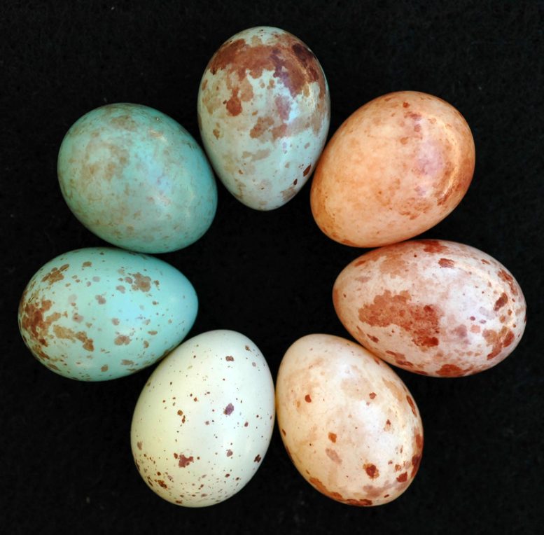
Cuckoo Finch Eggs Laid by Different Females

Cuckoo finch eggs laid by different females. Diversity of maternally inherited egg phenotypes within a single interbreeding species, the brood-parasitic cuckoo finch. Different cuckoo finch matrilines mimic the eggs of different host species (here, tawny-flanked prinia and red-faced cisticola) and have further diversified to approximate the range of variable egg “signatures” within each host species, an anti-parasite adaptation that aids host parents in recognizing their own eggs. Credit: Claire N. Spottiswoode

While many people around the world just finished unwrapping their Easter eggs, scientists have solved one of nature’s biggest criminal cases, an egg forgery scandal two million years in the making. Their findings suggest that the victims of this fraud may now be gaining the upper hand.

Many birds around the world side-step the costs of parenthood by laying their eggs in the nest of other species. Known as “brood parasitism,” this way of life has many advantages but also presents challenges such as how to convince the other species to accept a foreign egg.

Many brood parasites achieve this by mimicking the colors and patterns of their host’s eggs, but some exploit the care of several different host species whose eggs all look different.
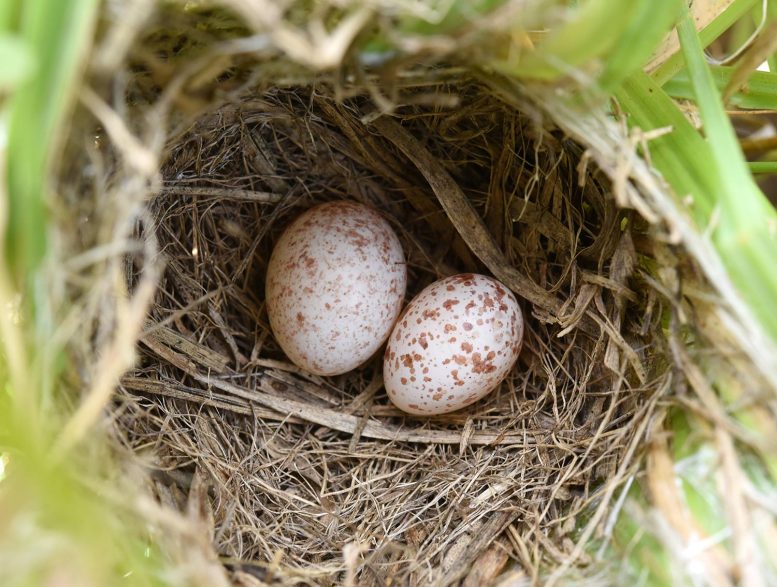
Cuckoo Finch Egg in Zitting Cisticola Nest

Cuckoo finch egg in zitting cisticola nest. Cuckoo finch eggs closely mimic the color and pattern of the eggs of each of their several host species, to trick host parents into accepting the parasitic egg as one of their own. Here a cuckoo finch has successfully had its egg (at left) accepted in the nest of a zitting cisticola (egg at right). Credit: Claire N. Spottiswoode

How can a single brood-parasitic bird species imitate the eggs of multiple different bird species at the same time in order to trick them into raising their young?

And how do these parasitic forgers pass this capability on to their young despite interbreeding between birds raised by different hosts?

These questions have been puzzling scientists for more than a century. Now genetic research by an international team led by Professor Claire Spottiswoode from the Cambridge’s Department of Zoology and the FitzPatrick Institute of African Ornithology, University of Cape Town; and Professor Michael Sorenson at Boston University, has made a major breakthrough, and their findings may be bad news for the egg forgers.

The study, published on April 11, 2022, in Proceedings of the National Academy of Sciences (PNAS), focuses on the genetics of egg mimicry in the cuckoo finch, a species that adopts a brood-parasitic lifestyle and exploits many species of warbler across Africa.
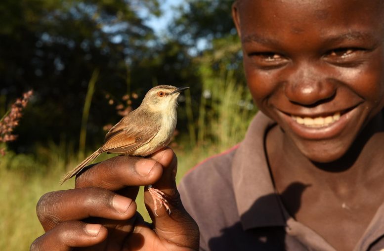
Tawny-Flanked Prinia With Field Assistant Tom Hamusikili in Zambia

A tawny-flanked prinia, a common host species of the cuckoo finch, captured in Zambia for genetic sampling with the help of field assistant Tom Hamusikili. Credit: Claire N. Spottiswoode

Female cuckoo finches inherit their capacity to imitate the look of their hosts’ eggs from their mothers, according to the research study, via the female-specific W chromosome (analogous to the male-specific Y chromosome in humans).

Such ‘maternal inheritance’ allows cuckoo finches to side-step the risk of inheriting the wrong mimicry genes from a father raised by a different host, and so has allowed distinct lineages of cuckoo finch females to evolve specialized egg mimicry of several different host species. Such mimicry dupes host parents into accepting a parasitic egg as their own rather than throwing it out of the nest, and so has been crucial to the success of these African birds.

But the researchers believe that this long-established ‘genetic architecture’ of maternal inheritance may come back to haunt the cuckoo finches. Dr. Spottiswoode said:

“In this particular coevolutionary arms race between species, natural selection has created a double-edged sword.”

“While maternal inheritance has allowed cuckoo finches to exploit multiple host species, it’s likely to slow their ability to evolve counter-adaptations as their hosts evolve new defenses.

“In particular, parasites face a daunting challenge because some host species have in return evolved an astonishing diversity of egg color and pattern ‘signatures’, that help hosts to distinguish their own eggs from parasitic mimics.”


Cuckoo finch mimicry of tawny-flanked prinia eggs

This photograph shows eggs of the cuckoo finch (middle circle) and one of its common host species, the tawny-flanked prinia (outer circle), revealing the diversity of host egg color and pattern “signatures” (an anti-parasite adaptation that aids host parents in recognizing their own eggs) that are mimicked by cuckoo finch egg “forgeries.”
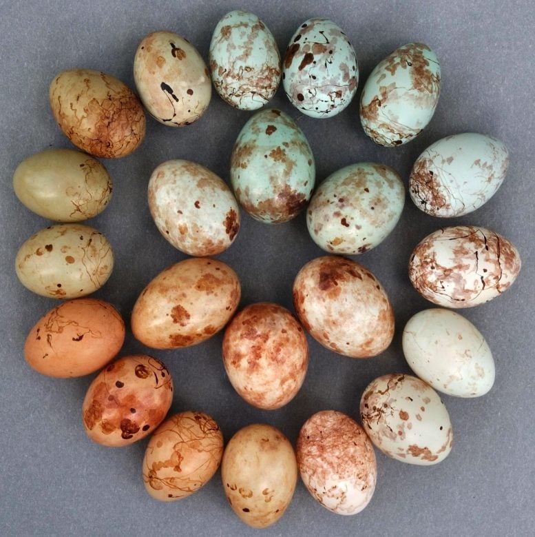
Cuckoo Finch Mimicry of Tawny-Flanked Prinia Eggs

The eggs of the cuckoo finch (middle circle) and a common host species, the tawny-flanked prinia (outer circle), are shown in this photograph, revealing the diversity of host egg color and pattern “signatures” that are mimicked by cuckoo finch egg “forgeries.” Credit: Claire N. Spottiswoode

However, cuckoo finches do not mimic the rich olive-green eggs (top left) laid by some tawny-flanked prinia females. The new study suggests that a genetic constraint in cuckoo finches may account for their apparent inability to mimic this host egg type.


The field data were collected at a study site in southern Zambia together with Dr. Wenfei Tong and Dr. Gabriel Jamie from the University of Cambridge and Ailsa Green, Silky Hamama, Ian Taylor, and Collins Moya from the surrounding community in Zambia.

Cuckoo finches in this area trick four different species of grass-warbler to devastating effect: if host parents fail to detect and remove a parasitic egg in their nest, the young cuckoo finch typically outcompetes the hosts’ own hatchlings, which soon starve to death.
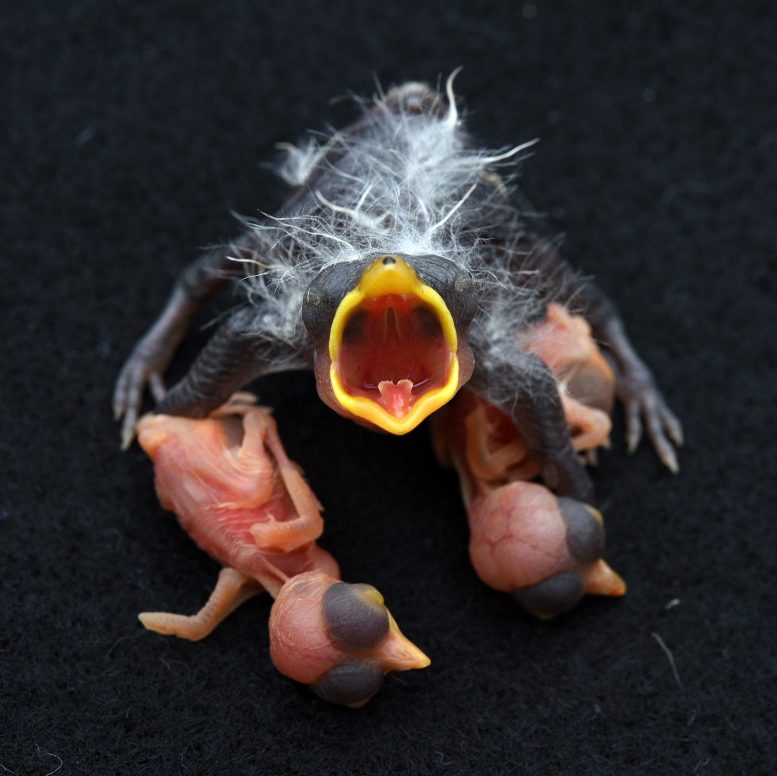
Cuckoo Finch and Host Chicks

Cuckoo finch and host chicks. Brood parasitism is costly for hosts because a cuckoo finch chick begs for food very vigorously as soon as it hatches, outcompeting the host parents’ own chicks (here zitting cisticolas) which typically soon die of starvation. Credit: Claire N. Spottiswoode

The team collected DNA samples from 196 cuckoo finches from 141 nests belonging to the four grass-warbler species and studied the majority by sequencing thousands of short segments across their genomes.

In their fightback against the forgers, grass-warblers have become skilled quality controllers, rejecting eggs that differ from their own in color and pattern, and all four species have evolved the ability to deposit unique ‘signatures’ onto their own eggs to enhance their detection of intruders. Tawny-flanked prinias, for example, lay eggs with blue, white, red, or olive-green backgrounds overlaid with a variety of patterns.

Cuckoo finches have responded not only by evolving mimicry of the eggs of their several host species, but have also further diversified to mimic at least some of the signature-like variation seen in the eggs of different females within each host species.

The team established that both abilities are handed down through maternal inheritance, finally validating a hypothesis first proposed in 1933 by ornithologists pondering how the common cuckoo in Europe was similarly able to mimic the eggs of several different host species.

Forgers facing an uncertain future?

The researchers believe that the cuckoo finches now face an uphill struggle because they cannot recombine the different forgery traits evolved by their separate family lines.

For example, two different lineages of cuckoo finch mothers have evolved eggs with either blue or red backgrounds, as an evolutionary response to similar diversity in their tawny-flanked prinia hosts, but there is no evidence that they can create the precise mixture of pigments needed to produce the olive-green eggs that some host females can produce.

Collins Moya Fieldfwork Zambia

Study co-author Collins Moya (left) carrying out fieldwork in the grasslands of southern Zambia, together with field assistant Kiverness Moono (right). Credit: Claire N. Spottiswoode

In a previous study, Professor Spottiswoode found that a growing proportion of eggs laid by tawny-flanked prinia hosts are olive-green, suggesting this is part of an accelerating evolutionary fightback. As expected, the team found that these host birds are passing down their anti-fraud ‘egg signature’ abilities through a different genetic process (bi-parental inheritance) to that used by the cuckoo finches. Spottiswoode said:

“Cuckoo finches are missing out on a powerful source of evolutionary novelty and that could prove costly in this ongoing arms race.”

She added: “The way they inherit their ability to mimic host eggs has a downside by likely making the grass-warblers’ defenses more effective, and constraining the parasite’s ability to respond.

“We may see the emergence of unforgeable egg signatures which could force cuckoo finches to switch to other naïve host species. Or the parasitic birds might become increasingly dependent on young host individuals that haven’t yet learned their own signatures and are bad at spotting mismatched eggs.”

The study argues that ‘selection from host defenses drove cuckoo finches to transfer control of egg appearance to the maternally inherited part of the genome’ at least 2 million years ago.

Reference: “Genetic architecture facilitates then constrains adaptation in a host–parasite coevolutionary arms race” by Claire N. Spottiswoode, Wenfei Tong, Gabriel A. Jamie, Katherine F. Stryjewski, Jeffrey M. DaCosta, Evan R. Kuras, Ailsa Green, Silky Hamama, Ian G. Taylor, Collins Moya, and Michael D. Sorenson, 11 April 2022, Proceedings of the National Academy of Sciences.
DOI: 10.1073/pnas.2121752119

The research was funded primarily by fellowships from the Biotechnology and Biological Sciences Research Council and the Royal Society, and by the National Science Foundation.

Genetic Mutations Across Animal Kingdom Shed New Light on Aging

DNA Transfer Concept

Despite enormous disparities in longevity and body mass, new research has revealed that the number of genetic mutations acquired by 16 species is similar across their lifetimes.

Quantity of mutations acquired is similar over the lifetime of 16 species, despite vast differences in lifespan and body mass.

The first study to evaluate the accumulation of mutations across a wide range of animal species has shed fresh light on long-standing debates regarding the role of genetic changes in aging and cancer. Despite vast differences in lifespan and size, researchers from the Wellcome Sanger Institute discovered that diverse animal species end their natural lives with similar numbers of genetic alterations.

The study, published on April 13, 2022, in the journal Nature, analyzed genomes from 16 species of mammal, from mice to giraffes. The authors confirmed that the longer the lifespan of a species, the slower the rate at which mutations occur, lending support to the long-standing theory that somatic mutations play a role in aging.

Giraffe Herd

The study looked at the genomes of 16 different mammal species, ranging from mice to giraffes. The research found that as a species’ longevity increases, the rate of mutations decreases, confirming the long-held idea that somatic mutations play a role in aging.

Genetic changes, known as somatic mutations, occur in all cells throughout the life of an organism. This is a natural process, with cells acquiring around 20 to 50 mutations per year in humans. Most of these mutations will be harmless, but some of them can start a cell on the path to cancer or impair the normal functioning of the cell.

Since the 1950s, some scientists have speculated that these mutations may play a role in aging. But the difficulty of observing somatic mutations has made it challenging to study this possibility. In the last few years, technological advances have finally allowed genetic changes to be observed in normal tissues, raising hopes of answering this question.[1]

Another long-standing question is Peto’s paradox. Since cancers develop from single cells, species with larger bodies (and therefore more cells) should theoretically have a much higher risk of cancer. Yet cancer incidence across animals is independent of body size. Animal species with large bodies are believed to have evolved superior mechanisms to prevent cancer. Whether one such mechanism is a reduction in the accumulation of genetic changes in their tissues has remained untested.

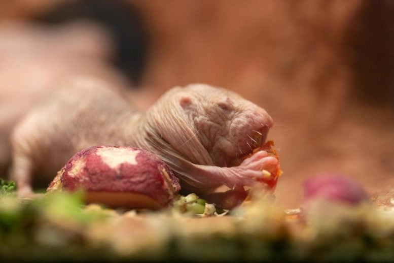
Naked Mole-Rat

The long-lived, highly cancer-resistant naked mole-rat was among the species studied in the research.

In this study, researchers at the Wellcome Sanger Institute set out to test these theories by using new methods to measure somatic mutation in 16 mammalian species, covering a wide range of lifespans and body masses.[2] This included species such as human, mouse, lion, tiger, giraffe, and the long-lived, highly cancer-resistant naked mole-rat, with samples provided by a number of organizations including the Zoological Society of London.

Whole-genome sequences were generated from 208 intestinal crypts[3] taken from 48 individuals, to measure mutation rates in single intestinal stem cells.

Analysis of the patterns of mutations (or mutational signatures) provided information on the processes at work. The researchers found that somatic mutations accumulated linearly over time and that they were caused by similar mechanisms across all species, including humans, despite their very different diets and life histories.

Evidence of a possible role of somatic mutations in aging was provided by the researchers’ discovery that the rate of somatic mutation decreased as the lifespan of each species increased.

Dr. Alex Cagan, a first author of the study from the Wellcome Sanger Institute, said: “To find a similar pattern of genetic changes in animals as different from one another as a mouse and a tiger was surprising. But the most exciting aspect of the study has to be finding that lifespan is inversely proportional to the somatic mutation rate. This suggests that somatic mutations may play a role in aging, although alternative explanations may be possible. Over the next few years, it will be fascinating to extend these studies into even more diverse species, such as insects or plants.”

The search for an answer to Peto’s paradox goes on, however. After accounting for lifespan, the authors found no significant association between somatic mutation rate and body mass, indicating that other factors must be involved in larger animals’ ability to reduce their cancer risk relative to their size.

Dr. Adrian Baez-Ortega, a first author of the study from the Wellcome Sanger Institute, said: “The fact that differences in somatic mutation rate seem to be explained by differences in lifespan, rather than body size, suggests that although adjusting the mutation rate sounds like an elegant way of controlling the incidence of cancer across species, evolution has not actually chosen this path. It is quite possible that every time a species evolves a larger size than its ancestors – as in giraffes, elephants, and whales – evolution might come up with a different solution to this problem. We will need to study these species in greater detail to find out.”

Despite vast differences in lifespan and body mass between the 16 species studied, the quantity of somatic mutations acquired over each animal’s lifetime was relatively similar. On average a giraffe is 40,000 times bigger than a mouse, and a human lives 30 times longer, but the difference in the number of somatic mutations per cell at the end of lifespan between the three species only varied by around a factor of three.

Dr. Simon Spiro, ZSL (Zoological Society of London) wildlife veterinary pathologist, said: “Animals often live much longer in zoos than they do in the wild, so our vets’ time is often spent dealing with conditions related to old age. The genetic changes identified in this study suggest that diseases of old age will be similar across a wide range of mammals, whether old age begins at seven months or 70 years, and will help us keep these animals happy and healthy in their later years.”

Understanding the exact causes of aging remains an unsolved question and an area of active investigation. Aging is likely to be caused by the accumulation of multiple types of damage to our cells and tissues throughout life, including somatic mutations, protein aggregation and epigenetic changes, among others. Comparing the rates of these processes across species with very different lifespans can shed light on their role in aging.

Dr. Inigo Martincorena, senior author of the study from the Wellcome Sanger Institute, said: “Aging is a complex process, the result of multiple forms of molecular damage in our cells and tissues. Somatic mutations have been speculated to contribute to aging since the 1950s, but studying them had remained difficult. With the recent advances in DNA sequencing technologies, we can finally investigate the roles that somatic mutations play in aging and in multiple diseases. That this diverse range of mammals end their lives with a similar number of mutations in their cells is an exciting and intriguing discovery.”

Notes

  1. Further information on the study of somatic mutation in healthy cells is available on the Sanger Institute website.
  2. The full list of species sequenced is: black-and-white colobus monkey, cat, cow, dog, ferret, giraffe, harbor porpoise, horse, human, lion, mouse, naked mole-rat, rabbit, rat, ring-tailed lemur, and tiger.
  3. Colonic crypts are anatomical structures in the epithelium of the colon. Because all of the cells in a crypt are descended from a single stem cell, they are ideal for studying the rates and patterns of somatic mutation.

Reference: “Somatic mutation rates scale with lifespan across mammals” by Alex Cagan, Adrian Baez-Ortega, Natalia Brzozowska, Federico Abascal, Tim H. H. Coorens, Mathijs A. Sanders, Andrew R. J. Lawson, Luke M. R. Harvey, Shriram Bhosle, David Jones, Raul E. Alcantara, Timothy M. Butler, Yvette Hooks, Kirsty Roberts, Elizabeth Anderson, Sharna Lunn, Edmund Flach, Simon Spiro, Inez Januszczak, Ethan Wrigglesworth, Hannah Jenkins, Tilly Dallas, Nic Masters, Matthew W. Perkins, Robert Deaville, Megan Druce, Ruzhica Bogeska, Michael D. Milsom, Björn Neumann, Frank Gorman, Fernando Constantino-Casas, Laura Peachey, Diana Bochynska, Ewan St. John Smith, Moritz Gerstung, Peter J. Campbell, Elizabeth P. Murchison, Michael R. Stratton and Iñigo Martincorena, 13 April 2022, Nature.
DOI: 10.1038/s41586-022-04618-z

What Do Bacteria Sound Like? Bacterial Soundtracks Revealed by Nanotechnology

Bacterium on Graphene Drum

A graphene drum can reveal the sound of bacteria.

Have you ever wondered if bacteria make distinctive sounds? If we could listen to bacteria, we would be able to know whether they are alive or not. When bacteria are killed using an antibiotic, those sounds would stop – unless of course, the bacteria are resistant to the antibiotic. This is exactly what a team of researchers from TU Delft, led by dr. Farbod Alijani, has now managed to do: they captured low-level noise of a single bacterium using graphene. Now, their research is published in the journal Nature Nanotechnology.

The sound of a single bacterium

Farbod Alijani’s team at Delft University of Technology (TU Delft) was originally investigating the fundamentals of the physical mechanics of graphene, when a curious idea struck them. They wondered what would happen if this extremely sensitive material came into contact with a single biological object. “Graphene is a form of carbon consisting of a single layer of atoms and is also known as the wonder material,” says Alijani. “It’s very strong with nice electrical and mechanical properties, and it’s also extremely sensitive to external forces.”


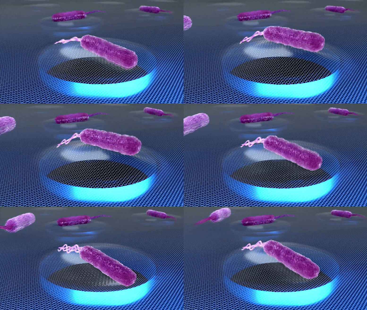
This animation shows how a graphene drum can reveal the sound of bacteria. The sound stops when a bacterium is killed by an antibiotic. Credit: Irek Roslon – TU Delft

Farbod Alijani’s team of researchers initiated a collaboration with the nanobiology group of Cees Dekker and the nanomechanics group of Peter Steeneken. Together with PhD student Irek Roslon and postdoc Dr. Aleksandre Japaridze, the scientists ran their first experiments using E. coli bacteria. Cees Dekker: “What we saw was striking! When a single bacterium adheres to the surface of a graphene drum, it generates random oscillations with amplitudes as low as a few nanometers that we could detect. We could hear the sound of a single bacterium!”

Punching a graphene drum with a bacterium

The extremely small oscillations are a result of the biological processes of the bacteria with main contribution from their flagella (tails on the cell surface that propel bacteria). “To understand how tiny these flagellar beats on graphene are, it’s worth saying that they are at least 10 billion times smaller than a boxer’s punch when reaching a punch bag. Yet, these nanoscale beats can be converted to sound tracks and listened to — and how cool is that,” Alijani says.

Bacterium on Graphene Drum

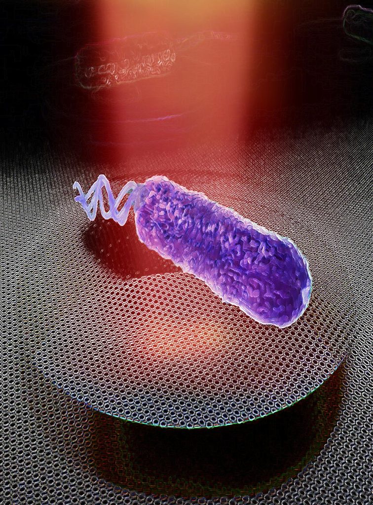
Artist’s impression of a graphene drum detecting nanomotion of a single bacterium. Credit: Irek Roslon, TU Delft

Graphene for fast detection of antibiotic resistance

This research has enormous implications for the detection of antibiotic resistance. The experimental results were unequivocal: If the bacteria were resistant to the antibiotic, the oscillations just continued at the same level. When the bacteria were susceptible to the drug, vibrations decreased until one or two hours later, but then they were completely gone. Thanks to the high sensitivity of graphene drums, the phenomenon can be detected using just a single cell.

Farbod Alijani: “For the future, we aim at optimizing our single-cell graphene antibiotic sensitivity platform and validate it against a variety of pathogenic samples. So that eventually it can be used as an effective diagnostic toolkit for fast detection of antibiotic resistance in clinical practice.” Peter Steeneken concludes: “This would be an invaluable tool in the fight against antibiotic resistance, an ever-increasing threat to human health around the world.”

Reference: “Probing nanomotion of single bacteria with graphene drums” by Irek E. Rosłoń, Aleksandre Japaridze, Peter G. Steeneken, Cees Dekker and Farbod Alijani, 18 April 2022, Nature Nanotechnology.
DOI: 10.1038/s41565-022-01111-6

Bacteria Generate Electricity From Methane: Generating Power While Purifying the Environment of Greenhouse Gases

Plasma Energy Generation Concept

Artist’s concept of microbial energy generation.

Generating power while purifying the environment of greenhouse gases should be achievable using bacteria. In a new publication, microbiologists from Radboud University have demonstrated that it is possible to make methane-consuming bacteria generate power in the lab.

The bacteria, Candidatus Methanoperedens, use methane to grow and naturally occur in fresh water such as ditches and lakes. In the Netherlands, the bacteria mostly thrive in locations where the surface and groundwater are contaminated with nitrogen, as they require nitrate to break down methane.

The researchers initially wanted to know more about the conversion processes occurring in the microorganism. In addition, they were also curious whether it would be possible to use it to generate power. “This could be very useful for the energy sector,” says microbiologist and author Cornelia Welte. “In the current biogas installations, methane is produced by microorganisms and subsequently burnt, which drives a turbine, thus generating power. Less than half of the biogas is converted into power, and this is the maximum achievable capacity. We want to evaluate whether we can do better using microorganisms.”

A kind of battery

Fellow microbiologists from Nijmegen have previously shown that it is possible to generate power using anammox bacteria that use ammonium during the process instead of methane. “The process in these bacteria is basically the same,” says microbiologist Heleen Ouboter. “We create a kind of battery with two terminals, where one of these is a biological terminal and the other one is a chemical terminal. We grow the bacteria on one of the electrodes, to which the bacteria donate electrons resulting from the conversion of methane.”

Through this approach, the researchers managed to convert 31 percent of the methane into electricity, but they aim at higher efficiencies. “We will continue focusing on improving the system,” Welte says.

Reference: “Methane-Dependent Extracellular Electron Transfer at the Bioanode by the Anaerobic Archaeal Methanotroph “Candidatus Methanoperedens”” by Heleen T. Ouboter, Tom Berben, Stefanie Berger, Mike S. M. Jetten, Tom Sleutels, Annemiek Ter Heijne and Cornelia U. Welte, 12 April 2022, Frontiers in Microbiology.
DOI: 10.3389/fmicb.2022.820989

Noninvasive Sound Technology Breaks Down Tumors, Kills Cancer Cells, and Spurs the Immune System

Histotripsy Ultrasound Array Transducer

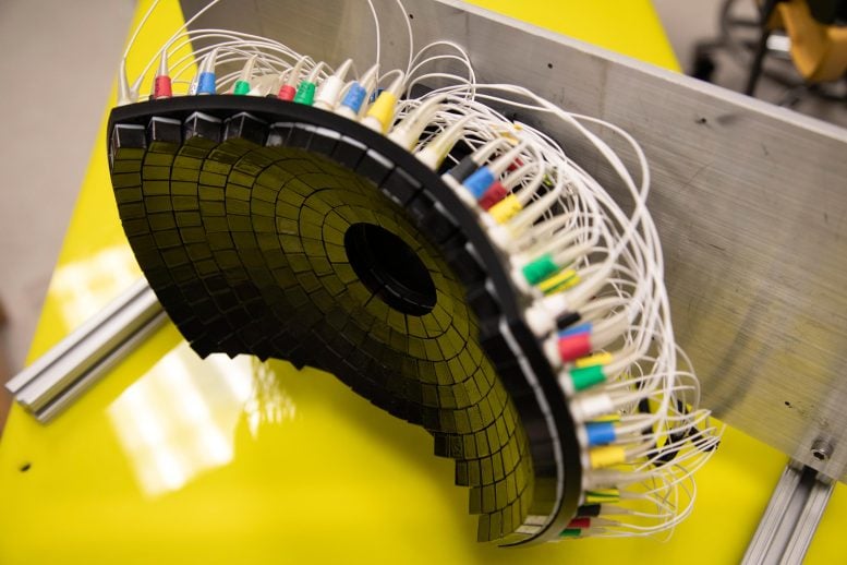
The 700kHz, 260-element histotripsy ultrasound array transducer used in Prof. Xu’s lab. Credit: Photo by Marcin Szczepanski/Lead Multimedia Storyteller, Michigan Engineering

Tumors Partially Destroyed With Sound Don’t Come Back

A new technique pioneered in rats at the University of Michigan could improve outcomes for cancer and neurological conditions.

Noninvasive sound technology developed at the University of Michigan breaks down liver tumors in rats, kills cancer cells, and spurs the immune system to prevent further spread—an advance that could lead to improved cancer outcomes in humans.

By destroying only 50% to 75% of liver tumor volume, the rats’ immune systems were able to clear away the rest, with no evidence of recurrence or metastases in more than 80% of the animals.

“Even if we don’t target the entire tumor, we can still cause the tumor to regress and also reduce the risk of future metastasis,” said Zhen Xu, professor of biomedical engineering at U-M and corresponding author of the study in Cancers.

Zhen Xu

Zhen Xu, Professor of Biomedical Engineering at the University of Michigan works in her office. Dr. Xu’s research focuses on developing new ultrasound technique for treatment of cancer, cardiovascular diseases, and neurological diseases. She and her colleagues have developed histotripsy, an ultrasound ablation technique via controlled cavitation. Histotripsy is the first image-guided ablation technique that is non-invasive, non-ionizing, and non-thermal. Dr. Xu’s work spans from basic science, device development, preclinical investigations, to clinical translation of histotripsy. Credit: Photo by Marcin Szczepanski/Lead Multimedia Storyteller, Michigan Engineering

Results also showed the treatment stimulated the rats’ immune responses, possibly contributing to the eventual regression of the untargeted portion of the tumor and preventing further spread of the cancer.

The treatment, called histotripsy, noninvasively focuses ultrasound waves to mechanically destroy target tissue with millimeter precision. The relatively new technique is currently being used in a human liver cancer trial in the United States and Europe.

In many clinical situations, the entirety of a cancerous tumor cannot be targeted directly in treatments for reasons that include the mass’ size, location or stage. To investigate the effects of partially destroying tumors with sound, this latest study targeted only a portion of each mass, leaving behind a viable intact tumor. It also allowed the team, including researchers at Michigan Medicine and the Ann Arbor VA Hospital, to show the approach’s effectiveness under less than optimal conditions.

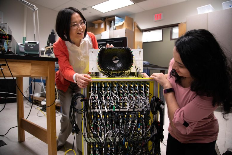
Zhen Xu and Tejaswi Worlikar

Zhen Xu,Professor of Biomedical Engineering at the University of Michigan (left) and Tejaswi Worlikar, Biomedical Engineering PhD student discuss the 700kHz, 260-element histotripsy ultrasound array transducer they use in Prof. Xu’s lab. Credit: Photo by Marcin Szczepanski/Lead Multimedia Storyteller, Michigan Engineering

“Histotripsy is a promising option that can overcome the limitations of currently available ablation modalities and provide safe and effective noninvasive liver tumor ablation,” said Tejaswi Worlikar, a doctoral student in biomedical engineering. “We hope that our learnings from this study will motivate future preclinical and clinical histotripsy investigations toward the ultimate goal of clinical adoption of histotripsy treatment for liver cancer patients.”

Liver cancer ranks among the top 10 causes of cancer related deaths worldwide and in the U.S. Even with multiple treatment options, the prognosis remains poor with five-year survival rates less than 18% in the U.S. The high prevalence of tumor recurrence and metastasis after initial treatment highlights the clinical need for improving outcomes of liver cancer.

Where a typical ultrasound uses sound waves to produce images of the body’s interior, U-M engineers have pioneered the use of those waves for treatment. And their technique works without the harmful side effects of current approaches such as radiation and chemotherapy.

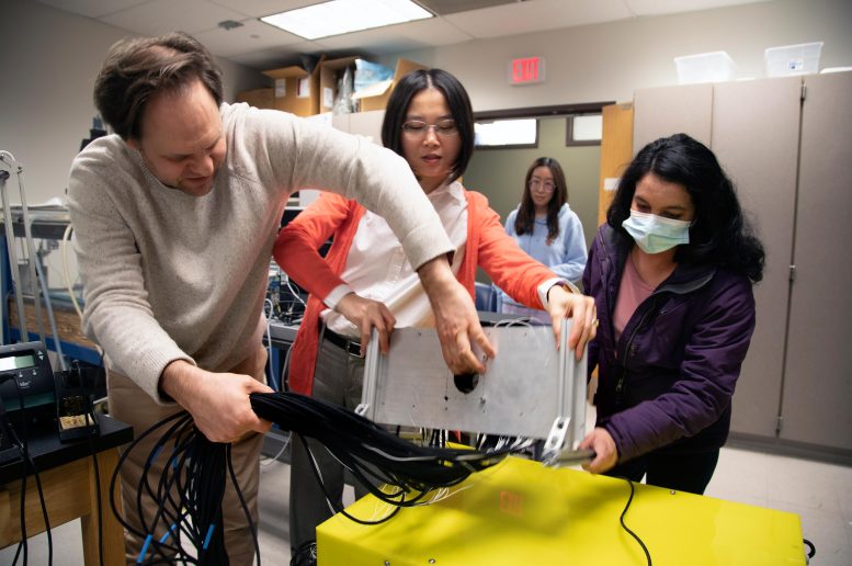
Moving Histotripsy Ultrasound Array Transducer

Zhen Xu, Professor of Biomedical Engineering at the University of Michigan (center) and Tejaswi Worlikar, Biomedical Engineering PhD student (right) move the 700kHz, 260-element histotripsy ultrasound array transducer they use in Prof. Xu’s lab. Credit: Photo by Marcin Szczepanski/Lead Multimedia Storyteller, Michigan Engineering

“Our transducer, designed and built at U-M, delivers high amplitude microsecond-length ultrasound pulses—acoustic cavitation—to focus on the tumor specifically to break it up,” Xu said. “Traditional ultrasound devices use lower amplitude pulses for imaging.”

The microsecond long pulses from UM’s transducer generate microbubbles within the targeted tissues—bubbles that rapidly expand and collapse. These violent but extremely localized mechanical stresses kill cancer cells and break up the tumor’s structure.

Reference: “Impact of Histotripsy on Development of Intrahepatic Metastases in a Rodent Liver Tumor Model” by Tejaswi Worlikar, Man Zhang, Anutosh Ganguly, Timothy L. Hall, Jiaqi Shi, Lili Zhao, Fred T. Lee, Mishal Mendiratta-Lala, Clifford S. Cho and Zhen Xu, 22 March 2022, Cancers.
DOI: 10.3390/cancers14071612

Since 2001, Xu’s laboratory at U-M has pioneered the use of histotripsy in the fight against cancer, leading to the clinical trial #HOPE4LIVER sponsored by HistoSonics, a U-M spinoff company. More recently, the group’s research has produced promising results on histotripsy treatment of brain therapy and immunotherapy.

The study was supported by grants from the National Institutes of Health, Focused Ultrasound Foundation, VA Merit Review, U-M’s Forbes Institute for Discovery and Michigan Medicine-Peking University Health Sciences Center Joint Institute for Translational and Clinical Research.

The Brain’s Alarm System To Suppress Intrusive Thoughts and Inhibit Unwanted Memories

Headache Confusion Obsession Dark Thoughts Illustration

New research shows that to suppress intrusive thoughts, the brain uses an alarm system that alerts other regions to inhibit them.

A brain region can proactively and reactively detect the need to inhibit unwanted memories.

Forget what you saw: a brain region detects when you are about to think of an unwanted memory and alerts other regions to suppress it, according to research published in JNeurosci today (April 18, 2022). JNeurosci is the Society for Neuroscience’s first journal.

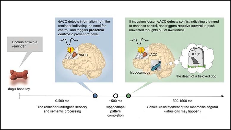
Brain Employs Alarm System to Suppress Intrusive Thoughts

A model of how the ACC proactively and reactively signals the need for thought suppression. Credit: Crespo García et al., JNeurosci 2022

Crespo García et al. measured participants’ brain activity with both EEG and fMRI while they completed a memory task. The participants memorized sets of words (i.e., gate and train) and were asked to either recall a cue word’s pair (see gate, think about train) or only focus on the cue word (see gate, only think about gate).

During proactive memory suppression, activity increased in the anterior cingulate cortex (ACC), a brain region involved in cognitive control, within the first 500 milliseconds of the task. The ACC relayed information to the dorsolateral prefrontal cortex (DLPFC), which then inhibited activity in the hippocampus, a key region for memory recall.

The activity levels in the ACC and DLPFC remained low for the rest of the trial, a sign of success — the memory was stopped early enough so no more suppression was needed. If the memory was not suppressed in time, the ACC generated a reactive alarm, increasing its activity to signal to the DLPFC to stop the intrusion.

Reference: “Anterior Cingulate Cortex Signals the Need to Control Intrusive Thoughts During Motivated Forgetting” by Maité Crespo García, Yulin Wang, Mojun Jiang, Michael C. Anderson and Xu Lei, 18 April 2022, JNeurosci.
DOI: 10.1523/JNEUROSCI.1711-21.2022 

Substance Derived From Licorice May Have Anti-Inflammatory and Anti-Cancer Effects

Licorice Root and Candy

Licorice root and black licorice candy. A licorice-derived substance may have anti-inflammatory and anti-cancer effects.

Researchers look to licorice for promising cancer treatments.

Licorice is more than a candy people either love or hate — it may play a role in preventing or treating certain types of cancer, according to researchers at the University of Illinois Chicago.

Gnanasekar Munirathinam and his research team are studying substances derived from the licorice plant Glycyrrhiza glabra to determine if they could be used to prevent or stop the growth of prostate cancer. Munirathinam is an associate professor in the department of biomedical sciences at the College of Medicine Rockford.

A research review into molecular insights of a licorice-derived substance called glycyrrhizin for preventing or treating cancer conducted by Dr. Munirathinam and student researchers suggests further research could lead to specific agents for clinical use.

The journal Pharmacological Research recently published the study titled “Oncopreventive and oncotherapeutic potential of licorice triterpenoid compound glycyrrhizin and its derivatives: Molecular insights.”

“When we look at the research out there and our own data, it appears that glycyrrhizin and its derivative glycyrrhetinic acid have great potential as anti-inflammatory and anti-cancer agents,” Munirathinam said. “More research is needed into exactly how these could best be used to develop therapies, but this appears to be a promising area of cancer research.”

Should everyone go out and eat a bunch of licorice? Probably not, because it may affect blood pressure, interact with certain medications, and cause serious adverse effects, including death, when used excessively. An occasional sweet treat of licorice candy or tea may be better options until more studies can show how to best harness the plant’s benefits.

“Very few clinical trials in humans have been conducted,” Munirathinam said. “We hope our research on prostate cancer cells advances the science to the point where therapies can be translated to help prevent or even cure prostate and other types of cancer.”

Reference: “Oncopreventive and oncotherapeutic potential of licorice triterpenoid compound glycyrrhizin and its derivatives: Molecular insights” by Rifika Jain, Mohamed Ali Hussein, Shannon Pierce, Chad Martens, Preksha Shahagadkar and Gnanasekar Munirathinam, 19 February 2022, Pharmacological Research.
DOI: 10.1016/j.phrs.2022.106138

College of Medicine Rockford students Rifika Jain, Mohamed Ali Hussein, Preksha Shahagadkar, Shannon Pierce and Chad Martens are co-authors of the review, which was partly supported by the National Institutes of Health (R0CA227218) and Brovember Inc.