It’s possible that I shall make an ass of myself. But in that case one can always get out of it with a little dialectic. I have, of course, so worded my proposition as to be right either way (K.Marx, Letter to F.Engels on the Indian Mutiny)
Friday, October 06, 2023
OBGYN
Advancements in assisted reproduction contribute to building a healthy society
A special issue of Fertility and Sterility® shares expert perspectives on what has been accomplished and what lies ahead in the field of reproductive endocrinology and infertility medicine
A SPECIAL ISSUE OF FERTILITY AND STERILITY SHARES EXPERT PERSPECTIVES ON THE CURRENT STATE OF THE FIELD OF ASSISTED REPRODUCTION, WHAT HAS BEEN ACCOMPLISHED, AND WHAT LIES AHEAD.
CREDIT: FERTILITY AND STERILITY, SEPTEMBER 2023 COVER
Philadelphia, October 5, 2023 – Reproductive endocrinology and infertility (REI) has finally come of age, moving beyond the “basics,” limited accessibility, and thorny initial issues. A special issue of Fertility and Sterility®, published by Elsevier, explores the past, present, and future of REI, nearly five decades after the first in vitro fertilization (IVF) baby, Louise Brown, was born in 1978.
Entitled Reproduction as the Foundation for a Healthy Society, the issue presents expert perspectives on achievements, opportunities, and challenges as this rapidly growing discipline advances its contribution to medical care and the building of heathy societies around the world. The contributions are openly available so that information and insights are accessible to all practitioners and patients.
Ten million overwhelmingly healthy babies have been born since IVF was introduced. In some countries as many as 9% of births have been achieved through IVF. The 1 in 8 people who face infertility are now able to realize their dreams of having children, however, access to REI care still remains a challenge. For REI to take its place as a core specialty in medicine, ethical, societal, legal, and economic implications of the technology need to be better understood to reduce barriers to broadening care to those who need it.
Guest Editor, Kurt T. Barnhart, MD, MSCE, Department of Obstetrics and Gynecology, Perelman School of Medicine, University of Pennsylvania, and Editor-in-Chief of Fertility and Sterility, explained, “We are now into the third decade in the 21st century, and a tension remains between societal obligations and the reproductive medicine we practice on a daily basis. This is evident by dramatically increasing media attention on REI’s impact on the society. It is only right the REI returns the favor through publication of this special issue.”
Guest Editor Ruben Alvero, MD, Fertility and Reproductive Health, Lucille Packard Children’s Hospital, Sunnyvale, California, and Department of Obstetrics and Gynecology, Stanford University School of Medicine, elaborated, “In recent years, many for whom parenthood was previously unachievable have become beneficiaries, including individuals facing gonadotoxic therapy or other medical complications, as well as those in the LGBTQ+ community. Still, access to care remains a challenge for varying populations within societies and for currently underserved regions of the world.”
The issue’s content focuses on important considerations and specific aspects of the field, with many articles presenting strategies to bridge the gap between societal obligations and the practice of reproductive medicine.
Important topics and recommendations that emerge from this collection include:
Significant collaboration among professionals, organizations, the World Health Organization, and policymakers has begun, but much more will be necessary to achieve these goals.
The alternatives to IVF are summarized including up-to-date evidence on effectiveness, safety, and cost-effectiveness (expectant management, intrauterine insemination, tubal flushing, in vitro maturation and intravaginal culture).
With rapidly growing demand, a supply-demand mismatch is anticipated for the REI workforce. The shortage of physicians, laboratory staff, and ancillary providers can be addressed through technology in the laboratory and the employment of advanced practice providers (APPs) in the clinic.
Low-cost assisted reproduction will continue to develop to help those most underserved. Technical advances in the IVF laboratory will be achieved as computational, nanotechnological, microfluidic, and process advances are made. This automation will facilitate access to care, although training technicians remains a challenge.
Better preconception care and interventions are explored as well as the value of preimplantation genetic testing to reduce the potential risk of passing on inherited medical conditions.
The impact of reproductive endocrinology and infertility medicine on the current and future health of babies and parents and the greater population is addressed.
Ovarian aging remains one of the most critical clinical challenges. Many would-be parents do not decide to reproduce until reaching the age when it is difficult or impossible -- without having preserved their eggs for a host of reasons.
The destiny of possibly up to a million cryopreserved embryos worldwide is an emerging concern as their originators pass out of the reproductive stages of their lives. How these embryos could aid in research and used for reproductive purposes for third-party individuals are considered, however, mechanisms for achieving these transfers are as yet poorly developed.
Robert J. Norman, MD, Robinson Research Institute, Adelaide Medical School, University of Adelaide, concluded, “REI has matured as a field and is now poised to broadly expand in a greater number of communities. While there were many reservations in lay and even some medical communities at the onset of assisted reproduction, acceptance of the field is now widespread, save for a relatively small number with continuing ethical concerns. Communities throughout the world have embraced the benefits of reproductive medicine in helping people achieve their goals and preserve their dreams of family.”
New research has shed light on how genetics influences the growth of the placenta, revealing a link to risk of disease in the mother.
Scientists from the University of Exeter worked with colleagues in Norway and Denmark to lead a largescale international collaboration which examined placental growth in the greatest detail yet. They caried out the first ever genome-wide association study of the weight of the placenta at birth, generating a number of revelations. Among the findings published inNature Genetics, the team concluded that faster growth of the placenta can contribute to risk of preeclampsia, and to earlier delivery of the baby.
The placenta is an organ which grows in the womb alongside the foetus, which is attached to it by the umbilical cord. The placenta provides oxygen and nutrients to the growing foetus and removes waste as the baby develops. A poorly functioning placenta is associated with pregnancy complications, and later risk of disease in the child.Despite its key role, little is yet known about how the growth of the placenta is regulated. Understanding placental growth is important, as babies with very small or large placentas are at higher risk of complications.
Professor Pål Njølstad, of the University of Bergen in Norway, who co-led the paper, said: “The placenta is such an important organ during pregnancy, providing an intricate and vital link between mother and baby. Our study has identified 40 variations in the genetic code linked to how big a placenta can grow, which improves our understanding of this vital organ in humans. Several of these genetic variations also influence the weight of the baby, but some appear to be predominantly concerned with placental growth.”
The team found that where the genetic code of the foetus meant it was more likely that the placenta would grow bigger, there was a higher risk of pre-eclampsia in the mother. This could be because the placenta grows too fast, which can upset the balance between the baby’s demand for resources and how much the mother is able to provide, which can be a factor in pre-eclampsia that occurs later in pregnancy.
Professor Rachel Freathy, of the University of Exeter Medical School, who is funded by Wellcome, is a co-lead on the paper. She said: “Pre-eclampsia is a condition that may develop in pregnancy, which causes high blood pressure. Some of the mother’s organs, such as the kidneys and liver, stop working properly. Detecting it early is essential to avoid severe health problems for mother and baby, yet how preeclampsia develops isn’t fully understood. Our study suggests that faster growth of the placenta contributes to a higher risk of preeclampsia in the mother. It seems specific to placenta growth because we did not find the same risk when we looked at the genetics of baby weight.”
Faster-growing placenta was also linked to shorter pregnancy. Senior researcher, group leader Bjarke Feenstra, of Copenhagen University Hospital and Statens Serum Institut, Denmark, who also co-led the study, said: “We found that babies with genetic code for a bigger placenta were more likely to be born earlier, which underscores the importance of investigating placental biology in studies of pregnancy duration and the timing of delivery.”
One key finding from the study related to insulin, which regulates blood sugar. The foetus produces insulin in response to glucose from the mother, which acts as a growth factor. The team found this insulin is also linked to the growth of the placenta, which helps to explain why placentas tend to be large in pregnancies where the mother has high blood glucose due to diabetes.
“While this is a great first step,” said Professor Stefan Johansson, also co-lead at the University of Bergen, “the final weight of a placenta can only tell us a limited amount about its function. Further studies are needed to examine the shape and development of placenta over the course of pregnancy. Our work is just the starting point for future research which could help us understand far more about the placenta’s role in the growth of the baby and risk of pregnancy complications.”
Genome-wide association study of placental weight identifies distinct and shared genetic influences between placental and fetal growth
Study shows prior exposure to common virus shields against birth defects and miscarriage
Study uncovers how pre-existing immunity to Cytomegalovirus (CMV) may significantly reduce the risk of birth defects and miscarriage during pregnancy, offering hope for the future vaccine
Researchers at Tulane University have shown for the first time that mothers are much less likely to transmit a common virus known to cause miscarriages and birth defects if they are exposed to the virus prior to becoming pregnant. The study marks a significant step toward the development of a vaccine that could protect mothers and their babies.
Cytomegalovirus (CMV) is a common herpesvirus that most women contract unknowingly before reaching child-bearing age. It's usually harmless except during pregnancy when, if passed on to the developing fetus, it is a leading cause of miscarriage and birth defects, including cerebral palsy and hearing loss.
Researchers have long known that the risk for complications is particularly high for women infected by CMV for the first time during pregnancy, but they haven’t fully understood why those who already carry the virus are less vulnerable.
The Tulane study reveals how pre-existing immunity to CMV effectively limits its transmission during pregnancy and protects against associated birth defects. The study, published in PLOS Pathogens, pinpoints the specific immune mechanisms responsible for that protection.
Researchers at the Tulane National Primate Research Center used a nonhuman primate model that closely mirrors human CMV infection and transmission. They observed that when pregnant mothers were initially infected with CMV during the first trimester, all of them transmitted the virus to their offspring, resulting in a high rate of miscarriage.
However, when nonhuman primates previously infected with CMV were reinfected during their pregnancies, their offspring were protected. The robust immune response observed in mothers upon reinfection resulted in only one out of five mothers passing the virus through the placenta, with no adverse health outcomes for any of the infants.
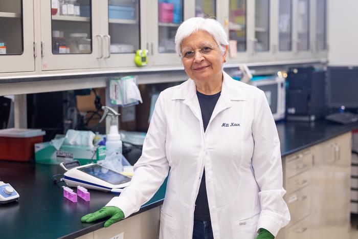
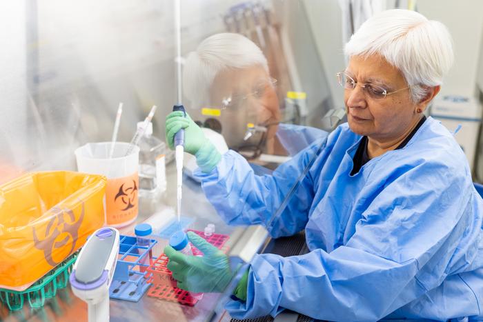
“Understanding how pre-existing immunity can protect against CMV transmission during pregnancy is crucial for developing an effective CMV vaccine that can safeguard all pregnant women and their unborn babies,” said Dr. Amitinder Kaur, principal investigator and professor of microbiology and immunology.
The findings show that if a mother already has CMV immunity before becoming pregnant, her immune system can effectively protect her baby from congenital CMV transmission if she is reinfected during pregnancy. This research could have highly significant implications for the development of a CMV vaccine to prevent infections in pregnant women, particularly in areas with a high prevalence of CMV.
This study was made possible with research provided by first author Matilda Moström, PhD, assistant director of flow cytometry core at Tulane National Primate Research Center, and resources supported by National Institutes of Health grants DP2HD075699, P01AI129859, and P51OD011104.
Dr. Amitinder Kaur, professor of microbiology and immunology at Tulane University, led the study that showed how pre-existing immunity to Cytomegalovirus effectively limits its transmission during pregnancy and protects against associated birth defects.
Under strict embargo: 19:00hrs BST Thursday 5 October 2023
Peer reviewed
Experimental study
Animals
Researchers at the Francis Crick Institute have shown that pregnancy hormones ‘rewire’ the brain to prepare mice for motherhood.
Their findings, published today in Science, show that both oestrogen and progesterone act on a small population of neurons in the brain to switch on parental behaviour even before offspring arrive. These adaptations resulted in stronger and more selective responses to pups.
It is well known that while virgin female rodents do not show much interaction with pups, mothers spend most of their time looking after young. It was thought that hormones released when giving birth are most crucial for this onset of maternal behaviour.
But earlier research also showed that rats who have given birth by Caesarean section, and virgin mice exposed to pregnancy hormones, still display this maternal behaviour, suggesting that hormone changes already during pregnancy may be more important.
In the current study, the researchers found that female mice indeed showed increased parental behaviour during late pregnancy, and that exposure to pups wasn’t necessary for this change in behaviour.
They found that a population of nerve cells (galanin-expressing neurons) in an area of the brain called the medial preoptic area (MPOA) in the hypothalamus, associated with parenting, was impacted by oestrogen and progesterone.
Brain recordings showed that oestrogen simultaneously reduced the baseline activity of these neurons and made them more excitable, whereas progesterone rewired their inputs, by recruiting more synapses (sites of communication between neurons).
Making these neurons insensitive to hormones completely removed the onset of parental behaviour during pregnancy. Mice failed to show parental behaviour even after giving birth, suggesting there is a critical period during pregnancy when these hormones take effect.
While some of these changes lasted for at least a month after giving birth, others seem to be permanent, suggesting pregnancy can lead to long-term rewiring of the female brain.
Jonny Kohl, Group Leader of the State-Dependent Neural Processing Laboratory at the Crick, said: "We know that the female body changes during pregnancy to prepare for bringing up young. One example is the production of milk, which starts long before giving birth. Our research shows that such preparations are taking place in the brain, too.
“We think that these changes, often referred to as ‘baby brain’, cause a change in priority – virgin mice focus on mating, so don’t need to respond to other females’ pups, whereas mothers need to perform robust parental behaviour to ensure pup survival. What’s fascinating is that this switch doesn’t happen at birth – the brain is preparing much earlier for this big life change.”
Rachida Ammari, postdoctoral fellow at the Crick, and first author along with PhD student Francesco Monaca, said: “We’ve demonstrated that there’s a window of plasticity in the brain to prepare for future behavioural challenges. These neurons receive a large number of inputs from elsewhere in the brain, so now we’re hoping to understand where this new information comes from.”
The researchers believe the brain may also be rewired in a similar way during pregnancy in humans, as the same hormonal changes are expected to impact the same areas of the brain. This could influence parental behaviour alongside environmental and social cues.
-ENDS-
For further information, contact: press@crick.ac.uk or +44 (0)20 3796 5252
Notes to Editors
Reference: Ammari, R., Monaca, F.et al. (2023). Hormone-mediated neural remodelling orchestrates parenting onset during pregnancy. Science. 10.1126/science.adi0576.
The Francis Crick Institute is a biomedical discovery institute dedicated to understanding the fundamental biology underlying health and disease. Its work is helping to understand why disease develops and to translate discoveries into new ways to prevent, diagnose and treat illnesses such as cancer, heart disease, stroke, infections, and neurodegenerative diseases.
An independent organisation, its founding partners are the Medical Research Council (MRC), Cancer Research UK, Wellcome, UCL (University College London), Imperial College London and King’s College London.
The Crick was formed in 2015, and in 2016 it moved into a brand new state-of-the-art building in central London which brings together 1500 scientists and support staff working collaboratively across disciplines, making it the biggest biomedical research facility under a single roof in Europe.
No comments:
Post a Comment