It’s possible that I shall make an ass of myself. But in that case one can always get out of it with a little dialectic. I have, of course, so worded my proposition as to be right either way (K.Marx, Letter to F.Engels on the Indian Mutiny)
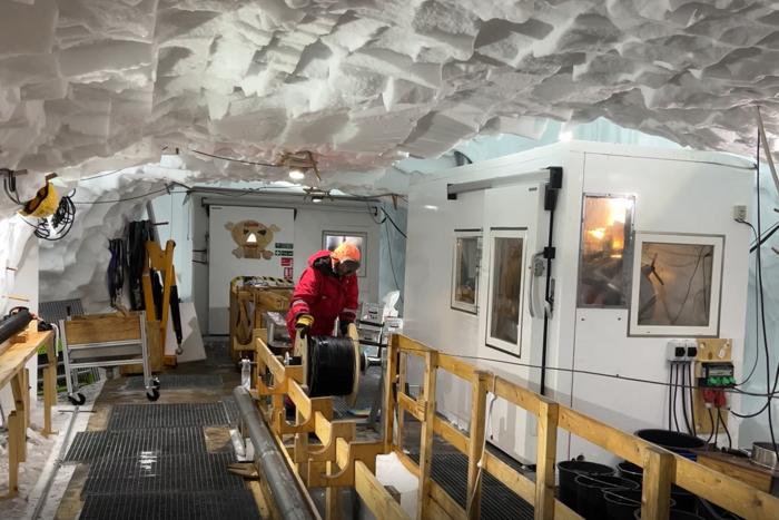
A researcher lowers a fibre-optic cable 1,500 metres into the borehole in or-der to record signals from inside the ice stream continuously for 14 hours.
• In Greenland, an international team of researchers led by ETH Zurich has discovered that countless tiny ice quakes take place deep inside ice streams.
• These quakes are responsible for the fact that ice streams also move with a continuous stick-slip motion and not only like viscous honey as previously considered.
• The researchers recorded seismic data from inside the ice stream using a fibre-optic cable in a 2,700-metre deep borehole.
The great ice streams of the Antarctic and Greenland are like frozen rivers, carrying ice from the massive inland ice sheets to the sea – and a change in their dynamics will contribute significantly to sea-level rise. In order to estimate just how much sea levels will rise, climate researchers rely on computer simulations of the ice streams. Until now, they have based these simulations on an assumption that the ice streams flow slowly but steadily into the sea like thick honey.
However, satellite measurements of the flow speed of ice streams show that such simulations are inaccurate and have shortcomings to correctly reflect reality. This leads to considerable uncertainties in estimates of how much mass the ice streams are losing and how quickly and how high sea levels will rise.
Ice streams both judder and flow
Now, a team of researchers led by ETH professor Andreas Fichtner has made an unexpected discovery: deep within the ice streams, there are countless weak quakes taking place that trigger one another and propagate over distances of hundreds of metres. This discovery helps to explain the discrepancy between current simulations of ice streams and satellite measurements, and the new findings should also impact the way ice streams are simulated in the future.
“The assumption that ice streams only flow like viscous honey is no longer tenable. They also move with a constant stick-slip motion,” says Fichtner. The ETH professor is confident that this finding will be integrated into simulations of ice streams, making estimates of changes in sea level more accurate.
Riddles relating to ice cores resolved
Moreover, the ice quakes explain the origin of numerous fault planes between ice crystals in ice cores obtained from great depths. These fault planes are the result of tectonic shifts and have been known to scientists for decades, although no explanation had been found for them until now.
“The fact that we’ve now discovered these ice quakes is a key step towards gaining a better understanding of the deformation of ice streams on small scales,” explains Olaf Eisen, Professor at the Alfred Wegener Institute and one of the study’s co-authors.
The study by this international research team led by ETH Zurich has just been published in the journal Science and also involved researchers from the Alfred Wegener Institute, Helmholtz Centre for Polar and Marine Research (AWI), the University of Strasbourg, the Niels Bohr Institute (NBI), the Swiss Federal Institute WSL and other universities.
Fire and ice are related
The fact that these ice quakes cannot be observed at the surface and have therefore remained undiscovered until now is due to a layer of volcanic particles located 900 metres below the surface of the ice. This layer stops the quakes from propagating to the surface. Analysis of the ice core showed that these volcanic particles originate from a massive eruption of Mount Mazama in what is now Oregon (USA) some 7,700 years ago. “We were astonished by this previously unknown relationship between the dynamics of an ice stream and volcanic eruptions,” Fichtner recalls.
The ETH professor also noticed that the ice quakes start from impurities in the ice. These impurities are also leftovers from volcanoes: tiny traces of sulphates that entered the atmosphere in volcanic eruptions and flew halfway around the world before being deposited on the Greenland ice sheet in snowfall. These sulphates reduce the stability of the ice and favour the formation of microfissures.
A 2,700-metre borehole in the ice
The researchers discovered the ice quakes using a fibre-optic cable that was inserted into a 2,700-metre-deep borehole and recorded seismic data from inside a massive ice stream for the first time. This borehole was drilled into the ice by researchers from the East Greenland Ice-core Project (EastGRIP), led by the Niels Bohr Institute and strongly supported by the Alfred Wegener Institute, resulting in the extraction of a 2,700-metre-long ice core. Once drilling work was complete, the researchers took the opportunity to lower a fibre-optic cable 1,500 metres into the borehole and record signals from inside the ice stream continuously for 14 hours.
The research station and borehole are located on the North East Greenland Ice Stream (NEGIS), around 400 kilometres from the coast. The NEGIS is the biggest ice stream of the Greenland ice sheet, whose retreat is a large contributor to current rising sea levels. In the area of the research station, the ice is moving towards the sea at a speed of around 50 metres per year.
As ice quakes occur frequently over a wide area in the researchers’ measurements, ETH researcher Fichtner believes it is also plausible that they occur in ice streams everywhere, all the time. To verify this, however, it will be necessary to take seismic measurements of this kind in other boreholes – and there are already plans to do just that.
###
References
Fichtner A, Hofstede C, Kennett B L N, Svensson A, Westhoff J, Walter F, Ampuero J-P, Cook E, Zigone D, Jansen D, Eisen O, Hidden cascades of seismic ice stream deformation. Science. 6.2.2025, DOI: 10.1126/science.adp8094
In this Special Issue of Science, 3 Reviews and a Policy Forum highlight research on Earth’s frozen places – from the Arctic to the Antarctic – and how it’s changing due to climate change and the geopolitical challenges this important work faces. In the first Review, Julienne Stroeve and colleagues provide a preview of what the Arctic region may look like in a warmer world. Without stronger climate action, global temperatures are set to rise +2.7°C above preindustrial levels, causing irreversible Arctic transformation. Under this future, Stroeve et al. show that nearly all days in the region would exceed past temperature extremes, summers would see an ice-free Arctic Ocean, Greenland’s melt zones would quadruple, and permafrost would shrink by half. While these changes would severely disrupt ecosystems, and damage Arctic communities and infrastructure, the authors note that stronger climate action could significantly mitigate these consequences, preserving the Arctic’s stability and resilience. In a second Review, Helen Fricker and colleagues focus on Antarctic ice, which plays a crucial role in regulating Earth’s climate, global sea levels, ocean circulation, and planetary reflectivity. Fricker et al. highlight key knowledge gaps in these processes, which create uncertainties in the stability of the Antarctic ice sheet and the potential impacts of its continued loss. According to Fricker et al., progress in these areas depends on high-resolution satellite monitoring, targeted field studies, improved modeling, and strengthened interdisciplinary collaboration to reduce uncertainties and refine future projections. A third Review focuses on Antarctic biodiversity. According to Luis Pertierra and colleagues, Antarctica hosts myriad unique life forms, yet much of the region’s biodiversity and ecological functioning remains poorly understood. Outside of a few well-known vertebrate species, much about the region’s invertebrate, plant, and microbial life remains unknown. Here, Pertierra et al. highlight the shortfalls in Antarctic biodiversity research, explain how these gaps hinder ecological insight and conservation in the region, and offer a framework for addressing them. Lastly, in a Policy Forum, Jennifer Spence and colleagues discuss the geopolitical challenges facing Arctic research. The Arctic is at the forefront of critical global issues, including the uneven impacts of climate change, the challenge of balancing economic development with conservation, and the complexity of governance across multiple boundaries. These issues are complicated further by rising populism, geopolitical tensions, and distrust in governmental institutions. Spence et al. highlight three key issues, including understanding the regional and global effects of climate change, ensuring research benefits Arctic communities, and maintaining international cooperation in the face of growing geopolitical pressures. However, despite these challenges, the Arctic region has pioneered innovative governance models that prioritize Indigenous engagement, as well as fostering international cooperation through institutions like the Arctic Council to promote sustainable development and environmental protection.
Coronal brain slice showing projections from different visual areas in the cerebral cortex to the ventrolateral geniculate nucleus (vLGN). These pathways are part of the circuit identified as mediating the suppression of instinctive fear responses.
Researchers at the Sainsbury Wellcome Centre (SWC) at UCL have unveiled the precise brain mechanisms that enable animals to overcome instinctive fears. Published today in Science, the study in mice could have implications for developing therapeutics for fear-related disorders such as phobias, anxiety and post-traumatic stress disorder (PTSD).
The research team, led by Dr Sara Mederos and Professor Sonja Hofer, mapped out how the brain learns to suppress responses to perceived threats that prove harmless over time.
"Humans are born with instinctive fear reactions, such as responses to loud noises or fast-approaching objects," explains Dr Mederos, Research Fellow in the Hofer Lab at SWC. "However, we can override these instinctive responses through experience – like children learning to enjoy fireworks rather than fear their loud bangs. We wanted to understand the brain mechanisms that underlie such forms of learning”.
Using an innovative experimental approach, the team studied mice presented with an overhead expanding shadow that mimicked an approaching aerial predator. Initially, the mice sought shelter when encountering this visual threat. However, with repeated exposure and no actual danger, the mice learned to remain calm instead of escaping, providing researchers with a model to study the suppression of fear responses.
Based on previous work in the Hofer Lab, the team knew that an area of the brain called the ventrolateral geniculate nucleus (vLGN) could suppress fear reactions when active and was able to track knowledge of previous experience of threat. The vLGN also receives strong input from visual areas in the cerebral cortex, and so the researchers explored whether this neural pathway had a role in learning not to fear a visual threat.
The study revealed two key components in this learning process: (1) specific regions of the visual cortex proved essential for the learning process, and (2) a brain structure called the ventrolateral geniculate nucleus (vLGN) stores these learning-induced memories.
“We found that animals failed to learn to suppress their fear responses when specific cortical visual areas where inactivated. However, once the animals had already learned to stop escaping, the cerebral cortex was no longer necessary,” explained Dr Mederos.
"Our results challenge traditional views about learning and memory," notes Professor Hofer, senior author of the study. "While the cerebral cortex has long been considered the brain's primary centre for learning, memory and behavioural flexibility, we found the subcortical vLGN and not the visual cortex actually stores these crucial memories. This neural pathway can provide a link between cognitive neocortical processes and ‘hard-wired’ brainstem-mediated behaviours, enabling animals to adapt instinctive behaviours.”
The researchers also uncovered the cellular and molecular mechanisms behind this process. Learning occurs through increased neural activity in specific vLGN neurons, triggered by the release of endocannabinoids – brain-internal messenger molecules known to regulate mood and memory. This release decreases inhibitory input to vLGN neurons, resulting in heightened activity in this brain area when the visual threat stimulus is encountered, which suppresses fear responses.
The implications of this discovery extend beyond the laboratory. “Our findings could also help advance our understanding of what is going wrong in the brain when fear response regulation is impaired in conditions such as phobias, anxiety and PTSD. While instinctive fear reactions to predators may be less relevant for modern humans, the brain pathway we discovered exists in humans too," explains Professor Hofer. "This could open new avenues for treating fear disorders by targeting vLGN circuits or localised endocannabinoid systems."
The research team is now planning to collaborate with clinical researchers to study these brain circuits in humans, with the hope of someday developing new, targeted treatments for maladaptive fear responses and anxiety disorders.
This research was funded by the Sainsbury Wellcome Centre core grant from the Gatsby Charity Foundation and Wellcome (090843/F/09/Z); a Wellcome Investigator Award (219561/Z/19/Z); an EMBO postdoctoral fellowship (EMBO ALTF 327-2021) and a Wellcome Early Career Award (225708/Z/22/Z).
Source:
Read the full paper in Science: ‘Overwriting an instinct: visual cortex instructs learning to suppress fear responses’
Media contact:
For more information or to speak to the researchers involved, please contact:
April Cashin-Garbutt, Head of Research Communications and Engagement, Sainsbury Wellcome Centre E: a.cashin-garbutt@ucl.ac.uk T: +44 (0)20 3108 8028
About the Sainsbury Wellcome Centre
The Sainsbury Wellcome Centre (SWC) brings together world-leading neuroscientists to generate theories about how neural circuits in the brain give rise to the fundamental processes underpinning behaviour, including perception, memory, expectation, decisions, cognition, volition and action. Funded by the Gatsby Charitable Foundation and Wellcome, SWC is located within UCL and is closely associated with the Faculties of Life Sciences and Brain Sciences. For further information, please visit: www.sainsburywellcome.org
Known for their powerful punch, mantis shrimp can smash a shell with the force of a .22 caliber bullet. Yet, amazingly, these tough critters remain intact despite the intense shockwaves created by their own strikes.
Northwestern University researchers have discovered how mantis shrimp remain impervious to their own punches. Their fists, or dactyl clubs, are covered in layered patterns, which selectively filter out sound. By blocking specific vibrations, the patterns act like a shield against self-generated shockwaves.
The study will be published on Friday (Feb. 7) in the journal Science.
The findings someday could be applied to developing synthetic, sound-filtering materials for protective gear as well as inspire new approaches to reducing blast-related injuries in military and sports.
“The mantis shrimp is known for its incredibly powerful strike, which can break mollusk shells and even crack aquarium glass,” said Northwestern’s Horacio D. Espinosa, the study’s co-corresponding author. “However, to repeatedly execute these high-impact strikes, the mantis shrimp’s dactyl club must have a robust protection mechanism to prevent self-damage. Most prior work has focused on the club’s toughness and crack resistance, treating the structure as a toughened impact shield. We found it uses phononic mechanisms — structures that selectively filter stress waves. This enables the shrimp to preserve its striking ability over multiple impacts and prevent soft tissue damage.”
An expert on bio-inspired materials, Espinosa is the James N. and Nancy J. Farley Professor in Manufacturing and Entrepreneurship and a professor of mechanical engineering at Northwestern’s McCormick School of Engineering, where he directs the Institute for Cellular Engineering Technologies. Espinosa led the study in partnership with M. Abi Ghanem of the Institute of Light and Matter, a joint research unit between Claude-Bernard-Lyon-I University and the Center for National Scientific Research in France.
A devastating blow
Living in shallow, tropical waters, mantis shrimp are armed with one hammer-like dactyl club on each side of its body. These clubs store energy in elastic, spring-like structures, which are held in place by latch-like tendons. When the latch is released, the stored energy, too, is released — propelling the club forward with explosive force.
With a single blow, mantis shrimp can slaughter prey or defend their territory from interloping competitors. As the punch rips through surrounding water, it creates a low-pressure zone behind it, causing a bubble to form.
“When the mantis shrimp strikes, the impact generates pressure waves onto its target,” Espinosa said. “It also creates bubbles, which rapidly collapse to produce shockwaves in the megahertz range. The collapse of these bubbles releases intense bursts of energy, which travel through the shrimp’s club. This secondary shockwave effect, along with the initial impact force, makes the mantis shrimp’s strike even more devastating.”
Protective patterns
Surprisingly, this force does not damage the shrimp’s delicate nerves and tissues, which are encased within its armor.
To investigate this phenomenon, Espinosa and colleagues used two advanced techniques to examine the mantis shrimp’s armor in fine detail. First, they applied transient grating spectroscopy, a laser-based method that analyzes how stress waves propagate through materials. Second, they employed picosecond laser ultrasonics, which provide further insights into the armor’s microstructure.
Their experiments revealed two distinct regions — each engineered for a specific function — within the mantis shrimp’s club. The impact region, responsible for delivering crushing blows, consists of mineralized fibers arranged in a herringbone pattern, giving it resistance to failure. Beneath this layer, the periodic region features twisted,corkscrew-like fiber bundles. These bundles form a Bouligand structure, a layered arrangement, in which each layer is progressively rotated relative to its neighbors.
While the herringbone pattern reinforces the club against fractures, the corkscrew arrangement governs how stress waves travel through the structure. This intricate design acts as a phononic shield, selectively filtering high-frequency stress waves to prevent damaging vibrations from propagating back into the shrimp’s arm and body.
“The periodic region plays a crucial role in selectively filtering out high-frequency shear waves, which are particularly damaging to biological tissues” Espinosa said. “This effectively shields the shrimp from damaging stress waves caused by the direct impact and bubble collapse.”
In this study, the researchers analyzed 2D simulations of wave behavior. Espinosa said 3D simulations are needed to fully understand the club’s complex structure.
“Future research should focus on more complex 3D simulations to fully capture how the club’s structure interacts with shockwaves,” Espinosa said. “Additionally, designing aquatic experiments with state-of-the-art instrumentation would allow us to investigate how phononic properties function in submerged conditions.”
The study, “Does the mantis shrimp pack a phononic shield?” was supported by the Air Force Office of Scientific Research, the Office of Naval Research and the National Science Foundation.
Researchers Johannes Scharwies and José Dinneny standing in front of corn plants grown to study root responses to moisture at the Stanford Greenhouse Facility.
A corn plant knows how to find water in soil with the very tips of its roots, but some varieties, including many used for breeding high-yielding corn in the U.S., appear to have lost a portion of that ability, according to a Stanford-led study. With climate change increasing droughts, the findings hold potential for developing more resilient varieties of corn.
The study, published in the journal Science, uncovers genetic mechanisms behind root “hydropatterning,” or how plant roots branch toward water and avoid dry spaces in soil. In particular, the researchers discovered that ethylene, a plant hormone known to help bananas ripen, also influences how roots grow to seek water.
“Plants are sophisticated in the way that they ‘see’ where water is in soil, and the genes that are responsible for that play an important role in helping the plant create a root system that is optimized for efficient water uptake,” saidJosé Dinneny, the study’s senior author and a professor of biology in Stanford’sSchool of Humanities and Sciences.
Essentially, the plants use the gaseous ethylene produced by their roots to sense where the air spaces are in the soil, Dinneny said. Then, they regulate the branching of roots downstream of that hormone.
Water-seeking roots
While the Dinneny lab hadpreviously revealed the fine sensitivity that corn roots have for detecting water, just how well a plant does this was found to heavily depend on the specific variety of corn.
For this study, the researchers developed a new, simplified way to study water sensitivity in roots, making it possible to analyze the responses of 250 corn varieties that reflect the genetic pool present in modern corn breeding. They found that corn varieties adapted to grow in tropical or subtropical areas like Mexico were very good at making new root branches toward water and avoiding dry areas. In contrast, the varieties adapted to temperate regions of North America frequently grew roots in many directions without distinguishing between dry and wet areas in the soil. This work was made possible through an international collaboration of several research groups contributing expertise in quantitative genetics, evolution, and root development.
The development of modern corn grown in the U.S., a major crop often grown on highly fertile agricultural lands, may have weakened the plants’ water-seeking root response, the researchers said. They also noted that comparisons to field studies showed that stronger hydropatterning was linked to greater root depth.
“Interestingly, the plants that are better at sensing where the water is are also making deeper root systems,” said lead author Johannes Scharwies, a postdoctoral scholar in Dinneny’s lab. “One hypothesis could be that if the plant doesn’t waste time growing root branches into places where it doesn’t find any water and nutrients, then it has more energy to grow deeper down where water is more likely.”
Targeting drought resilience
The researchers found through genetic analyses that two plant hormones, auxin and ethylene, play a role in how corn roots respond to water. While auxin was already known to help control this process, ethylene’s involvement was a new discovery. In experiments with thale cress (Arabidopsis thaliana) – a model plant often used in research – the researchers found that the genetic signaling pathways of the two hormones complement each other: Auxin signaling promotes root branch development toward water, while ethylene suppresses branching when the root is exposed to air.
Further research is needed to better understand the interaction of these genetic pathways before corn varieties can be developed with more drought-resilient root systems, but the findings underscore the importance of studying these localized responses at root tips, Scharwies said.
“Each root tip acts like a sensor in the soil. They forage for water and nutrients and control in which direction new root branches should be grown,” he said. “We need to spend more time looking at these very local root responses to understand what the whole plant does, and then we can use that to develop plants that are more resilient against drought.”
Corn roots growing in a custom-designed assay to measure root branching responses to moisture, a phenomenon known as hydropatterning.
Credit
LiPo Ching, Stanford University
Additional co-authors on the study include life sciences technician Taylor Clarke, lab manager Andrea Dinneny, and postdoctoral scholar Héctor Torres-Martínez from Stanford as well as researchers from the Howard Hughes Medical Institute, Iowa State University, Norwegian University of Life Sciences, University of Oslo in Norway, University of Nottingham in the U.K., and the University of North Carolina, Chapel Hill.
In a new study, researchers at Texas A&M have evaluated a low-cost yet effective technology called photodynamic inactivation using curcumin to curb bacterial resistance
In 2017, a tragic incident unfolded in a Nevada hospital. A woman, admitted for pneumonia, tragically succumbed to multiple organ failure and sepsis. The culprit? A strain of bacteria that had developed resistance to a staggering 26 different antibiotics. These superbugs, or antibiotic-resistant bacteria, stand as one of the most pressing public health threats globally.
Joining the effort to fight these deadly pathogens, researchers at Texas A&M have now shown that curcumin, the compound that gives turmeric its characteristic bright yellow color, can potentially be used to reduce antibiotic resistance.
The researchers showed that when curcumin is intentionally given to bacteria as food and then activated by light, it can trigger deleterious reactions within these microbes, eventually killing them. This process, they demonstrated, reduces the number of antibiotic-resistant strains and renders conventional antibiotics effective again.
Before antibiotics, infectious diseases were the leading cause of death and disability around the world. With the advent of these life-saving medications, the human lifespan has increased by 23 years on average. In the last several decades, while the discovery of novel antibiotics has plateaued, antibiotic resistant bacteria have simultaneously become more common, ushering in the era of superbugs, such as methicillin-resistant Staphylococcus aureus (MRSA), vancomycin-resistant enterococcus, and pneumonia, which are all extremely hard to treat. In fact, infectious diseases are projected to be the main causes of human mortality once again, claiming up to 10 million lives annually.
“When bacteria start becoming resistant to conventional antibiotics, we have what we call an antibiotic catastrophe,” said Dr. Vanderlei Bagnato, professor in the Department of Biomedical Engineering and senior author on the study. “To overcome this challenge, we need alternative ways to either kill the superbugs or find a novel way to modify natural processes within the bacteria so that antibiotics start to act again.”
Bacteria display natural variation within a given population. This heterogeneity introduces variations in cell behaviors, including response to antibiotics, which can directly contribute to treatment resistance if some strains survive antimicrobial medication and continue replicating. Thus, the researchers wanted to curb bacterial heterogeneity to control bacterial resistance.
Photodynamic inactivation, a technique that has shown promise in combating bacterial resistance, uses light and light-sensitive molecules, called photosensitizers, to produce reactive oxygen species that can kill microorganisms by disrupting their metabolic processes. In their experiments, the team used curcumin, which is also a natural food for bacteria. They tested this technique on strains of Staphylococcus aureus that are resistant to amoxicillin, erythromycin, and gentamicin.
The researchers exposed the bacteria to many cycles of light exposure and then compared the minimum concentration of antibiotics needed to kill the bacteria after light exposure versus those that did not get light exposure.
“When we have a mixed population of bacteria where some are resistant, we can use photodynamic inactivation to narrow the bacterial distribution, leaving behind strains that are more or less similar in their response to antibiotics,” said Bagnato. “It’s much easier now to predict the precise antibiotic dose needed to remove the infection.”
The team noted that photodynamic inactivation using curcumin has tremendous potential as an adjuvant or additional therapy with antibiotics for diseases, like pneumonia, caused by antibiotic-resistant bacteria.
“Photodynamic inactivation offers a cost-effective treatment option, which is crucial for reducing medical expenses not only in developing countries but also in the United States,” said Dr. Vladislav Yakovlev, professor in the Department of Biomedical Engineering and author on the study. “It also has potential applications in military medicine, where this technology could be used to treat battlefield wounds and prevent the development and spread of antimicrobial resistance, a significant concern in combat situations."
Contributors to the research include Dr. Jennifer Soares, who is the primary author on the paper, and Dr. Kate Blanco from Institute of Physics of São Carlos, University of São Paulo, Brazil.
This research was financially supported by São Paulo Research Foundation, National Council for Scientific and Technological Development, Cancer Prevention and Research Institute of Texas, Governor’s University Research Initiative, the Air Force Office of Scientific Research, and the National Institutes of Health.