Renewed Bruce 3 back in service
Just days after it was reconnected to the grid, Canada's Bruce unit 3 has officially returned to service - more than seven months ahead of schedule.
_46020.jpg)
The Major Component Replacement (MCR) which the Candu unit has undergone saw robotic tools used on a reactor face to rebuild a Candu reactor for the first time. The project also saw Bruce Power and its partners set a Candu refurbishment record for calandria tube removal by completing it 11 days ahead of schedule.
Unit 3 began its Major Component Replacement outage in March 2023 and was originally scheduled to return to service in January 2027. It was reconnected to the grid in the early hours of 3 June, since when Bruce Power continued with power ascension and the final testing and approvals required for commercial operation. Its early completion "is the most expedient refurbishment in Ontario to date and reinforces the province's position as a global leader in nuclear energy", according to Ontario's Ministry of Energy and Mines.
"The Unit 3 MCR project was delivered safely and successfully, continuing Ontario's track record of delivering nuclear refurbishments on time, on budget and with quality by a skilled workforce, industry partners and a robust Made-in-Canada supply chain," Bruce Power said.
The refurbishment means the unit's life has been extended by more than three decades.
The Major Component Replacement projects are part of Bruce Power's Life-Extension Program to refurbish Bruce units 3-8, to enable the site to operate into the 2060s (units 1 and 2 have already been refurbished). Unit 6's MCR was completed ahead of schedule and under budget in 2024, and, with unit 4's MCR already under way, this represents the midway point for the programme, the company said. Each MCR builds on those that have gone before: unit 3's successful return to service, with key phases completed ahead of schedule, has been supported by innovation and continuous improvement; record-setting execution in critical work programmes, reflecting advancements in tooling, planning and workforce expertise; and ongoing improvements in efficiency and quality driven by lessons learned from earlier refurbishments.
Provisions built into Bruce Power's refurbishment agreement with Ontario's Independent Electricity System Operator will ensure that Ontario's citizens benefit from savings realised during the Life-Extension Program and operation of the Bruce Nuclear Generating Station. Bruce said it is expecting to return about CAD150 million (about USD108 million) to the Independent Electricity System Operator as a result of its performance.
"With unit 3 now back in service and providing safe, clean, reliable and affordable electricity to the province, we continue to demonstrate that large-scale nuclear projects in Ontario can be delivered safely, efficiently, and with real long term financial benefits for ratepayers," said Eric Chassard, President and Chief Executive Officer, Bruce Power. "This achievement reflects the dedication of our workforce, our skilled trades partners, and the strength of our made-in-Canada nuclear industry."
Bruce Power's Life‑Extension Program directly and indirectly supports some 22,000 jobs annually and contributes billions of dollars each year to Ontario's economy.
"When Ontario successfully completed the world's largest nuclear refurbishment at Darlington ahead of schedule and under budget, critics said it couldn't be done again. Yet again, we are proving them wrong," the province's Minister of Energy and Mines Stephen Lecce said.
Australian thorium to fuel Ampera energy system
_59969.jpg)
Thorium is a slightly radioactive element that is more than three times as abundant in the Earth's crust as uranium. Although not fissile (capable of sustaining a nuclear chain reaction in the same way that uranium-235 does in a conventional nuclear reactor), it is 'fertile' - upon absorbing a neutron, it transmutes to fissile uranium-233 - so could be used to 'breed' uranium-233 in reactor fuel.
Ampera says it is developing subcritical thorium-based microreactor systems that are energy dense and do not require refuelling. Through its proprietary TRISO fuel platform, neutron-source technology and advanced additive manufacturing, it aims to deliver scalable, factory-built, rapidly deployable, emission-free power for data centres, defence, industrial and maritime applications.
In February, Ampera formed Ampera Australia Pty Ltd to expedite the procurement and import of thorium to the USA. This followed the October 2025 announcement by the governments of the USA and Australia of a framework for securing supply in the mining and processing of critical minerals and rare earths.
"Our strategy is to secure thorium directly at the source and vertically integrate the entire fuel value chain, from mineral supply through advanced fuel production," said Ampera founder and CEO Brian Matthews. "Thorium offers a compelling combination of abundance, energy potential, economics, and safety, making it an ideal fuel for Ampera's advanced microreactors and a promising resource for the broader nuclear industry."
The company says its broad fuel platform is built on "proprietary processes protected by trade secrets and more than 60 patents for nuclear fuel manufacturing, including proprietary jetting technology used to produce high-quality safe tri-structural isotropic (TRISO) fuel kernels."
"Thorium is the future for ultra-safe, clean power production," Matthews said. "By producing TRISO thorium kernels in the United States, we can ensure ample access to the needed fuel supply as we scale up and also minimize price volatility risk."
In February, Ampera submitted a formal letter to the US Nuclear Regulatory Commission indicating its desire to begin the pre-application process for its factory-fabricated, containerised microreactor, and in April, it entered into a strategic collaboration with Monaco-based shipping company Scorpio Tankers Inc to jointly develop and commercialise advanced microreactors for marine, shipping and related maritime applications. The same month, Ampera opened its global headquarters in Florida. It has said it plans to produce TRISO thorium kernels at another location in the state.
Dummy fuel successfully loaded in Akkuyu 1
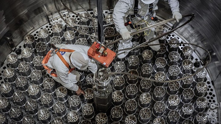
The dummy fuel is designed to be an exact replica of nuclear fuel in design, weight and dimensions, and its loading is key to checking systems for loading the real fuel as well as confirming readiness for the next stage of commissioning operations.
The dummy fuel does not contain any nuclear materials and its loading precedes the cold and hot running tests of reactor plant equipment during the commissioning process for new units, before the reactor starts up.
![]()
(Image: Akkuyu Nuclear)
![]()
(Image: Akkuyu Nuclear)
The loading of the fuel dummies was carried out under the supervision of Turkey's Nuclear Regulatory Authority.
Sergei Butckikh, CEO of Akkuyu Nuclear, said: "The completion of loading of dummy fuel assemblies at Akkuyu NPP Unit 1 is a full rehearsal for loading nuclear fuel. Using the dummies, we work out procedures for handling nuclear fuel in conditions as close to operational as possible, and confirm the readiness of equipment and personnel for the next pre-launch stage."
Background
Akkuyu, in the southern Mersin province, is Turkey's first nuclear power plant. Rosatom is building four VVER-1200 reactors, under a so-called BOO (build-own-operate) model. According to the terms of the 2010 Intergovernmental Agreement between the Russian Federation and the Republic of Turkey, the aim was for the commissioning of the first power unit of the nuclear power plant to take place within seven years from receipt of all permits for the construction of the unit.
The licence for the construction of the first unit was issued in 2018, with construction work beginning that year. The first steam generators were shipped to the site - for unit 1 - in August 2020. Nuclear fuel was delivered to the site in April 2023. The aim is for unit 1 to begin supplying Turkey's energy system during 2026.
When the 4,800 MWe plant is completed, it is expected to meet about 10% of Turkey's electricity needs.
![]()
Work is taking place on all four units - first concrete for unit 4 (right) was poured in August 2023 (Image: Akkuyu Nuclear)
Turkey has plans for a second nuclear power plant, at Sinop, and has also been in talks with China about plans for a third plant, in the Thrace region in the country's northwest.
The country is also developing plans for small modular reactors, with the aim of adding 5 GWe of capacity by 2050 - which would mean the equivalent of at least 16 individual SMRs.
SMRs to be considered at Romanian port
_88875.jpg)
"As ports electrify and grow, DP World sees access to reliable, low-carbon energy as critical to future competitiveness," the company said. "Rising demand from electrified equipment, shore power, AI data centres, residential heating and industrial activity is placing greater pressure on existing energy systems, driving demand for stable and scalable power. Nuclear energy, including SMRs, has the potential to provide consistent, low-carbon electricity for port operations and wider industrial use."
DP World has signed an agreement with French research organisation Commissariat à l’Energie Atomique et aux Energies Alternatives (CEA) and strategy specialist TerraWater Institute to launch a feasibility study into the use of SMRs at the Port of Constanța. At the mouth of the Danube-Black Sea Canal, the port links sea routes into Eastern and Central Europe, with deep-water access for larger vessels.
The study will model projected energy demand at the port from 2030 to 2050, evaluate integrated low-carbon energy systems, and assess the technical, strategic and economic feasibility of nuclear-based solutions. It will also examine safety standards and considerations for surrounding communities, drawing on CEA's expertise in SMR design and nuclear safety.
DP World said the study is intended to inform future decision-making on how best to meet long-term energy needs for the port and the wider economy. Any future development would be subject to further technical assessment, regulatory review and stakeholder engagement, it noted.
"DP World sees the transition to a net-zero economy not only as an environmental imperative, but as a driver of future growth across global trade," said Nicholas Mazzei, VP Sustainability – Europe, DP World. "Nuclear SMRs are not just energy projects for our ports, they are a competitive infrastructure differentiator. This study will help us better understand how nuclear energy can strengthen operational resilience and help meet rising demand. Across Europe, nuclear energy is increasingly recognised as a resilient and cost-effective solution with the potential to underpin the next generation of industrial activity and the supply chains."
Myrto Tripathi, General Director, TerraWater Institute, added: "Ports sit at the intersection of industry, energy systems, and communities. This study is about understanding how future low-carbon energy systems could be designed to meet complex and evolving demands, while maintaining high standards of safety and environmental performance. For energy as for everything, offer should not shape demand and should provide opportunities rather than dictate terms. Industries' needs have to be understood, assessed and met, while decarbonising. This is the only energy paradigm we should strive for and what we are aiming to demonstrate with this study, thanks to nuclear."
"This study brings together expertise in nuclear technology and energy systems to assess how small modular reactors could be integrated into a real port environment," said Stéphane Sarrade, Directeur des Programmes Énergies at CEA. "By working with DP World and TerraWater, we are applying advanced modelling and analysis to better understand how these solutions could support reliable, low-carbon energy for ports."
In September last year, DP World signed a memorandum of understanding with US-based micro-nuclear technology developer Last Energy to establish the world's first port-centric micro-nuclear power plant at London Gateway in the UK. A proposed PWR-20 microreactor - to begin operations in 2030 - would supply London Gateway with 20 MWe of electricity to power the logistics hub, with additional capacity exported to the grid.
Fuel manufactured for Kudankulam 4's initial loading
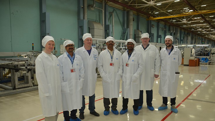
Under the contract agreed in 2024 with the Russian state nuclear corporation, TVEL will supply fuel for the lifetime of the VVER-1000 units, which comprise units 3 and 4 at the plant.
The Kudankulam site, near the southern tip of India, is already home to two Russian VVER-1000 pressurised water reactors - owned and operated by the Nuclear Power Corporation of India Ltd - which have been in commercial operation since 2014 (Kudankulam unit 1) and 2017 (unit 2). Four more are currently under construction in two phases: construction of units 3 and 4 began in 2017, with the work on units 5 and 6 beginning in 2021. Two further units - Kudankulam 7 and 8, larger AES-2006 units with VVER-1200 reactors - have been proposed as a fourth phase of the plant.
The first nuclear fuel was delivered for unit 3 in December. It was manufactured at Rosatom's Novosibirsk Chemical Concentrates Plant.
Rosatom says that during operation of the first two units, its specialists, together with Indian specialists "have significantly improved their efficiency through the introduction of advanced nuclear fuel and extended fuel cycles. Since 2022, the Kudankulam NPP has been supplied with advanced TVS-2M nuclear fuel. It ensures more reliable and cost-effective operation of the power units due to its rigid structure, a next-generation anti-debris filter, and a higher uranium mass".
It has also led to the time between refuelling shutdowns being extended from 12 months to 18 months. Units 3 and 4 will operate with 18-month fuel cycles from the start.
According to World Nuclear Association information, India currently has 24 operable nuclear reactors totalling 7,943 MW of capacity, with eight reactors - 4,768 MW - under construction. A further 10 units - some 7 GW of capacity - are in pre-project stages.
India has a target to expand its nuclear energy capacity to 100 GW by 2047. It plans to achieve this by a two-pronged approach, with the deployment of large-capacity reactors as well as small modular reactors (SMRs). In August last year Minister of State Jitendra Singh outlined to the country's Parliament the three types of SMR that are being designed and developed by the Bhabha Atomic Research Centre for demonstration: the 200 MWe Bharat Small Modular Reactor (sometimes referred to as BSMR-200); a 55 MWe small modular reactor (SMR); and a 5 MWt high-temperature gas-cooled reactor for hydrogen production by coupling with suitable thermochemical process for hydrogen production.
Orano starts construction at Mongolia uranium project
A ceremony at the Zuuvch Ovoo site marked the start of the construction phase for implementation of the project in Mongolia.
_97026.jpg)
"Yesterday, in the presence of Mr Batjargal Ochirpurev, Governor of the Dornogobi province, Mr Ganburen Gansukh, Governor of the Ulaanbadrakh sum and Mr Manlaijav Gun-Aajav, Secretary of the Nuclear Energy Commission, we celebrated a decisive milestone in the implementation of this strategic project led by Orano and its subsidiary Badrakh Energy, alongside our Mongolian partners," Orano Chairman Claude Imauven said on LinkedIn.
"As our two countries celebrated the 60th anniversary of their diplomatic relations in 2025, Zuuvch Ovoo illustrates our shared commitment to developing a strategic project that creates sustainable value for Mongolia and the Dornogobi Province," he added, before thanking the Mongolian authorities, Orano's partners, and the teams at Badrakh Energy and Orano "for their commitment to this exemplary cooperation".
Mongolia has substantial uranium resources - as of 2023, according to World Nuclear Association's information library, its 144,600 tU of uranium resources put it 10th in the world. Although it has been mined there in the past - in conjunction with Russian interests - no uranium has been mined in Mongolia since the mid-1990s when mining at the Dornod mine, operated by a subsidiary of Russia's Priargunsky Industrial Mining & Chemical Union, ceased.
_f716ec8f.jpg)
Image: Orano
Orano Mining has been present in Mongolia for more than 25 years, and has been carrying out exploration in the Gobi Desert since 1997, according to information from the company. The Zuuvch Ovoo deposit was discovered in 2010. In January 2025, Orano and the Government of Mongolia signed an investment agreement to develop and operate the project, in the south-eastern Dornogovi province.
The project will use in-situ leach (ISL, also known as in-situ recovery, or ISR) methods, demonstrated in pilot operations in 2021-2022. Development is planned to take 4 years. The project will have a nominal production capacity of about 2,500 tU per year for a 30-year estimated lifespan, creating 1,600 direct and indirect jobs.
Under the terms of the investment agreement, more than 51% of the direct benefits generated by the project will be received by the Mongolian state.
KHNP says EC has dropped foreign subsidy probe into Czech project
_73546.jpg)
The Czech government selected Korea Hydro & Nuclear Power (KHNP) as its preferred bidder in July 2024 for two new units near the current Dukovany nuclear power plant, about 200 kilometres southeast of Prague. Two more units at the Temelín nuclear power plant are also being considered. The engineering, procurement and construction contract was signed in June 2025, for two APR-1000 units at a projected cost of CZK407 billion (USD18.6 billion). The aim is to start construction in 2029.
France's EDF, which had been eliminated from the bidding process, launched legal challenges against the contract decision. The company's objections to the tender process included the belief that the KHNP offer price and the inclusion of a guarantee that the construction would not be delayed or become more expensive, would be "unfeasible without illegal state aid given the prices in the nuclear industry". EDF said that if their rival bidder had state support it would breach European Union rules. KHNP rejected EDF's claims and said "we emphasise that we have not received any subsidies that could damage or distort fair competition in relation to the project".
In response, the European Commission (EC) launched a preliminary review of KHNP and 'Team Korea' - the winning consortium of Korean companies that includes KHNP - in February 2025 to independently examine matters related to the new nuclear power plant project in the Czech Republic. The EU Extraterritorial Subsidy Regulation is a system designed to assess whether financial contributions provided to companies by non-EU countries distort competition in the EU market.
"KHNP and Team Korea faithfully cooperated with the preliminary review process by submitting relevant materials and explaining necessary matters in accordance with the request of the EC," KHNP said. "As a result, the EC completed the preliminary review and finally notified KHNP on 5 June that it had decided not to initiate an in-depth investigation."
"This decision is an official judgement made by the EU after directly reviewing the relevant issues," Industry Minister Kim Jung-kwan was quoted as saying by The Chosun Daily. "It is a result of confirming that KHNP and Team Korea have faithfully complied with international norms and EU laws and systems while pursuing the project."
Czech Industry and Trade Minister Karel Havlíček said on social media platform X, that the EC decision "to close the preliminary review under the Regulation on distortive foreign subsidies affecting the internal market ... is good news for this project and for the development of the nuclear industry and the future assurance of energy security in the Czech Republic and the European Union".
There has been a separate EC review taking place relating to the Czech new nuclear plan - in April 2024 the EC approved the original Czech government funding plan for a single new nuclear reactor at the Dukovany nuclear power plant site, but in October last year the Czech Republic officially notified the EC it had expanded its plans to two new nuclear units. The following month, the EC announced it had launched an inquiry into Czech funding plan for new nuclear. At the time it said it had doubts about whether it was fully in line with EU State aid rules and wanted to ensure that "no more aid than necessary is ultimately granted. In particular, the Commission has doubts on whether the proposed package achieves an appropriate balance between reducing risks to enable the investment and maintaining incentives for efficient behaviour, while avoiding excessive risk transfer to the State".
Ceremony to mark first concrete for Uzbekistan SMR
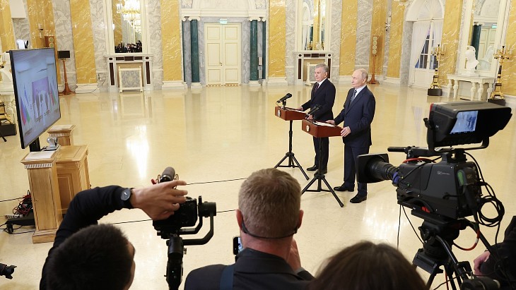
The presidents of Uzbekistan and Russia, meeting in St Petersburg, joined the event via video link, with International Atomic Energy Agency Director General Rafael Mariano Grossi among those attending in person.
Azim Akhmedkhadzhaev, Director of the Uzbekistan Atomic Energy Agency, said: "Today, we are not simply laying the first concrete for the nuclear power plant's foundation. We are laying the foundation for a bright and sustainable future for the Republic of Uzbekistan. This integrated nuclear power plant will symbolise a new technological stage for our country - a stage of energy independence, industrial growth, and environmental security."
"Uzbekistan is confidently moving to the forefront of the global energy sector, strengthening its sovereignty and opening new horizons for innovative development. We are building more than just a power plant - we are laying the foundation for a new era of prosperity, technological leadership, and well-being for future generations of Uzbeks."
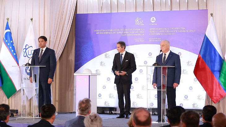
The IAEA's director general was at the ceremony (Image: Uzatom)
First concrete followed the Committee for Industrial, Radiation, and Nuclear Safety under the Cabinet of Ministers of the Republic of Uzbekistan issuing a licence on 4 June to the general contractor for the construction of the nuclear power plant unit's first unit, a Russian-made RITM-200N.
The planned plant
A contract signed in May 2024, during a visit to the country by Russian President Vladimir Putin, was originally for the construction of a 330 MW capacity nuclear power plant featuring six units of the RITM-200N water-cooled small modular reactor (SMR), which is adapted from nuclear-powered icebreakers' technology, with thermal power of 190 MW or 55 MWe and with an intended service life of 60 years. The first unit was scheduled to go critical in late 2029 with units commissioned one by one.
In 2025 a supplemental agreement to the contract for the new nuclear power plant - in the Jizzakh region - covered the decision to change its contents to two gigawatt-scale VVER-1000 units and two SMRs. This increased the proposed capacity to more than 2,100 MWe, compared with the previous 330 MWe.
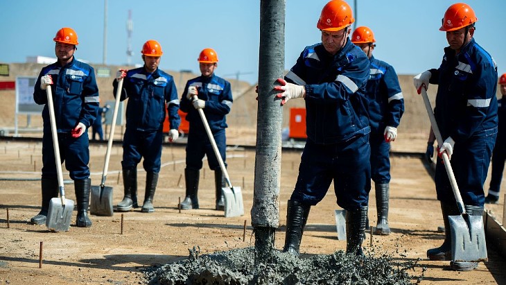
Concrete work at the site began in March (Image: Rosatom)
Excavation work began in October last year for the pit for the first of the SMRs at the site. About 1.5 million cubic metres of soil were excavated during the digging of a pit 13 metres deep. In March this year, Rosatom said that about 900 cubic metres were being poured during the concrete foundation work for the reactor building. That was due for completion in April and it said that the foundation has since been levelled and waterproofed before the pouring of the first concrete for the reactor building's foundation slab.
What the presidents said
President Putin said: "The fact that Russia and Uzbekistan are implementing such a truly flagship high-tech project is a shining example of the friendship and alliance between our two countries ... the project will provide related orders for many Uzbek companies: new jobs will be created, and local contractors will be actively involved in installation, material supply, transportation, and other services. In total, approximately 15,000 people are expected to be employed at the construction site.
"Importantly, Russia will not only build the nuclear power plant but also provide its Uzbek partners with a preferential export loan and support throughout the plant's entire lifecycle. This includes commitments for long-term reactor fuel supplies, servicing and maintenance, and spent nuclear material management. Essentially, with our country's assistance, a national high-tech nuclear industry is being developed in Uzbekistan."
President Shavkat Mirziyoyev said: "Today, we are launching not just the next stage of an infrastructure project, but are participating in an historic event. We are ushering in a new era of technological, industrial, and scientific development for our country. In Uzbekistan, the foundations are being laid for the development of a new field - modern nuclear energy - an industry that symbolises advanced scientific capabilities, cutting-edge engineering expertise, and a strategic vision for the future.
"It is important to note that this project ... is unique in the world; it combines the latest advances in small-scale nuclear generation and large-scale baseload energy."
The IAEA's Grossi noted the uniqueness of the project - which features the first export order for any SMR - and added: "I see investors from other countries here, and they're interested in this project. This project will also contribute to the development of the digital economy, data centres, and other opportunities."
Andrey Petrov, First Deputy Director General for Nuclear Energy at Rosatom, said: "Uzbekistan is embarking on a path of accelerated high-tech development, and Rosatom is honoured to be part of this historic process. Once operational, the nuclear power plant will be able to meet up to 14% of the country's energy needs. Moreover, the nuclear city project we proposed to Uzbekistan will create a new community. The nuclear power plant will be more than just a small town; it will be a true science city - a showcase for cutting-edge nuclear and related technologies."