It’s possible that I shall make an ass of myself. But in that case one can always get out of it with a little dialectic. I have, of course, so worded my proposition as to be right either way (K.Marx, Letter to F.Engels on the Indian Mutiny)
Monday, April 20, 2026
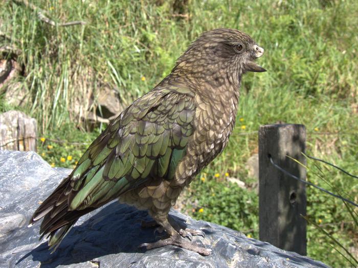
Disabled parrot is undefeated alpha male of his group thanks to novel “beak jousting”
A study reported in the Cell Press journal Current Biology on April 20 shows how physical disabilities in the animal world can be overcome through behavioral innovation. The report features an endangered kea parrot in captivity at New Zealand’s Willowbank Wildlife Reserve named Bruce who is missing his entire upper beak. While earlier reports had described his unique use of pebbles as self-care tools, the new findings show how he uses a novel beak jousting technique to turn his disability into social dominance.
“Bruce is the alpha male of his group,” says study first author Alexander Grabham of Te Whare Wānanga o Waitaha | University of Canterbury (UC) in New Zealand. “He achieved this status by himself with the aid of a completely novel fighting technique—a jousting thrust with his exposed lower beak—that beak-intact kea cannot replicate.”
Compared to other kea using their beaks during fights, the researchers found that Bruce not only used jousting more frequently but also targeted different body areas in different ways. His jousting was also more effective than when he kicked. His innovative fighting technique led him to win every single male dominance interaction that the researchers recorded.
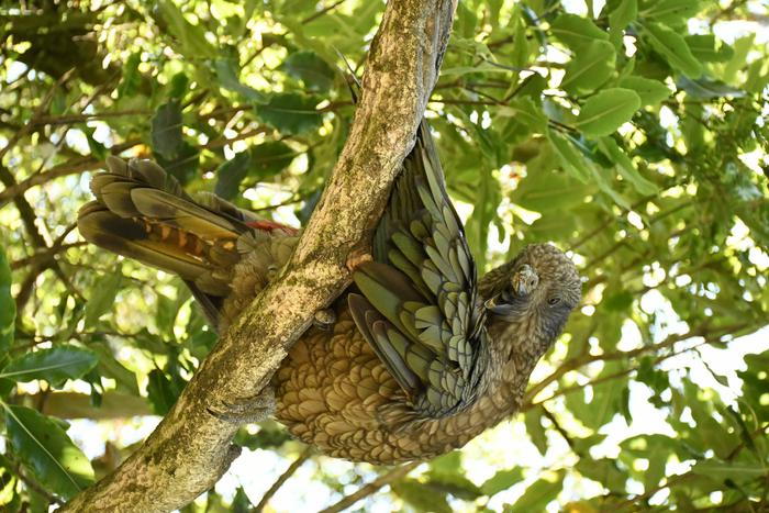
His winning record apparently led to other health benefits. Bruce had the lowest levels of corticosterone hormone metabolites levels, which is a sign of reduced stress compared to his peers. He enjoyed priority access to feeders and was the only male to be allopreened by other males, including beak cleaning.
Bruce had already earned some fame before, offering the first recorded case of self-care tool use in a kea. Grabham and colleagues noticed that Bruce fought other kea in a way they had never seen before. They wanted to learn more about what he was doing exactly and what it meant for his social position and the rest of his group.
Overall, the researchers have recorded 227 agonistic interactions from the Willowbank kea, including 9 males and 3 females. Out of 162 interactions between males, Bruce came out on top, winning all 36 interactions he was part of. The findings confirmed Bruce as the clear winner and dominant alpha male of the group.
The researchers describe how he uses his exposed lower beak in jousting thrusts, both at close range and from afar. Bruce uses his beak up close by extending his neck. He also would run or jump to propel his beak at opponents. They found that 73% of the time, his jousting behaviors, which other parrots don’t replicate, displaced opponents immediately. Their observations show he dominates not only in agonistic interactions but also socially during feeding and allopreening.
The findings highlight the remarkable behavioral flexibility and intelligence of endangered kea. But they also have broader implications about physical disabilities and what’s possible, according to the researchers.
“Bruce shows us that behavioral innovation can help bypass physical disability, at least in species with the cognitive flexibility to develop new solutions,” Grabham says. “Previous research has shown links between large brains, behavioral flexibility, and survival at the species level. Bruce demonstrates how those links play out in a single individual, on traits that matter day-to-day, like social dominance. Our findings also raise an important welfare question: if a disabled animal can innovate its way to success, well-intentioned interventions like prosthetics might not always improve their quality of life. Sometimes the animal can do better without help.”
###
This work was supported by the Templeton World Charity Foundation, an ERC Consolidator Grant UNIPROB, a Robert C. Bates Postgraduate Fellowship, and a Gordon Grant Postgraduate Fellowship.
Current Biology (@CurrentBiology), published by Cell Press, is a bimonthly journal that features papers across all areas of biology. Current Biology strives to foster communication across fields of biology, both by publishing important findings of general interest and through highly accessible front matter for non-specialists. Visit: http://www.cell.com/current-biology. To receive Cell Press media alerts, contact press@cell.com.
Bruce perched in a tree on one leg preening himself
A migratory bird brain, the Eurasian blackcap (Sylvia atricapilla), has been mapped for the first time using high-resolution light microscopy. The open-source software tools developed, and the detailed processes published, form a foundation for new brain atlases to be built for any species, providing a valuable resource for neuroscience worldwide. Created by a team from the Sainsbury Wellcome Centre at UCL and the University of Oldenburg, Germany, a paper describing the atlas has been published today (20 April 2026) in Current Biology.
Brain atlases - digital, high-resolution, 3D maps of brain structures - are transforming neuroscience. They improve the ability of researchers to interpret their own data, they enable cross-validation between and within experiments, and they foster collaboration - driving forward studies into learning, memory and cognition.
“A digital open-source brain atlas allows researchers to directly align their own experimental multimodal data to the common coordinate space of the atlas. It enables consistency, meaning researchers around the world can speak the same language when it comes to the brain. We are delighted to bring this resource to the community, and even more excited about building many more atlases for other research communities in the future,” said Dr Simon Weiler, Senior Research Fellow at the Sainsbury Wellcome Centre at UCL, and lead author of the study.
The team is already working on creating a similar digital 3D brain atlas of the zebra finch (Taeniopygia guttata), a bird used to study vocal learning.
The new Eurasian blackcap atlas is freely accessible via BrainGlobe for the neuroscience research community and will advance studies of magnetoreception, migration and navigation. The technology means that any brain sample, even historic histology samples that have been stored for years on glass slides, for example, can be mapped onto the atlas.
Birds are among nature’s foremost navigators, using the Earth’s magnetic field to orient themselves and travel between breeding and wintering grounds. Many species travel thousands of miles with centimetres of precision. In the same publication, the team has revealed a previously unknown direct link between magnetosensitive areas in the brain and the decision-making centre, the nidopallium caudolaterale (equivalent to the prefrontal cortex in mammals), demonstrating how the atlas can assist in characterising novel brain pathways.
"To me, this is a key tool that the migration, navigation, and magnetoreception community has been lacking for decades. It will greatly improve consistency and comparability between studies and related species and will significantly accelerate our understanding of underlying neuronal mechanisms,” said Professor Henrik Mouritsen, University of Oldenburg, an author of the study.
To create the atlas, the team at SWC used serial two-photon (STP) tomography to image eight male Eurasian blackcap brains. This advanced imaging technique results in well-aligned 2 x 2 x 5 μm voxel size images of entire brains. The individual 3D images from different brains were then iteratively aligned and averaged to create a representative brain template. Following this, experts at the University of Oldenburg manually annotated the template. This resulted in 44 segmented brain areas, including principal brain compartments, prominent anatomical subdivisions shared across all bird species, regions of the song system, and sensory regions implicated in magnetic field processing.Finally, the atlas was incorporated into the BrainGlobe ecosystem and automatic registration, cell detection and object mapping were demonstrated on experimental data.
“The core aim of BrainGlobe is to democratise computational neuroanatomy. Creating novel atlases is a step in achieving this. All parts of the pipeline are open-source, and over the coming months we will be improving it so that we, and anyone else, can rapidly create new atlases,” said Dr Adam Tyson, Head of the Neuroinformatics Unit at the Sainsbury Wellcome Centre at UCL and lead of the BrainGlobe Initiative.
While the team used state-of-the-art STP tomography, other microscopies, including light-sheet images are also suitable for creating atlases. Future advances in whole-brain labelling procedures, paired with STP tomography, will further guide brain area subdivision based on region-specific identification of marker genes or proteins, and the atlas will be regularly updated to incorporate new data.
ENDS
This research was funded by the Gatsby Charitable Foundation, Wellcome, the Alexander von Humboldt Foundation, the Chan Zuckerberg Initiative DAF, the European Research Council and the Deutsche Forschungsgemeinschaft.
Source:
Read the full paper in Current Biology: ‘An open-source three-dimensional digital brain atlas of a migratory bird, the Eurasian blackcap’
The Sainsbury Wellcome Centre (SWC) brings together world-leading neuroscientists to generate theories about how neural circuits in the brain give rise to the fundamental processes underpinning behaviour, including perception, memory, expectation, decisions, cognition, volition and action. Funded by the Gatsby Charitable Foundation and Wellcome, SWC is located within UCL and is closely associated with the Faculties of Life Sciences and Brain Sciences. For further information, please visit: www.sainsburywellcome.org
About the University of Oldenburg
Carl von Ossietzky University was founded in 1973, making it one of Germany's younger universities. Its goal is to find answers to the big questions facing society in the 21st century through cutting-edge interdisciplinary research and teaching.
Researchers and administrative staff work hand in hand and across disciplines. Many are involved in research – for example, in collaborative research centers, research groups, European projects, or the three clusters of excellence NaviSense, Hearing4all.connects, and Ocean Floor.
The university works closely with more than 300 international cooperation partners and universities. It also has links with non-university institutions in research, education, culture, and business. The research location is further strengthened by the establishment of the Helmholtz Institute for Functional Marine Biodiversity, Max Planck Research Groups, and Fraunhofer working groups.
The university prepares around 15,000 students for professional life. The spectrum ranges from the humanities and cultural sciences to economics, law, and social sciences to mathematics, computer science, the natural sciences, and medicine.
A new study published in Landscape Ecology shows how fast-growing poplar plantations can improve functional connectivity for forest birds in fragmented agricultural landscapes, provided they are strategically located and species have moderate to high dispersal capacity. The findings suggest that managed forests may contribute not only to biomass supply, but also to biodiversity conservation in highly human-modified regions.
Using spatial connectivity models in two European river sub-catchments in Spain and France, researchers examined how existing forest patches, both within and outside Natura 2000 areas, and poplar plantations interact to support movement across the landscape for three forest bird species with contrasting dispersal abilities.
“Plantations can act as stepping stones between forest patches, although their effectiveness depended strongly on their location within the landscape,” says Sara Pineda-Zapata, a Doctoral Researcher at the University of Eastern Finland and the lead author of the study. “We wanted to understand whether plantations, often viewed only through the lens of wood and biomass production, could also support ecological processes in fragmented landscapes,” she continues.
In Spain, plantations generated connectivity gains that were greater than their area would suggest, with some patches playing an important role in maintaining ecological connectivity for forest networks, including Natura 2000 areas. In France, plantation patches were more isolated and contributed less effectively. The strongest benefits were observed for species capable of moving over longer distances; for short-distance dispersers like the common chaffinch, plantations had a more limited effect unless they were located very close to an existing forest habitat, suggesting that even narrow gaps can remain major barriers for less mobile species.
“Plantations are often assessed only in terms of production, but when strategically located, they can provide much more than wood. They can contribute to landscape structure, help maintain ecological flows and complement conservation efforts in intensively used agricultural regions. The key message is that location matters, and that planning matters,” says Professor Blas Mola at the University of Eastern Finland.
Professor Alejandra Morán of the University of Basel in Switzerland highlighted that the results are relevant beyond bird movement: “Connectivity influences how species move, persist and respond to environmental change. When we think about ecosystem services, we should consider not only what land use produces in one place, but how it shapes ecological processes across the wider landscape.”
Rémi Duflot, from the University of Jyväskylä, emphasised the broader implications: "Birds are particularly informative because they respond quickly to landscape fragmentation. However, we caution that plantations cannot replace natural forests in ecological quality, and that increasing tree cover may reduce habitat for open-habitat species, while forest specialists often require more complex structures than plantations provide.”
“What our results show is that in fragmented landscapes, well-placed plantations can become part of the solution, opening up interesting possibilities for designing productive landscapes that are also more supportive of biodiversity,” concludes Pineda-Zapata.
A new study has revealed cocaine pollution changed how wild fish moved through their environment, with juvenile Atlantic salmon swimming farther and dispersing more widely.
The international study, led by researchers from Griffith University, the Swedish University of Agricultural Sciences, Zoological Society of London and Max Planck Institute of Animal Behaviour, is the first to demonstrate the effects of cocaine contamination on fish behaviour in the wild rather than in laboratory conditions.
To understand how these pollutants influenced animal movement, the researchers used slow-release chemical implants and acoustic telemetry tracking to monitor 105 juvenile Atlantic salmon over eight weeks in Lake Vättern, Sweden.
The fish were assigned to one of three treatment groups: a control group, a group exposed to cocaine, and a group exposed to benzoylegonine, the primary metabolite of cocaine that is commonly detected in wastewater.
The team found fish exposed to benzoylecgonine swam up to 1.9 times farther per week than unexposed fish and dispersed up to 12.3km farther across the lake.
These changes became more pronounced over time, indicating that exposure altered how fish used space in a complex natural ecosystem
Co-author Dr Marcus Michelangeli, from Griffith University’s Australian Rivers Institute, said the findings were important because movement played a central role in how animals interacted with their environment.
“Where fish go determines what they eat, what eats them, and how populations are structured,” he said.
“If pollution is changing these patterns, it has the potential to affect ecosystems in ways we are only beginning to understand.”
Cocaine and its metabolites were increasingly detected in rivers and lakes around the world, primarily entering waterways through wastewater systems that were not designed to fully remove these compounds.
While previous research has shown cocaine could affect animal behaviour, those studies had been limited to laboratory settings.
This study provides the first evidence that these effects also occurred in the wild, where animals experienced far more complex environmental conditions.
The researchers also found the cocaine metabolite benzoylecgonine had a stronger effect on fish movement than cocaine itself.
This was significant because risk assessments typically focused on the parent compound, even though metabolites were often more common in waterways, suggesting current approaches may overlook important biological effects.
The team emphasised the findings did not indicate a risk to people consuming fish.
The exposure levels reflected those already found in polluted waterways, the compounds break down over time, and the fish studied were juveniles well below legal-catch size.
Dr Michelangeli said the study highlighted a broader issue about the types of pollutants entering aquatic ecosystems.
“The idea of cocaine affecting fish might seem surprising, but the reality is that wildlife is already being exposed to a wide range of human-derived drugs every day,” he said.
“The unusual part is not the experiment, it’s what’s already happening in our waterways.”
Future research would aim to determine how widespread these effects were, identify which species were most at risk, and test whether altered movement patterns translated into changes in survival and reproduction.
The paper ‘Cocaine pollution alters the movement and space use of Atlantic salmon (Salmo salar) in a large natural lake’ has been published in Current Biology.
How primitive plants evolved to survive Earth’s most catastrophic extinction event
Earth responded to its most severe past warming event by evolving a new and bizarre type of photosynthesis that allowed a group of primitive plants to survive.
Research led by the University of Leeds has revealed how lycophytes - a type of ancient plant - not only survived a mass extinction 250 million years ago but then came to dominate the recovering landscapes.
During the Permian-Triassic mass extinction, which is also known as the “Great Dying,” global temperatures rose dramatically with most forests collapsing under extreme heat and vast areas of land becoming barren.
The researchers believe lycophytes may have been the first plants to use this mechanism revealing a biological innovation that was able to keep Earth’s biosphere active with the plants able to remove carbon from the atmosphere, ultimately combating the effects of the warming event.
Today, plants using CAM photosynthesis make up only a small proportion of global vegetation and are most common in hot and dry environments such as deserts.
Lead author of the study, Dr Zhen Xu from Leeds’ School of Earth and Environment, said: “Our results suggest that under future warming, plants with CAM photosynthesis traits could become far more important.
“If the world experiences sustained extreme heat, plant communities may shift toward species that are better able to tolerate high temperatures and water stress.”
Lycophytes are spore-bearing vascular plants (a type of plant characterised by the presence of tissues for transporting water and nutrients). There are more than 1,200 species of the plant still in existence. They can survive in many places but are most diverse in tropical regions.
To understand how lycophytes survived when so many other plants perished, the researchers first studied their evolutionary relationships to find their closest relatives, such as the quillworts that can still be found around the world, including in Scotland. They then studied carbon isotopes (variants of carbon atoms) in fossil plants from South China from the late Permian to the Middle Triassic period. Different types of photosynthesis leave different carbon isotope signatures, so this can reveal how ancient plants functioned.
They found that lycophytes had carbon isotope values that were noticeably different from other plants during the Permian–Triassic extinction period. This difference became smaller once environmental conditions had improved.
The team then compared where the lycophyte fossils were found with climate model simulations. The results suggest that these plants lived in places where surface temperatures likely exceeded 50 °C.
The researchers believe increased knowledge about Earth’s geological past can help to inform predictions about future climate resilience, something which they say is becoming increasingly important in a warmer world.
Co-author of the study, Professor Barry Lomax of the University of Nottingham, said: "The analysis pulled together many separate scientific disciplines to test how this group of enigmatic plants not only survived the great dying but also how they were able to thrive in a highly stressed environment.
“By linking these data together, we are able to further understand plant adaptation to past climate emergencies deepening our understanding of the resilience of the Earth system to climate perturbations."
Professor Benjamin Mills from Leeds’ School of Earth and Environment added: “Understanding how plants’ diverse physiological strategies shaped ecosystems in the past helps us to anticipate how vegetation might reorganize in the future, and because plants are the base of terrestrial food webs, changes in dominant plant strategies can alter the functioning of the entire Earth system.”
Images two and three show lycophyte reproductive cones, belonging to the genus Lepacyclotes.
For media enquiries, please contact the University of Leeds press office via pressoffice@leeds.ac.uk
University of Leeds
The University of Leeds is one of the largest higher education institutions in the UK, with more than 40,000 students from about 140 different countries. We are renowned globally for the quality of our teaching and research.
We are a values-driven university, and we harness our expertise in research and education to help shape a better future for humanity, working through collaboration to tackle inequalities, achieve societal impact and drive change.
The University is a member of the Russell Group of research-intensive universities, and is a major partner in the Alan Turing, Rosalind Franklin and Royce Institutes www.leeds.ac.uk
An international study published in Current Biology presents the results of the analysis of ancient mitochondrial DNA obtained from eight Neanderthal teeth discovered in Stajnia Cave, Poland. For the first time, the research reconstructs the genetic profile of a small group of Neanderthals from the same site, north of the Carpathians, who lived during the same ancient chronological phase.
“This is an extraordinary result because, for the first time, we are able to observe a small group of at least seven Neanderthals from Central-Eastern Europe who lived around 100,000 years ago,” says Andrea Picin, professor at the University of Bologna and coordinator of the research. “In most cases, Neanderthal genetic data come from single fossils or from remains scattered across different sites and periods. At Stajnia, by contrast, it has been possible to reconstruct a small group of individuals, providing for the first time a coherent genetic picture of Neanderthals in this part of Europe.”
“We had known for some time that Stajnia Cave preserved exceptional evidence, but these results exceeded our expectations,” say Wioletta Nowaczewska of the University of Wrocław and Adam Nadachowski of the Institute of Systematics and Evolution of Animals of the Polish Academy of Sciences, co-authors of the study. “Being able to identify such an ancient small group of Neanderthals in such a complex site is an important achievement for Polish research and for the study of Neanderthals in Europe.”
The discovery also helps us better understand the distribution of a particular Neanderthal maternal lineage in western Eurasia. The mitochondrial DNA of the Stajnia Neanderthals falls within the same branch as that of other individuals found in the Iberian Peninsula, south-eastern France, and the northern Caucasus, suggesting that this genetic component was widely distributed before being replaced by those typical of more recent Neanderthals.
“A particularly fascinating aspect is that two teeth belonging to juvenile individuals and one belonging to an adult share the same mitochondrial DNA,” adds Mateja Hajdinjak, co-author of the article and researcher at the Max Planck Institute for Evolutionary Anthropology. “This suggests that these individuals might be closely related to each other.”
Another important aspect of the study concerns the comparison with the Neanderthal fossil Thorin, discovered in Mandrin Cave in France, which carries a mitochondrial genome similar to that of the Stajnia Neanderthals and has so far been assigned to a chronology of around 50,000 years ago. “Our study is a reminder that the oldest chronologies must be treated with great caution,” explains Sahra Talamo, professor at the University of Bologna and co-coordinator of the study. “When radiocarbon values approach the limit of calibration, it is essential not to assign more precision than the data can actually support. In such cases, the comparison between archaeology, radiocarbon dating, and genetics becomes crucial.”
From an archaeological point of view, the discovery reinforces the idea that Central-Eastern Europe was not a marginal periphery in Neanderthal history, but rather a key area for understanding population movements, biological connections, and the spread of technological traditions during the Middle Paleolithic. Stajnia Cave and southern Poland thus become a privileged observatory for reconstructing not only the biology of Neanderthals, but also their movements and the connections between groups distributed across wide areas of Europe.
For the first time, the research reconstructs the genetic profile of a small group of Neanderthals from the same site, north of the Carpathians, who lived during the same ancient chronological phase.
Credit
Max Planck Institute for Evolutionary Anthropology
Journal
Current Biology
Archaeogenetics: Neanderthals and the bottleneck theory
FAU prehistorians contribute finds from the University’s own Sesselfelsgrotte cave to an international study
The first Neanderthals emerged approximately 300,000 years ago. They settled in large parts of Europe and spread as far as southern Siberia. “We still don’t have a comprehensive understanding of Neanderthal population history, nor of the demographic processes that led to their extinction,” says Prof. Dr. Thorsten Uthmeier, Chair of Prehistoric Archaeology and the Archaeology of Prehistoric Hunter-Gatherers at FAU. “Maps of archaeological sites suggest that an event occurred during the last glacial period that caused a rapid decline in the geographic distribution and genetic diversity of the early population. It was believed that only a small group survived and that all later Neanderthals descended from this group. In genetics, such processes are referred to as ‘bottlenecks’.”
DNA samples provide new insights
To test and refine the bottleneck theory, an international research team led by the University of Tübingen analyzed ten new mitochondrial DNA sequences (mtDNAs) from Neanderthal remains found at six archaeological sites in Belgium, France, Germany, and Serbia and compared them with 49 previously published mtDNAs. “The mtDNA samples do not come from the cell nucleus, but rather from the mitochondria – single-celled structures that regulate a cell’s energy metabolism and have their own DNA,” explains Uthmeier. Mitochondrial DNA is used in archaeological research because it is more stable, occurs in greater quantities, and is easier to analyze than nuclear DNA. One of the new mtDNA samples included in the study comes from a Neanderthal fetus that was discovered in 1968 by FAU researchers in the Sesselfelsgrotte cave in the Altmühl Valley near Kelheim, Germany. The preparatory work at FAU was conducted as part of the project “SHARP – Testing hypotheses on the transition from Neanderthals to Homo sapiens at the Paleolithic site of Sesselfelsgrotte,” which began in 2023 and is funded by the National Geographic Society.
From the samples, researchers can identify lineages. “mtDNA mutates much less frequently than nuclear DNA, which plays a key role in determining our appearance and physical constitution, among other things,” says Thorsten Uthmeier. “However, the degree of diversification in the mtDNA samples provides an insight into how closely related the Neanderthal groups – from which the bone and tooth fossils originate – were to one another.” Innovative analytical methods, such as decoding genetic information by breaking DNA down into individual gene sequences, made it possible to include samples in the study that had previously proven impossible to analyze. By comparing the newly decoded mtDNA with existing mtDNA, researchers have now been able not only to establish kinship but also to estimate ages based solely on genetic data. The ability to determine the age of samples in this way is an important development, as conventional dating methods have proven unsuitable for dating a great many samples. The combination of these methods has made it possible to reconstruct temporal and spatial patterns in the distribution of late Neanderthals.
A refugium in France 65,000 years ago
The study’s findings suggest that the last bottleneck event – which is believed to have played a major role in the extinction of the Neanderthals – occurred around 65,000 years ago. According to Uthmeier, “as recently as 130,000 years ago, Neanderthals were widespread throughout Western Eurasia, predominantly in what is now northern Germany and Belgium. There were isolated groups in the Caucasus, and even one in the Altai Mountains in southern Siberia.” Over the course of just tens of thousands of years, both genetic diversity and the range of the species declined, and its core population shifted increasingly toward southwestern France. “We suspect that the climatic conditions 65,000−60,000 years ago, a very cold and dry period, triggered the retreat to this refugium and the extinction of the remaining Neanderthal lineages,” explains Uthmeier. Subsequently, the Neanderthals began to populate a much wider area again, with virtually all later Neanderthals descendants of the group originally based in southwestern France.
There is, however, one exception: During excavations in the heart of the refugium, in Mandrin Cave in the Rhône Valley, the skeleton of a Neanderthal was found and named Thorin. There is evidence that he lived past the bottleneck, but his mtDNA differs significantly from that of the other survivors and should actually have become extinct. “Until recently, it was thought that Thorin belonged to an isolated group that had remained in a very small area,” says Uthmeier. “However, the genetic analysis now conducted has shown that the fetus from the Sesselfelsgrotte in the Altmühl Valley, whose remains date from a similar period, was also related to this group. The Thorin line was apparently more widespread than previously thought. This finding really surprised us.”
The researchers were also able to provide data to answer the question of when and why Neanderthals ultimately became extinct. “The combination of DNA analysis and age dating has revealed that a sharp decline in population size began around 45,000 years ago,” says Thorsten Uthmeier. It is still unclear exactly what led to their extinction about 3,000 years later. In addition to significant differences in the size and density of social networks, it is possible that parts of the last Neanderthal population were absorbed by groups of Homo sapiens sapiens, who, having come from Africa, spread across increasingly larger areas of Europe. Uthmeier: “Modern humans and Neanderthals were capable of interbreeding, which is why we still carry a small percent of Neanderthal DNA in us today.”