Saturday, July 18, 2026


Sensing warm and cool: how the body detects temperature changes


University of Queensland
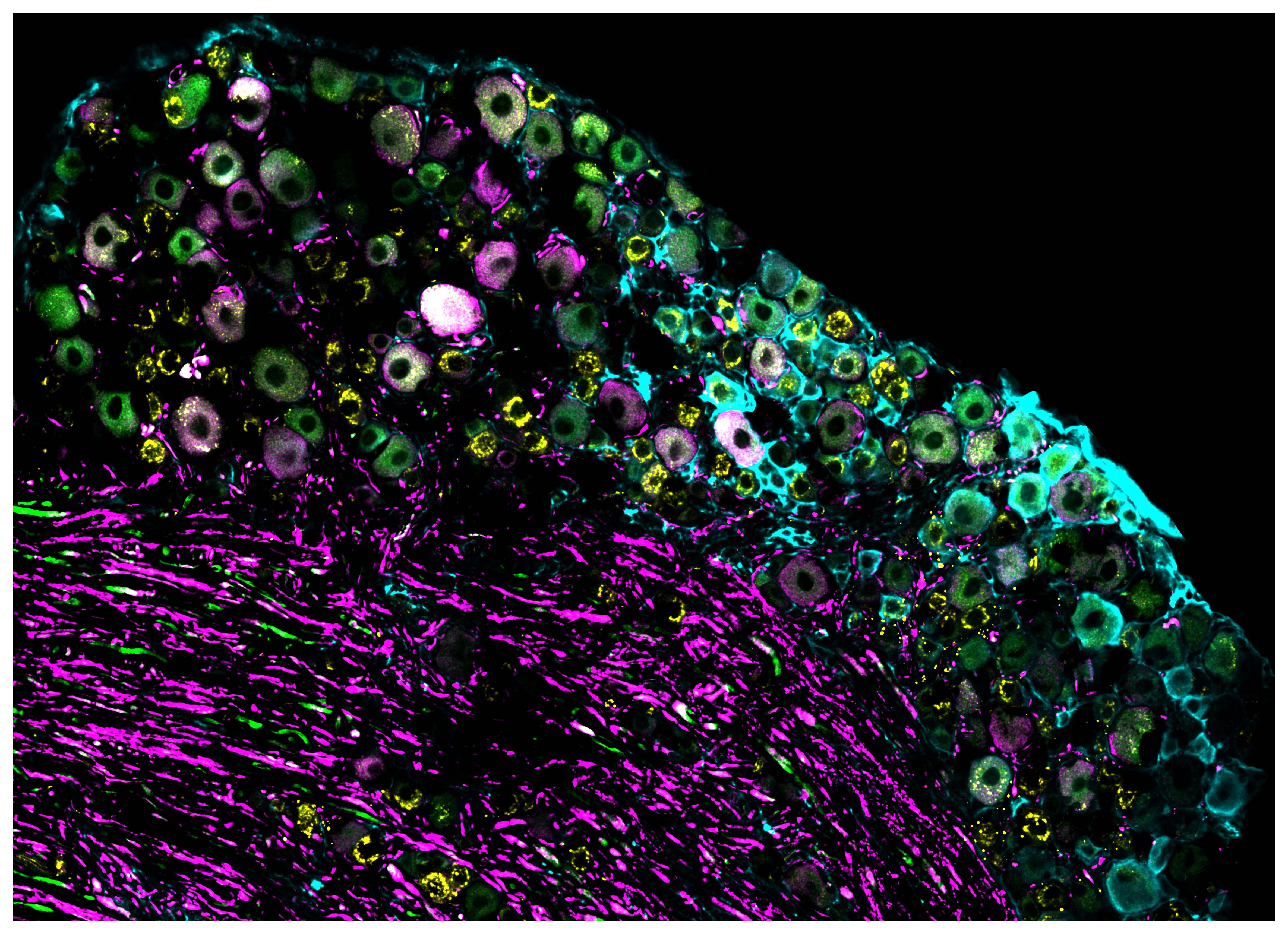
Researchers examined how thousands of thermoreceptor nerve cells responded to cool and warm temperatures. 

image: 

Researchers examined how thousands of thermoreceptor nerve cells responded to cool and warm temperatures.

view more 

Credit: Dr Phill Bokiniec, The University of Queensland.


New research has challenged what scientists understand about how the body’s nervous system senses warm and cool temperatures. 

University of Queensland researchers found most of the skin’s temperature sensitive nerve cells can sense both cool and warmth, challenging a widely accepted view that skin relies on separate nerve cells to detect each temperature.

Dr Clarissa Whitmire, from UQ’s Queensland Brain Institute , said these specialised nerve cells, called thermoreceptors, are critical to human survival.

“Thermoreceptors are the body’s first responders, detecting and relaying to the brain what is happening at the body’s surface,” Dr Whitmire said.

“Our study shows that rather than relying on 2 separate nerve cells to sense warmth and cool, the body’s thermoreceptors can signal both sensations to the brain – increasing activity in cooler conditions and decreasing when temperatures rise. 

“These findings could help explain what happens when the body’s thermoreceptors become impaired in ageing and disease.”

Using advanced imaging in mice models, researchers tracked how thousands of thermoreceptor cells responded to cool and warm temperatures.

The study focused on every day, ordinary non-painful temperatures such as entering cold rooms, or warm bathwater.

Dr Phill Bokiniec from UQ’s Queensland Brain Institute, said this new understanding of the body’s thermoreceptors could inform treatments for people struggling with thermal dysfunction.

“Humans tightly regulate their core body temperature, making accurate temperature sensing critical to homeostasis – the body's ability to maintain a stable internal environment,” Dr Bokiniec said.

“People living with spinal cord injury, multiple sclerosis, diabetes or peripheral neuropathy can lose aspects of thermal sensors, making it difficult to respond to environmental temperature changes. 

“Ageing is also a significant concern – older adults are at risk in heat waves and climate change - and disrupted thermal sensors may contribute to why they struggle to regulate temperature.”

Dr Whitmire said researchers further wanted to understand if impaired thermoreceptors were an early indicator of degeneration in the body, similarly to how hearing loss has been linked to dementia.

“Our hope is our research will change the way the body’s thermoreceptors are understood, which is critical to developing effective therapies,’’ she said.

“This is important because if treatments target the wrong nerve cells or pathways, they simply won’t work.”

Read the research in Neuron


Researchers discover the eye’s hidden cleanup system


Newly identified lymphatic network could fundamentally shift how we understand and treat leading causes of blindness


University of British Columbia


Many of the world’s leading causes of irreversible blindness, including glaucoma and age-related macular degeneration, share a common problem: the buildup of fluid, waste and inflammatory debris in the back of the eye. 

However, for decades, scientists have lacked a clear understanding of how the eye clears this waste away. 

New research from the University of British Columbia and the University of Toronto has identified what appears to be a missing piece of the puzzle: a previously unknown waste drainage system at the back of the eye. The hidden circulatory pathway, called the posterior ocular lymphatic outflow, or POLO pathway, provides a route for fluid and waste to exit the eye and enter the body’s lymphatic system.  

“The retina is one of the most metabolically active parts of the body, constantly generating byproducts that need to be cleared,” said Dr. Neeru Gupta, professor and head of UBC’s department of ophthalmology and visual sciences. “This discovery helps explain how the eye flushes this waste and promises to transform how we think about and treat a range of eye conditions.” 

A foundation for future therapies 

Diseases like glaucoma, macular degeneration and retinal disorders are all associated with fluid buildup, accumulation of metabolic waste, and tissue stress and inflammation. Age-related macular degeneration alone affects approximately 2.5 million Canadians.  

The discovery of the POLO pathway suggests there is a natural system responsible for clearing this material and opens new opportunities to harness this system to treat disease.   

“This gives us a whole new framework for understanding these diseases and a potential target for therapeutics,” said Dr. Gupta. “The question now is: How can we enhance or exploit this cleanup system to treat or even prevent disease.” 

Hiding in plain sight 

For more than a century, the eye was thought to lack a lymphatic system, which is present in nearly every other organ and helps to regulate fluid, remove waste and support immune function. 

Dr. Gupta and Dr. Yeni Yücel, professor and director of ophthalmic pathology at the University of Toronto, began challenging that idea in 2009 with the discovery of a lymphatic-related drainage pathway at the front of the eye, termed the “uveolymphatic” pathway. Yet the back of the eye, where many blinding diseases are rooted, has remained largely unexplored.  

“The retina is responsible for vision and it’s also where many of the most serious vision-loss diseases occur,” said Dr. Yücel. “Understanding how this part of the eye maintains a balanced environment and flow of materials is critical.”  

To uncover the POLO pathway, the team used advanced imaging in mice, combining MRI, near-infrared fluorescence and microscopic analysis. They introduced fluorescent tracer molecules into a thin space at the back of the eye and tracked their movement in real time. 

This allowed them to identify small lymphatic vessels in the choroid, a thin layer beneath the retina. Fluid drained from the back of the eye into surrounding orbital tissue and, within minutes, reached nearby lymph nodes that link the eye to the broader lymphatic system. 

“Because lymphatic vessels in the choroid were thought not to exist, the team used multiple techniques to demonstrate both their presence and function,” added Dr. Yücel, a pathologist-scientist at St. Michael’s Hospital. “We were surprised to see such a direct route for fluid to leave the eye and connect with the lymphatic system. It suggests the back of the eye has an active clearance pathway, which could play an important role in removing fluid, proteins and inflammatory material that build up in disease.”  

Further research is needed to understand how the lymphatic pathway operates in humans and how it could be targeted with therapeutics.  

But the researchers say it could one day lead to improved drug delivery to the back of the eye, new therapies that enhance fluid clearance, and deeper insight into how pressure, inflammation and fluid dynamics contribute to vision loss. 

“This is a foundational discovery that shows the eye is not as closed a system as we previously thought,” said Dr. Gupta. “It gives us a new map, a new mechanism and a new set of questions to explore.” 

The study was published today in Translational Vision Science & Technology [LINK].  

The research was supported by the Canadian Institutes of Health Research, Glaucoma Research Society of Canada, Henry Farrugia Ophthalmology Research Fund, Canadian Space Agency, Dorothy Pitts Chair, Stephen M. Drance Chair, Thor and Nicky Eaton Research Fund and Canada Foundation for Innovation Leaders Opportunity Fund. 


New computational imaging method cuts X-ray dose while preserving high resolution


Ultra-low-light ghost imaging technique lays the groundwork for safer X-ray-based medical diagnostics


Optica

Experimental results 

image: 

Researchers have shown that it’s possible to take clear, high-resolution X-ray images using very little radiation. An image acquired using the new method (a) is compared with conventional radiology (b) under identical photon counts.

view more 

Credit: Tiqiao Xiao, Shanghai Advanced Research Institute, Chinese Academy of Sciences


WASHINGTON — Researchers have shown that it’s possible to take clear, high-resolution X-ray images using very little radiation. With more development, the new approach could eventually make medical X-ray diagnostics less risky and more accessible.

“While traditional X-ray imaging relies on enough X-ray photons reaching a detector to form a clear image, our approach uses computational techniques to reconstruct an image from fewer photons,” said research team leader Tiqiao Xiao from the Shanghai Advanced Research Institute, Chinese Academy of Sciences. “We were able to show the low-dose potential of this approach by achieving megapixel radiology with ultra-low-light.”

In Optica, Optica Publishing Group ’s journal for high-impact research, the researchers demonstrate X-ray ghost images with nearly 2-megapixel resolution using only ‌0.48%‌ of the X-ray photons typically required for X-ray imaging. The proof-of-concept study suggests that comparable X-ray image quality may eventually be achievable with far lower radiation doses than are used today.

“Our technology could be combined with routine hospital imaging equipment such as chest X-rays and CT scans,” said Xiao. “It would make medical X-ray imaging‌ safer, which is especially important for children, pregnant patients and people needing frequent scans.”

High-resolution with less radiation

Ghost imaging is a computational imaging approach that works by correlating two beams, in this case, X-rays. One beam encodes a random pattern that acts as a reference and never directly probes the sample while the other passes through the sample. Because very little X-ray power comes into contact with the object being imaged, ghost imaging has the potential to reduce radiation exposure when used for medical imaging.

Although various research teams have been working to implement X-ray ghost imaging, it hasn’t been fully demonstrated experimentally. The primary challenge has been obtaining high-resolution images while keeping X-ray exposure extremely low. The researchers overcame this limitation by reengineering the imaging system to maximize both image quality and dose efficiency.

In the new approach, which the researchers call ultra-low-light X-ray ghost imaging, the X-ray beam is split into a weak beam that interacts with the sample and a much stronger reference beam, allowing more information to be gathered from every X-ray photon. This was accomplished using highly uniform and strongly correlated X-ray crystal splitting, which enabled synchronous signal acquisition in both beams.

The researchers also incorporated detectors that are optimized for each beam — one designed to capture faint signals and another designed to record fine spatial details. Another key was a synthetic-aperture imaging approach that uses an intelligent algorithm to reconstruct a full high-resolution image from ‌dozens of measurements‌, far fewer measurements than conventional ghost imaging methods require.

“Together, these advances balance three key performance indicators simultaneously: a large field of view, ultra-low photon consumption and high imaging resolution,” said Xiao. “This provides a more practical and effective solution for X-ray ghost imaging.”

Demonstrating ultra-low-dose imaging

The researchers demonstrated their new ghost imaging method in a controlled comparative experiment using the X-ray test beamline (BL09B) at the Shanghai Synchrotron Radiation Facility. Using identical ultra-low-photon conditions, they imaged a sample area using the new method and conventional direct X-ray radiology. The results showed that the new method delivered much higher quality than direct imaging.

The new technique was able to produce X-ray images measuring 1992 × 944 pixels with the same contrast-to-noise ratio (CNR) as conventional radiology while using 0.48% of the photons. It also achieved the maximum CNR attainable with conventional radiology using 100 times fewer photons.

The researchers note that further development is needed before the new imaging method can be used clinically. They plan to improve the image quality and demonstrate the method with laboratory X-ray sources, such as X-ray tubes, a key step toward translating the technology from synchrotron-based experiments to real-world medical imaging.

Paper: C. Zhao, H. Zhang, J. Tang, N. Zhao, J. Wu, Z. Li, T. Xiao, “Ultra-low-light megapixel X-ray ghost imaging” 


Experimental setup 

The researchers show that X-ray ghost imaging can achieve nearly 2-megapixel resolution using only ‌0.48%‌ of the X-ray photons typically required for X-ray imaging. The experimental setup and key components of the ghost imaging system are shown.

Credit

Tiqiao Xiao, Shanghai Advanced Research Institute, Chinese Academy of Sciences


Brain signal linked to communication challenges in autism


University of Virginia Health System
Brain signal linked to communication challenges in autism 

image: 

Jack Van Horn, left, of the University of Virginia School of Data Science, and Kevin Pelphrey, of UVA's School of Medicine, combine data science and neuroscience to reveal brain activity patterns that were previously difficult to detect. 

view more 

Credit: University of Virginia


Why do some children with autism communicate more easily than others, even when they hear the same words?

Researchers from the University of Virginia believe the answer may lie in the brain’s electrical activity. In a new study published in Scientific Reports, they found that subtle patterns in brain activity while children listened to speech were linked to how well autistic youths communicate in everyday life. 

The findings offer new clues about the biology behind autism and could one day help researchers objectively measure communication challenges and evaluate new therapies.

The research analyzed brain activity in more than 300 children and adolescents while they listened to speech. The findings suggest subtle differences in brain electrical activity may help explain why some autistic youths have greater difficulty with verbal communication than others.

The study included researchers from the University of Virginia’s schools of Medicine and Data Science, along with colleagues from Seattle Children’s Research Institute, the University of Washington, Yale University, UCLA and several other institutions.

“This is an important step toward understanding the neural mechanisms underlying communication in autism,” UVA neuroscientist Kevin Pelphrey, a coauthor of the study, said. “If we can identify reliable biological markers, they could eventually help researchers evaluate interventions more objectively and understand why communication abilities differ so widely across the autism spectrum.”

Researchers have long known that many autistic individuals experience challenges with language and communication, but the underlying brain mechanisms have remained difficult to measure. Most clinical assessments rely on behavioral observations, rather than biological indicators.

To investigate those mechanisms, the research team recorded brain activity from 306 participants aged 7 to 18, including 162 youths with autism and 144 typically developing peers. Participants wore high-density electroencephalography, or EEG, caps equipped with 128 sensors while listening to streams of spoken nonsense words designed to measure how the brain processes speech.

Rather than focusing solely on traditional brain wave patterns, the researchers examined a newer measure of overall neural activity, known as the brain’s “aperiodic” signal. The signal reflects the balance between excitation and inhibition, two fundamental processes that help the brain distinguish meaningful information from background activity. 

The study found that autistic participants showed altered patterns in these signals, consistent with increased neural “noise,” suggesting the brain may process speech less efficiently.

More importantly, youths whose brain activity appeared noisier also tended to score lower on measures of everyday verbal communication. Those same brain signals were not associated with traditional language skills, such as vocabulary or grammar.

The researchers caution that the findings do not represent a diagnostic test for autism. Instead, they point to a promising biological marker that could eventually help researchers monitor changes in communication abilities over time, or measure whether therapies are affecting underlying brain function.

The work also highlights the growing role of advanced data science techniques in neuroscience, allowing researchers to uncover subtle patterns in complex brain data that were previously difficult to detect.

“The human brain generates an incredible amount of data every second,” said Jack Van Horn, a coauthor and professor in UVA’s School of Data Science. “The challenge isn’t collecting it anymore; it’s making sense of it. Advances in computational analysis are allowing us to separate meaningful signals from background activity in ways that weren’t possible just a few years ago.”

Although the study included one of the largest EEG datasets of its kind, researchers say additional work is needed before the findings could influence clinical care. Most participants had average or above-average verbal abilities, and future studies will need to determine whether the results extend to minimally verbal individuals with autism. 

The authors also note that EEG provides an indirect measure of brain activity and should ultimately be combined with other imaging techniques to better understand the underlying biology.

Still, the findings move scientists closer to a longstanding goal in autism research: developing objective biological measures that complement behavioral evaluations.


Heavy TV watching associated with smaller brain structures, study finds


Reduced volume was found in areas of the brain connected to memory formation, indicating a potential link between TV watching and higher dementia risk


University of Southern California


“Turn off that TV, it’ll rot your brain!” has been a household refrain for decades. While “rot” might be too strong a term, researchers are finding that the overall sentiment could have some merit.

A study published recently in Alzheimer’s and Dementia: Journal of the Alzheimer’s Association revealed that those who reported watching TV “very often” in midlife later exhibited reduced volume in areas of the brain associated with memory, smaller frontal and occipital lobes, and areas of damage in the brain’s white matter that are associated with aging, stroke risk, cognitive decline and dementia. 

“For years we’ve focused on how much people sit. Our findings suggest we should also pay attention to what they’re doing while they’re sitting,” says David Raichlen, professor of biological sciences and anthropology at the USC Dornsife College of Letters, Arts and Sciences and a senior author of the study. 

The findings weren’t just due to TV viewing’s sedentary nature. The study found that other types of sedentary activities did not have the same associations, indicating that what one does while sitting may matter much more than previously thought.

Watching changes

The researchers analyzed data from about 1,700 adults, average age 53, who enrolled in the Atherosclerosis Risk in Communities (ARIC) Study between 1987 and 1989. ARIC is a long-running study of the U.S. population designed to investigate cardiovascular and brain health. 

Participants were asked how frequently, on a scale ranging from “never/seldom” to “very often,” they watched television during their leisure time and how much of their workday they spent sitting.

More than two decades later, participants underwent brain MRI. Compared with people who reported “never” or “seldom” watching TV, those who watched TV “very often” showed widespread structural differences across the brain.

The researchers found smaller volumes in areas associated with early signs of Alzheimer’s disease and more white matter hyperintensity volumes, an indicator of cerebral small blood vessel disease associated with cognitive decline and dementia. These participants also had smaller occipital and frontal lobes, regions associated with visual processing and executive functioning.

Differences persisted even when the researchers controlled for factors such as physical activity, diabetes, body mass index, smoking, and alcohol use.

Of note, the researchers relied on self-reported data for TV consumption, which can be less precise than timed tracking. Study participants also did not undergo a baseline MRI. Future research could begin with a baseline MRI to more concretely demonstrate changes over time.

Not all sitting is made the same

Strikingly, the sedentary element of TV watching didn’t appear to be the main driver for these changes.

Those who reported high amounts of sitting at work actually had larger frontal and occipital lobes, as well as reduced white matter hyperintensity volumes, indicating better brain health than among those who sit to watch TV. This could be due to the intellectually stimulating nature of many sit-down jobs, say the study authors. 

Men appeared to be particularly vulnerable to these changes. When the MRI scans were separated by sex, researchers found that most of the changes to the brain, both from TV watching and occupational sitting, were seen in men.

Such findings indicate there is still more research to be done on this complex topic. However, we might eventually see a different approach to health guidance around sedentary activities. Rather than just directing their patients to move more, for example, physicians might recommend they reduce television time and add cognitively engaging activities for when they do sit.  

“We frequently encourage the public not to spend too much time sitting down, but experts may want to expand that recommendation to encompass the activities done while sitting, since those seems to have distinct impacts on brain health,” says study corresponding author Natan Feter, postdoctoral scholar in the Human and Evolutionary Biology program at USC Dornsife. 

About this study

In addition to Raichlen and Feter, study authors include Anamika Nanda, Mark Lai, Rand Wilcox and Sarah Hourihan of USC Dornsife; Daniel Aslan of Harvard University; Jayne Feter of Universidade Federal do Rio Grande do Sul; M. Katherine Sayre of University of California Santa Barbara; Pradyumna Bharadwaj, Madeline Ally, Hyun Son, Yann Klimentidis, Amit Arora and Gene Alexander of the University of Arizona; Silvio Maltagliati of USC Dornsife and Université Bretagne Sud.

This research was supported by National Institutes of Health grants P30AG072980, P30AG019610, R56AG067200, R01AG064587, and R01AG072445 and funding from the state of Arizona, the Arizona Department of Health Services and the McKnight Brain Research Foundation.