
The EU got access to Anthropic's most powerful model, three months later
 Copyright AP Photo/Patrick Sison
The EU's cybersecurity agency ENISA has gained access to Anthropic's Mythos 5, one of the most powerful AI technologies with disruptive cyber capabilities, three months after it was released to selected partners.
Brussels has finally obtained access to Anthropic's most powerful model, whose availability had been restricted due to its advanced hacking capabilities. But the request took over three months to bear fruit.
"Following our constructive engagement with Anthropic, we can confirm that the EU's cybersecurity agency ENISA has been granted access to Mythos 5 and is testing it now," said the European Commission's spokesperson for tech sovereignty, Thomas Regnier.
In April, the leading AI lab launched a preview of Mythos 5, a model capable of identifying and exploiting cyber vulnerabilities with unprecedented speed, making it a formidable hacking tool.
Given its cybersecurity implications, Anthropic restricted access to the model to a select number of partners via Project Glasswing, run in collaboration with the US government.
The programme started with around 50 organisations in April before Anthropic expanded it by some 150 more in June, bringing the total to roughly 200 partners. The US government subsequently introduced export control measures barring access to non-American users worldwide — including Anthropic's own employees outside the US.
The move prompted the European Commission to question whether it was discriminatory towards a trusted partner, fuelling concern that Washington was both capable and willing to trigger a "kill switch" on the latest US technologies.
Brussels continued pressing Anthropic for access, which was finally granted on Thursday. The Commission holds significant regulatory powers under the EU AI Act, powers that may make tech companies wary of sharing information that could later surface in infringement proceedings.
"ENISA has previously received access to OpenAI's GPT-5.6-Cyber model. Access was also granted to the latest OpenAI model, GPT-6 Astra," Regnier said.
Brussels has finally obtained access to Anthropic's most powerful model, whose availability had been restricted due to its advanced hacking capabilities. But the request took over three months to bear fruit.
"Following our constructive engagement with Anthropic, we can confirm that the EU's cybersecurity agency ENISA has been granted access to Mythos 5 and is testing it now," said the European Commission's spokesperson for tech sovereignty, Thomas Regnier.
In April, the leading AI lab launched a preview of Mythos 5, a model capable of identifying and exploiting cyber vulnerabilities with unprecedented speed, making it a formidable hacking tool.
Given its cybersecurity implications, Anthropic restricted access to the model to a select number of partners via Project Glasswing, run in collaboration with the US government.
The programme started with around 50 organisations in April before Anthropic expanded it by some 150 more in June, bringing the total to roughly 200 partners. The US government subsequently introduced export control measures barring access to non-American users worldwide — including Anthropic's own employees outside the US.
The move prompted the European Commission to question whether it was discriminatory towards a trusted partner, fuelling concern that Washington was both capable and willing to trigger a "kill switch" on the latest US technologies.
Brussels continued pressing Anthropic for access, which was finally granted on Thursday. The Commission holds significant regulatory powers under the EU AI Act, powers that may make tech companies wary of sharing information that could later surface in infringement proceedings.
"ENISA has previously received access to OpenAI's GPT-5.6-Cyber model. Access was also granted to the latest OpenAI model, GPT-6 Astra," Regnier said.
AMD unveils ‘personal super computer’ for new era of computing
 Copyright AP Photo
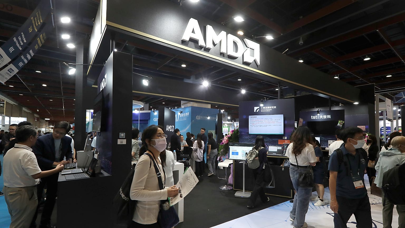
The American chipmaker launched what they have described as a ‘personal supercomputer’ in Berlin on Friday, designed and made for a “completely new era of computing” driven by AI.
The supercomputer, called the Threadripper Halo Station, was unveiled at the German tech event Internationale Funkausstellung by AMD Senior Vice President Jack Huynh.
The Threadripper Halo is an ultra-high-performance workstation equipped with up to two terabytes of system memory, up to 576 gigabytes of high bandwidth memory (HBM3E), along with 96 cores of Threadripper Pro, two MI350P data centre accelerators, all supported by liquid cooling.
Huynh said the new supercomputer is the “most powerful workstation in the world”, capable of running AI models with more than a trillion parameters.
The AMD executive gave a brief demonstration of its potential at the event by generating an entire 3D world and flight simulator from a single prompt.
The company’s pitch is to bring data-centre level AI performance and capacity to a desktop format, as demand for AI rapidly grows around the world.
The supercomputer is slated for release early next year, however AMD are yet to reveal how much it will cost. It is expected that it could exceed €100,000, geared towards corporate and institutional budgets rather than as personal computers as originally pitched.
Yet it will give its consumers a platform to deploy large-language models and agentic AI systems themselves rather than rely on increasingly expensive cloud computing.
The Threadripper Halo Station’s direct competitor is the NvidiaDGX Station, with both able to run large AI models in a desktop form, however they differ in how they approach this challenge.
AMD’s supercomputer dominates on memory capacity and bandwidth, offering more than three times as much memory than Nvidia’s, allowing it to run on trillion parameter AI models locally.
However, Nvidia’s offering more seamless integration when moving code from a local workstation to the cloud and, crucially, it is already available, retailing at around €100,000.
Industry analysts see AMD's announcement as a potential catalyst for reshaping the competitive landscape of the local AI computing market.
With cloud AI subscription costs adding up for many institutions and businesses, demand for computers such as the Threadripper Halo Station to run large models on their own infrastructure will continue.
Therefore, AMD's approach of delivering data-centre-class performance using standardised components to lower the barrier to entry could be a major turning point in the expansion of the AI and personal computing sectors.
The supercomputer, called the Threadripper Halo Station, was unveiled at the German tech event Internationale Funkausstellung by AMD Senior Vice President Jack Huynh.
The Threadripper Halo is an ultra-high-performance workstation equipped with up to two terabytes of system memory, up to 576 gigabytes of high bandwidth memory (HBM3E), along with 96 cores of Threadripper Pro, two MI350P data centre accelerators, all supported by liquid cooling.
Huynh said the new supercomputer is the “most powerful workstation in the world”, capable of running AI models with more than a trillion parameters.
The AMD executive gave a brief demonstration of its potential at the event by generating an entire 3D world and flight simulator from a single prompt.
The company’s pitch is to bring data-centre level AI performance and capacity to a desktop format, as demand for AI rapidly grows around the world.
The supercomputer is slated for release early next year, however AMD are yet to reveal how much it will cost. It is expected that it could exceed €100,000, geared towards corporate and institutional budgets rather than as personal computers as originally pitched.
Yet it will give its consumers a platform to deploy large-language models and agentic AI systems themselves rather than rely on increasingly expensive cloud computing.
The Threadripper Halo Station’s direct competitor is the NvidiaDGX Station, with both able to run large AI models in a desktop form, however they differ in how they approach this challenge.
AMD’s supercomputer dominates on memory capacity and bandwidth, offering more than three times as much memory than Nvidia’s, allowing it to run on trillion parameter AI models locally.
However, Nvidia’s offering more seamless integration when moving code from a local workstation to the cloud and, crucially, it is already available, retailing at around €100,000.
Industry analysts see AMD's announcement as a potential catalyst for reshaping the competitive landscape of the local AI computing market.
With cloud AI subscription costs adding up for many institutions and businesses, demand for computers such as the Threadripper Halo Station to run large models on their own infrastructure will continue.
Therefore, AMD's approach of delivering data-centre-class performance using standardised components to lower the barrier to entry could be a major turning point in the expansion of the AI and personal computing sectors.
AI D-Day weather forecast falters under pressure
University of Reading
Today's best weather computers would still have struggled to accurately predict the weather for the D-Day landings, University of Reading research shows.
The new research, accepted in the journal Weather ahead of the release of D-Day blockbuster Pressure, used AI models to produce 50 different forecasts of the storm that famously pushed back the Allies’ invasion of Normandy in June 1944.
The researchers checked how confident the AI model was about the weather Group Captain James Stagg had to forecast on 5 and 6 June. For 5 June, the day originally planned for the invasion, the model got the storm's position right and correctly predicted poor conditions over Normandy, matching Stagg's own analysis. For 6 June, the model predicted the storm would move towards Norway, bringing calmer weather, albeit much quicker than what really happened.
Professor Andrew Charlton-Perez, lead author of the study at the University of Reading, said: "Captain James Stagg had one of the hardest jobs in history. Get the forecast wrong, the invasion could have failed and the Second World War could have been lost.
“Some Allied meteorologists were insistent the weather would be fine for the invasion to take place as planned on 5 June, after weeks of hot weather. Our research shows Stagg would likely have defied his colleagues and made the same call to delay if he had the AI forecast to support him. The AI pointed to a real risk of dangerous winds on 5 June, enough to justify delaying the invasion by a day.
“The AI incorrectly predicted 6 June would be much calm and clearer than it really was, as soldiers faced more treacherous weather. Captain Stagg believed the conditions were fair enough on 6 June, leading to Eisenhower to give his famous order, "OK, we'll go."
“AI weather forecasts can process huge amounts of data in seconds, but they can still make mistakes. Having a trained meteorologist to make sense of all of the forecast data and make forecasts based on their knowledge and experience remains just as critical today as it was for the D-Day landings."
Where the model went wrong
The extraordinary story of how Captain James Stagg had to determine if the weather would be good enough to let 160,000 troops cross the Channel and storm the beaches of Normandy on 5 June is now the subject of a major Hollywood film called Pressure starring Andrew Scott as Stagg and Brendan Fraser as Eisenhower.
In the film, Stagg has to balance his advice between different forecasting teams who disagree about how a storm in the North Sea will affect the invasion.
Researchers compared the model against what actually happened over the Channel and the Normandy beaches, using weather records from the Copernicus Climate Change Service.
The model showed the storm clearing away from Britain too quickly on the 6th June. This wrongly suggested that 6 June would be calm and clear, when on the day of the invasion some of the invading forces faced high winds and stormy seas.
Underneath that overall confidence, the wind numbers told a different story. The model still showed a 30% chance the wind would break the safety limit for landing craft on 6 June, a real risk hidden behind its reassuring overall picture.
The model also predicted clearer skies than actually happened on 6 June, adding to the false sense that conditions would be fine.
Filling in the gaps
Weather records from 1944 were limited, so the researchers also compare the forecasts with new paper weather logs for the Faroe Islands recently found in an archive at the Met Office. These islands sit close to the path the storm took on its way past Britain, so the readings kept there in 1944 capture the storm at a point the main records miss.
Turning these old paper logs into digital data made it possible to check the storm's strength more closely and showed it may have been slightly stronger than current reconstructions suggest. Digitising more of these historic logs from archives across Europe could let scientists test storms like this one even more precisely in future.
Pressure releases in UK cinemas on Wednesday, 9 September.
Notes to editors:
Professor Andrew Charlton-Perez is available for interview. Contact the University of Reading Press Office on 0118 378 5757 or pressoffice@reading.ac.uk
Images via Studio Canal.
Full reference: Charlton-Perez, A., Burt, S., Cloke, H., Hawkins, E., Lee, R., Simmons, A. and Volonte, A. (2026), Pressure: reimagining the D-Day forecast through machine learning. Weather, 81: 284-293. https://doi.org/10.1002/wea.70123
Journal
Weather
Article Title
Pressure: reimagining the D-Day forecast through machine learning
Article Publication Date
2-Sep-2026
Augmentation not abdication, when using AI in research
An editorial by Charles Branas and Bruce Levine proposes norms for the use of AI in science. The authors distinguish between using AI tools for non-generative tasks, such as data classification or copyediting, and using AI tools to generate scientific ideas or seed research questions. While the former is broadly acceptable, they argue, the latter deserves more scrutiny and should be disclosed in scientific manuscripts. The use of generative AI for the core intellectual, creative, ethical, interpretive, or accountability-bearing work of research, where AI replaces human interpretation and judgement, should be disallowed, according to the authors. Additionally, the authors propose that researchers should not use AI to generate their own initial research ideas, research questions, or to make ethical determinations, risk assessments, or policy recommendations.
AI can reduce science’s happy accidents and human insights if everyone is relying on the same tools with similar inputs, the authors argue. They note that science should not be unthinkingly optimized, as if it were a manufacturing workflow producing standardized outputs. Science requires a wide diversity of inputs, approaches, and ideas. Generative AI is trained to produce probable outputs based on known patterns and trained on existing data, whereas scientific breakthroughs often require departing from prior assumptions and breaking free of typical patterns. Serendipity counts in science.
According to the authors, the originating research question and final evidentiary judgment in every scientific publication should be produced by human minds.
Journal
PNAS Nexus
Article Title
AI and authorship: Norms and uses to preserve human-led science
Article Publication Date
8-Sep-2026
Weeks of work in a few hours: AI tool reads cell membranes
One membrane to teach it, thousands to find - AI detects membranes and their proteins in 3D cell images, cutting weeks of analysis to hours.
Cell membranes and the proteins within them control many vital processes and play a key role in health and disease. But studying them in 3D images of cells has so far meant slow, manual work. A team from Helmholtz Munich, the Technical University of Munich (TUM) and the Biozentrum of the University of Basel has developed MemBrain v2, an AI tool that automates this task – cutting work that once took weeks down to a few hours. The freely available software finds membranes, locates specific membrane proteins and analyzes how these are spatially arranged, showing how cellular processes are organized at the molecular level. Depending on the application, the AI requires little or no additional training data to do this. That lets researchers around the world study how cells work in detail – faster and on a much larger scale. The tool is presented in the journal Nature Methods.
Cryo-electron tomography (cryo-ET) is a special microscopy technique that lets researchers look inside cells – in three dimensions and at very high resolution. Because the cells are flash-frozen for this, they are preserved almost unchanged, from whole cell structures down to individual molecules. Membranes, however, have so far been difficult to analyze in this kind of data.
"One challenge is that cryo-ET images can contain gaps in information due to technical limitations of the imaging process. As a result, certain membrane orientations are difficult or partly impossible to see. This is exactly where MemBrain v2 comes in, automating the process," explains first author Lorenz Lamm.
Optimizing a Key Technology in Cell Biology
MemBrain-seg detects membranes directly, without requiring users to provide additional annotations or training data. MemBrain-pick also requires only a small amount of training data: In one test, researchers manually annotated the positions of protein complexes on just a single membrane. Based on these annotations, the tool localized the corresponding protein complexes on additional membranes with an F1 score of 91 percent. Until now, this 3D image data had to be labeled painstakingly by hand, and the results could rarely be reused for new datasets. Existing programs usually handled only single parts of the analysis – for example outlining the membranes or locating the proteins within them.
Three Tools in One Software – and Little Data Suffices
For the first time, MemBrain v2 combines three steps in a single AI tool: it finds membranes (MemBrain-seg), locates the proteins embedded in them (MemBrain-pick) and measures how these proteins are arranged (MemBrain-stats).
In several applications, the tool matched the results of painstaking manual analyses, but was considerably faster. It is also easy to use and can be applied to other research questions without major adjustments. Because all components are open source, the membrane-detection module is already widely used around the world and has, for example, been applied across datasets from the Chan Zuckerberg Imaging Institute.
“By making these analyses faster and accessible to research groups worldwide, we can study cellular processes across much larger datasets. This can ultimately help us better understand how cells function – and what changes when disease develops,” says senior author Dr. Tingying Peng.
Larger Datasets and Finer Distinctions Ahead
MemBrain v2 has already contributed to new biological insights: In a separate study, the tool showed that important photosynthesis proteins are spatially separated within the membrane – challenging previous models of their organization. In the future, it is set to distinguish different protein types even more precisely.
“I’m especially pleased that MemBrain v2 is now being used in many further studies, where it simplifies demanding analyses or makes them possible in the first place – making a concrete contribution to new biological insights,” says first author Lorenz Lamm.
Original Publication
Lamm et al., 2026: MemBrain v2: an end-to-end tool for the analysis of membranes in cryo-electron tomography. Nature Methods. DOI: 10.1038/s41592-026-03178-8
Software: https://github.com/CellArchLab/MemBrain-v2
About the Researchers:
Lorenz Lamm, first author, PhD researcher in the groups of Dr. Tingying Peng (Helmholtz Munich) and Benjamin Engel (Biozentrum, University of Basel), corresponding author.
Dr. Tingying Peng, head of the Helmholtz AI group “AI for Microscopy Image Analysis” (Helmholtz Munich) and TU Munich, senior and corresponding author.
Prof. Benjamin D. Engel, group leader at the Biozentrum of the University of Basel, corresponding author.
About Helmholtz Munich
Helmholtz Munich is a leading biomedical research center. Its mission is to develop breakthrough solutions for a healthier society in a rapidly changing world. Interdisciplinary research teams focus on environmentally driven diseases, in particular the therapy and prevention of diabetes, obesity, allergies and chronic lung diseases. Using artificial intelligence and bioengineering, the researchers work to translate their findings to patients more quickly. Helmholtz Munich has more than 2,555 employees and is headquartered in Munich/Neuherberg. It is a member of the Helmholtz Association, the largest scientific organization in Germany, with more than 48,000 employees and 18 research centers. More about Helmholtz Munich (Helmholtz Zentrum München Deutsches Forschungszentrum für Gesundheit und Umwelt GmbH): www.helmholtz-munich.de
Journal
Nature Methods
Article Title
MemBrain v2: an end-to-end tool for the analysis of membranes in cryo-electron tomography.
Article Publication Date
8-Sep-2026
AI model decodes the language cells use to communicate
image:
The developing branching lung in an in vitro culture system.
view moreCredit: Whitehead Institute
As an embryo develops from a small cluster of stem cells, those once “blank slate” cells begin to take on more specialized roles like brain, liver, or muscle cells, and organize themselves into three-dimensional structures such as tissues and organs.
The fate of each cell — what type of specialized cell it will become — depends on which genes are turned on or off in the cell. These patterns of gene activity shape the cell’s structure and function, enabling it to take on a specific role in the body.
But this decision isn’t up to individual cells. They constantly send and receive chemical signals to and from neighboring cells, which help them understand where they are, what stage of development they are in, and what they should become. These messages spread through multi-step sequences called signaling pathways, which translate external signals into specific changes in gene activity in the cell.
For researchers, being able to retrace the sequence of instructions a cell has received would offer a powerful way to understand how tissues develop and how these processes go awry in disease. But this has been difficult to achieve because scientists have long assumed that the effects of signaling pathways vary widely across cell types, meaning they would need to map each pathway separately in each cell type — an arduous and painstaking process.
Now, in a new study led by Whitehead Institute Member Pulin Li and graduate student Nicholas Hutchins, researchers have discovered that each signaling pathway leaves behind a unique “fingerprint” — a distinctive pattern of gene activity that reflects the particular signals the cell has encountered.
Importantly, these fingerprints are consistent across different cell types for the same signaling pathway, which means that instead of mapping each cell type separately, scientists can reconstruct signaling histories across many cell types using these pathway-specific fingerprints.
This discovery was made possible through a machine learning model called IRIS. This model can detect fingerprints of different signaling pathways and pinpoint which signals a cell received at different stages of development inside an embryo, even for cell types it hasn’t encountered before.
This AI-driven approach marks a major advance over traditional methods, which require researchers to experimentally test every pathway in every possible cell type, and opens up the possibility to comprehensively map the signaling histories of every cell inside a mouse or human embryo at an unprecedented scale.
“Think of voice recognition systems like Siri, which are trained mainly in English, but then use that training to help them recognize other languages,” says Li, who is also an assistant professor of biology at the Massachusetts Institute of Technology (MIT). “This is called transfer learning, and this is why IRIS can work across many different cell types.”
The researchers’ detailed findings, published in the journal Nature Methods on Sept. 8, could accelerate stem cell engineering for regenerative medicine and improve the creation of organoids — miniature, 3-D models that mimic real organs — for studying disease mechanisms and testing new drugs. This is because once researchers learn the pattern of signals that drives a stem cell to become a specific cell type, they can recreate those signals to control the fate of stem cells in a lab or medical setting.
IRIS is a neural network-based model, which, in essence, is an AI-driven system designed to recognize patterns in complex data, similar to how our brains spot patterns. It examines a cell’s overall gene activity and estimates which signaling pathways were likely “on” at specific times during development.
Li and Hutchins trained IRIS on a large experimental dataset that measured how thousands of human embryonic stem cells responded to dozens of combinations of six major signaling pathways at multiple stages of development. This created a comprehensive map, or atlas, of how signaling combinations influence cell behavior.
The team then tested IRIS using single cells from mouse embryos during gastrulation, a stage when cells are rapidly branching into different fates. IRIS could accurately predict when and where specific signaling pathways would activate in cells destined to become part of heart, gut, muscle, and spinal cord tissue. This variation in signaling molecules is what guides cells to form the right structures in the right places.
With this approach, scientists can not only begin to understand the fundamentals of cell-to-cell communication, but also gain a practical roadmap for guiding stem cells into specific, functional cell types in the lab.
When the researchers employed IRIS to identify signals needed to create a cell type critical for lung development, the model predicted that activating a specific signaling pathway would encourage lung-specific development. Experiments in mouse embryos confirmed the model’s prediction. By identifying the precise combinations of signals that drive lung cell development, they can more reliably generate accurate, lab-grown models of lung tissue.
“In these ways, IRIS is helping us decode the language cells use to talk to each other at a much faster rate than we could realistically achieve through experiments,” Hutchins says.
These improved models would allow researchers to study diseases like asthma, lung cancer, and pulmonary fibrosis, in which lung tissue becomes scarred, often without a known cause. They can then use these models to test potential therapies, and ultimately design regenerative treatments that can repair the damaged tissue.
About Whitehead Institute:
Whitehead Institute is a nonprofit, independent biomedical research institute founded in 1982. The institute advances pioneering research in cancer, developmental biology, genetics, genomics, and related fields, with a mission to pursue bold, curiosity-driven science that deepens our understanding of life and improves human health. Led by 24 principal investigators and a global community of trainees and scholars, Whitehead Institute maintains a teaching affiliation with Massachusetts Institute of Technology (MIT) but is fully independent in its research programs, governance, and finances.
Journal
Nature Methods
Article Publication Date
8-Sep-2026

No comments:
Post a Comment