Artificial hibernation reveals secrets of long-term memory
Challenging long-held assumptions on how memory works, scientists uncover the importance of engram architecture to long-term memory retention.
image:
An illustration of the inner workings of a mouse brain during artificial hibernation. Workers prune the irrelevant dendritic spines from brain cells, leaving only the synapses necessary for long-term memory retention.
view moreCredit: Luo-chu Yang
How do we remember the distant past? For decades, scientists have believed synaptic potentiation — the adaptive strengthening of our brain’s cellular connections — to be the key to memory retention. Published in Science, research from the Okinawa Institute of Science and Technology (OIST) and collaborators including the University of Tsukuba, the Exploratory Research Center on Life and Living Systems (ExCELLS), and National Institutes of Physiological Sciences, challenges these long-held beliefs. Their new paper demonstrates the importance of higher-order synaptic architecture, suggesting that specific clustered patterns of connections between brain cells may be key to retaining long term memory.
Professor Kazumasa Tanaka, head of OIST’s Memory Research Unit, says. “Previously, synaptic strengthening was thought to be key to memory recall, and that stronger synapses with larger dendritic spines were fundamental to long-term memory retention. Here, we show that not every synapse matters, and demonstrate instead the vital importance of engram architecture. The study indicates that small clusters of engram-engram synapses are preserved to enable accurate recall even after hibernation.”
The physical forms of memories
Much like computer storage, we humans require a physical memory trace to be stored in the brain. This physical trace, known as an engram, is encoded through a dedicated network of brain cells undergoing changes at their synapses, the junctions where they meet. When connecting brain cells repeatedly fire together, their synapses strengthen, increasing neurotransmitter release and triggering structural changes, such as larger dendritic spines, which expand the contact area between the two cells. Conversely, when a particular connection isn’t very active, the synaptic bonds between the cells weaken, and may eventually disappear altogether.
The synapses with larger, more stable dendritic spines have traditionally been seen as key for memory. While we might expect these connections to stay consistent over the course of a memory, recent studies have found that the structures and numbers of cells involved in a particular engram can change over time, without affecting recall. So, what exactly is it with our engrams that let us remember memories?
Artificial hibernation as a research tool
To investigate, the researchers turned to hibernation. In a hibernation state, a decreased metabolism enables creatures to survive harsh, wintery conditions with little food. It also causes brain activity to dramatically reduce.
In 2020, a team led by Professor Takeshi Sakurai at the International Institute for Integrative Sleep Medicine (WPI-IIIS), Tsukuba Institute for Advanced Research (TIAR), University of Tsukuba, co-author of this current study, induced artificial hibernation for the first time in mice. Their research uncovered the brain circuitry necessary for inducing hibernation, unlocking a new tool for neuroscience research. They later reached out to Tanaka to collaborate, who eagerly agreed. “Our brains are incredibly complex. If hibernation can reduce and simplify brain activity and structure, it could make studying these convoluted systems a bit easier. That’s why I wanted to use artificial hibernation techniques to study memories,” he says.
By imaging mouse brains before, during and after artificial hibernation, the researchers found that more than half of synapses in the hippocampus region of the brain disappeared in hibernation, and neuronal firing rate, a measure of brain activity, reduced by roughly 70%. Synapse elimination didn’t seem to be influenced by dendritic spine size, with synapses involving both large and small spines equally likely to be removed.
The surprise came in behavioral tests after artificial hibernation. Interestingly, the animals’ memory recall remained the same, or even improved in some instances.
First author of the paper Dr. Yu-Ju Lin says, “It was astonishing. Logically, if all our engram synapses were essential in memory retention as traditionally thought, memory should have massively deteriorated.”
Shining a light on brain structure
To investigate why memory retention may have stayed intact or improved, the researchers used a technique called CLEM — correlative light and electron microscopy. Technician Ai Takahashi explains how this works, “CLEM combines fluorescence microscopy with high-resolution electron microscopy. By labelling samples with fluorescent tags, we can see different parts of the cell or organism in different colors. This can, for example, help us to pinpoint important proteins. Then using electron microscopy, we can zoom in on those same areas, to examine substructures in much higher detail.”
This study is the first to use CLEM to observe engrams, a significant achievement given how small and sparse engram synapses are. “Successfully correlating the light microscopy and electron microscopy datasets to image engrams is a very technically challenging feat,” adds Tanaka. “We hope our contributions to developing this method may provide new platforms for studying other important neuroscientific questions in future.”
The team fluorescently labelled the synapses thought to be involved in a particular memory trace and examined these before and after artificial hibernation. They found a significant decrease in synapses. However, certain clusters of synapses seemed to be spared.
“This suggests that for long-term memory, only particular clusters of synapses matter — the rest may be dispensable,” says Tanaka. “Interestingly, dendritic spine size, which has been shown to increase in initial memory encoding, doesn’t seem to play a factor in memory retention.”
Lin adds, “We’ve observed this interesting correlation between clusters and memory retention, but not yet proven a causal link. As technology develops, it will be interesting to study these clusters in more detail, to answer these remaining questions.”
The mechanisms of memory retention
Looking forward, the researchers hope to continue their studies on memories, with the aim of understanding the mechanisms by which these core clusters are protected.
“We’ve unlocked some insights into the architecture needed for memory retention,” says Tanaka. “But there are many more questions to explore. How does the brain maintain this structure over time? How do different memories interplay? We have so much left to learn.”
The researchers also plan to study other aspects of artificial hibernation. “This work focused on mice, but the same neuronal circuitry for hibernation is well-conserved across many mammals, including humans,” says Tanaka. “Therefore, through artificial hibernation studies, we may be able to discover new insights or applications that can translate to human health and neuroscience.”
Journal
Science
Method of Research
Experimental study
Subject of Research
Animals
Article Title
Artificial hibernation reveals synaptic engram architecture associated with memory retention.
Article Publication Date
13-Aug-2026
COI Statement
Y.J.L. and K.Z.T. are inventors on pending patent application US 18/879,494, related to therapeutic applications of artificial hibernation.
Related News Releases
- Artificial hibernation reveals secrets of long-term memory
(Okinawa Institute of Science and Technology (OIST) Graduate University)
Video shows when mice learned to find the reward location (sugar pellets were located at the end of the north arm) from a randomly selected starting arm before hibernation. See paper and supplementary files for more details.
Credit
Supplementary material within Lin et al., Science, 2026, DOI: 10.1126/science.aee7004
Shown on the left is a typical synapse, where one brain cell connects to another (the axon of one connecting to the dendrites of the other cell). Each dendrite is covered in tiny protrusions known as dendritic spines, which house receptors for neurotransmitters, chemical messengers that transmit signals between cells. Electrical signals in the axon trigger the release of these neurotransmitters across the synapse.
When a particular synapse is very active, it can lead to physical changes such as more neurotransmitter release and larger dendritic spines with a higher density of receptors.
The opposite can also occur if a connection isn’t very active for a long time; fewer neurotransmitters will be released, and dendritic spines can shrink or even disappear.
This diagram shows two characteristic patterns found within the study. On the left, we see a multi-synaptic bouton (MSB), where one presynaptic terminal joins to multiple different dendritic spines on different cells. The researchers found that MSBs were more likely to be conserved following hibernation, suggesting their essential role in memory retention.
On the right, we see clustered engram patterns. Dendritic spines that are close together but that connect to various axons of neurons in a different section of the brain are active within the same engram. Again, the researchers found that clustered engram synapses were preferentially conserved after artificial hibernation, which suggests their importance in memory retention.
On the right, we see clustered engram patterns. Dendritic spines that are close together but that connect to various axons of neurons in a different section of the brain are active within the same engram. Again, the researchers found that clustered engram synapses were preferentially conserved after artificial hibernation, which suggests their importance in memory retention.
Credit
Luo-chu Yang
Luo-chu Yang
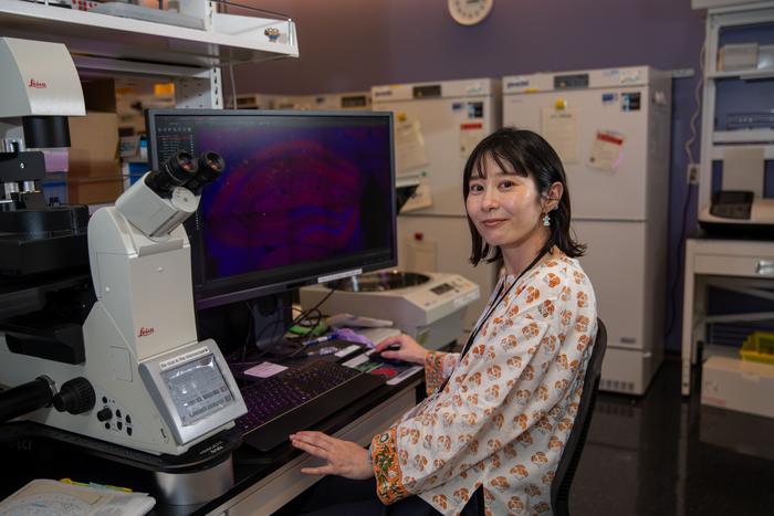
Technician Ai Takahashi was able to use CLEM to observe engrams, in a technical breakthrough for microscopy.
Electron microscopy cannot be used to track changes in the living brain. It is only able to capture brain structures at a single point in time. Therefore, identifying engrams – networks of specific cells and synapses involved in a memory – represents a real challenge. By adding fluorescent tags to engrams and studying brain samples with fluorescent light microscopy, the researchers were able to pinpoint the relevant connections involved, to then study these particular regions in more detail with electron microscopy methods.
By carefully correlating the light microscopy and electron microscopy data sets, the team became the first researchers in the world to identify and image engram synapses by this technique.
Technician Ai Takahashi was able to use CLEM to observe engrams, in a technical breakthrough for microscopy.
Electron microscopy cannot be used to track changes in the living brain. It is only able to capture brain structures at a single point in time. Therefore, identifying engrams – networks of specific cells and synapses involved in a memory – represents a real challenge. By adding fluorescent tags to engrams and studying brain samples with fluorescent light microscopy, the researchers were able to pinpoint the relevant connections involved, to then study these particular regions in more detail with electron microscopy methods.
By carefully correlating the light microscopy and electron microscopy data sets, the team became the first researchers in the world to identify and image engram synapses by this technique.
Credit
Andrew Scott/OIST
Andrew Scott/OIST

No comments:
Post a Comment